Cop
yright
© ABE&M t
odos os dir
eit
os r
eser
vados
.
Expression of SMAD proteins,
TGF-beta/activin signaling
mediators, in human thyroid tissues
Expressão de proteínas SMAD, mediadores da sinalização de TGF-beta/activina, em tecidos de tiroide humana
Sílvia E. Matsuo1, Ana Paula Z. P. Fiore1, Simone M. Siguematu1, Kátia N. Ebina1,
Celso U. M. Friguglietti1, Maria C. Ferro2, Marco A. V. Kulcsar1,3, Edna T. Kimura1
absTRacT
Objective: To investigate the expression of SMAD proteins in human thyroid tissues since the inactivation of TGF-β/activin signaling components is reported in several types of cancer. Phosphorylated SMAD 2 and SMAD3 (pSMAD2/3) associated with the SMAD4 induce the signal transduction generated by TGF-β and activin, while SMAD7 inhibits this intracellular signaling. Although TGF-β and activin exert antiproliferative roles in thyroid follicular cells, thyroid tumors express high levels of these proteins. Materials and methods: The protein expression of SMADs was evaluated in multinodular goiter, follicular adenoma, papillary and follicular carcinomas by immunohistochemistry. Results: The expression of pSMAD2/3, SMAD4 and SMAD7 was observed in both benign and malignant thyroid tumors. Although pSMAD2/3, SMAD4 and SMAD7 exhibited high cytoplasmic staining in carcinomas, the nuclear staining of pSMAD2/3 was not different between benign and malignant lesions. Conclusions: The inding of SMADs expression in thyroid cells and the presence of pSMAD2/3 and SMAD4 proteins in the nucleus of tumor cells indicates propagation of TGF-β/activin signaling. However, the high expression of the inhibitory SMAD7, mostly in malignant tumors, could contribute to the attenuation of the SMADs antiproliferative signaling in thyroid carcinomas. Arq Bras Endocrinol Metab. 2010;54(4):406-12
Keywords
SMAD2/3; SMAD4; SMAD7; thyroid cancer; TGF-β; activin
Resumo
Objetivo: Investigar a expressão de proteínas SMAD em tecidos de tiroide humana desde que a inativação dos componentes da sinalização de TGF-β/activina é relatada em diversos tipos de câncer. SMAD 2 e SMAD3 fosforilados (pSMAD2/3) associados com SMAD4 induzem a trans-missão do sinal gerado por TGF-β e activina, enquanto SMAD7 inibe essa sinalização intracelu-lar. Embora TGF-β e activina exerçam efeitos antiproliferativos nas células foliculares da tiroide, tumores de tiroide expressam altos níveis dessas proteínas. Materiais e métodos: A expressão proteica de SMADs foi avaliada em bócio multinodular, adenoma folicular, carcinomas papilífero e folicular por imuno-histoquímica. Resultados: A expressão de pSMAD2/3, SMAD4 e SMAD7 foi observada tanto em tumores benignos como malignos da tiroide. Embora pSMAD2/3, SMAD4 e SMAD7 exibissem alta positividade citoplasmática em carcinomas, a positividade nuclear de pSMAD2/3 não foi diferente entre lesões benignas e malignas da tiroide. Conclusões: O achado da expressão de SMADs em células tiroidianas e a presença das proteínas pSMAD2/3 e SMAD4 no núcleo de células tumorais indicam propagação da sinalização TGF-β/activina. Contudo, a alta expressão de SMAD7 inibitório, principalmente em tumores malignos, poderia contribuir para atenuação da sinalização antiproliferativa de SMADs em carcinomas de tiroide. Arq Bras Endocrinol Metab. 2010;54(4):406-12
Descritores
SMAD2/3; SMAD4; SMAD7; câncer de tiroide; TGF-β, activina
1 Departamento de Biologia
Celular e do Desenvolvimento, Instituto de Ciências Biomédicas, Universidade de São Paulo (ICB-USP), São Paulo, SP, Brazil
2 Departamento de Morfologia e
Patologia, Pontifícia Universidade Católica (PUC), Sorocaba, SP, Brazil
3 Departamento de Cirurgia
de Cabeça e Pescoço, USP, São Paulo, SP, Brazil
Correspondence to: Edna T. Kimura
Departamento de Biologia Celular e do Desenvolvimento,
Instituto de Ciências Biomédicas, Universidade de São Paulo Av. Prof. Lineu Prestes, 1524 05508-000 − São Paulo, SP, Brazil [email protected]
Cop
yright
© ABE&M t
odos os dir
eit
os r
eser
vados
.
inTRoDucTion
T
hyroid tumors are the most common neoplasms of the endocrine system (1). They are classiied as benign tumors (adenomas and multinodular goiters) and malignant tumors, which are allocated as differen-tiated (papillary and follicular carcinomas) and poorly differentiated (anaplastic carcinoma) (1). The thyroid malignant transformation is dependent on molecular alterations that result in a disruption of several intra-cellular signaling, including the unbalance of TGF-β/ activin inhibitory pathway, by a mechanism not yet clariied (2,3).The TGF-β superfamily is comprised of related proteins including the TGF-βs, activins and bone morphogenetic proteins which affect a wide variety of biological processes, regulating cell proliferation and differentiation, apoptosis and development (4,5). TGF-β acts by binding to speciic serine/threonine kinase transmembrane receptors, which induce intra-cellular signaling mediated by SMAD proteins. The receptors complexed to TGF-β phosphorylate and activate the receptor-regulated SMADs (R-SMAD), SMAD2 and SMAD3. The activation of SMAD2/ SMAD3 allows their interaction with common-me-diator SMAD (co-SMAD), SMAD4, and then this complex moves to the nucleus where it regulates gene transcription. Although activin binds to its own spe-ciic receptors, the same set of SMADs is recruited for its signal transduction (6-10). This TGF-β /acti-vin signaling is attenuated by SMAD7, an inhibitory SMAD (I-SMAD) that prevents phosphorylation of SMAD2/3 and interferes with the formation of com-plex SMAD2/3 and SMAD4 (11,12).
TGF-β and activin are proliferation-inhibitory fac-tors for epithelial cells, such as hepatic, intestinal, mam-mary, pancreatic and prostatic cells including thyroid follicular cells (13-19). Although both TGF-β and acti-vin exert inhibitory effects on the proliferation of nor-mal thyroid follicular cells, we have previously repor-ted that thyroid tumors express the TGF-β isoforms, TGFβ-1, TGFβ-2 and TGFβ-3 as well as activin iso-forms, activin A and activin B (20,21). An impairment of signal transduction has been identiied in the deve-lopment and progression of several epithelial cancers (17,22). Therefore, in this study, we investigated the integrity of TGF-β/activin signaling by analyzing the expression of SMAD2/3, SMAD4 and SMAD7 pro-teins in thyroid tumors.
maTeRiaLs anD meTHoDs
We analyzed the expression of SMAD4, phosphoryla-ted SMAD2/3 and SMAD7, in normal and tumoral thyroid tissue by immunohistochemical method.
Parafin sections from human thyroid of normal tis-sue (N, n = 12), multinodular goiter (MNG, n = 10), follicular adenoma (FA, n = 5), follicular carcinoma (FC, n = 4) and papillary carcinoma (PC, n = 9), were submitted to deparafinization in xylene and hydration through a series of decreasing alcohol concentrations. The immunohistochemical procedure was performed by an indirect 3-stage immunoenzymatic method as described previously (23). Briely, after endogenous peroxidase activity was blocked with 3% hydrogen pe-roxide for 15 minutes, tissues were washed in phospha-te-buffered saline (PBS) and were incubated with goat polyclonal anti-phosphorylated SMAD2/3 antibody (sc-11769, Santa Cruz Biotechnology, Santa Cruz, CA) or rabbit polyclonal anti-SMAD4 (sc-7154, Santa Cruz Biotechnology) or goat polyclonal anti-SMAD7 antibody (sc-7004, Santa Cruz Biotechnology) over-night. The primary antibodies were diluted at 1:100 in Tris-buffered saline and 0.05% albumin. Between the antibodies incubation the tissues were washed in PBS. After incubation with biotin-streptavidin-peroxidase, the reaction was revealed by a mixture of 3,3’-diamino-benzidine with hydrogen peroxide. The sections were then counterstained with Gill’s hematoxylin. The im-munopositivity of the reaction was detected as brown staining observed by light microscopy. The intensity of immunostaining of follicular cells was evaluated and classiied as weak, moderate and strong. Due to variable intensity of positivity seen even in the same tissue, we considered the staining intensity present in more than 50% of positive cells. The percentage of pSMAD2/3 immunopositive cells was determined by counting 3 areas randomly chosen in the tissue in a total of 900 cells. The percentage of nuclear stained cells for pS-MAD2/3 was also determined.
The negative control was performed omitting the primary antibody (SMAD4) and incubating tissues with primary antibody preabsorbed with corresponding pep-tide, pSMAD2/3 (sc-11769 P, Santa Cruz Biotechno-logy) and SMAD7 (sc-7004 P, Santa Cruz BiotechnoBiotechno-logy) peptides (antibody/peptide: 1/5) overnight.
Cop
yright
© ABE&M t
odos os dir
eit
os r
eser
vados
.
followed by the Student Newman-Keuls test to compa-re compa-results between samples. Differences were conside-red signiicant at P< 0.05.
This study was approved by the Ethical Committee for Human Research of the Biomedical Sciences Insti-tute, University of Sao Paulo, Brazil.
ResuLTs
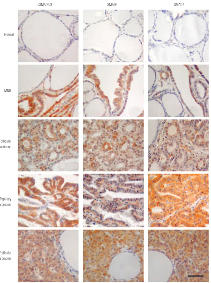
The expression pattern of SMADs in the neoplastic cells of papillary carcinoma was predominantly homo-geneous and strong. In MNG, heterohomo-geneous positi-vity of SMADs was observed in the same tissue with a given follicle displaying positive and negative cells. Phosphorylated SMAD2/3 was detected in all tissues, however its expression is stronger in carcinomas (folli-cular and papillary) (Figure 1). The analysis of immu-nohistochemical staining in the normal and neoplastic tissues is summarized in table 1.
To further investigate the integrity of SMAD signa-ling in thyroid tumors, we evaluated the percentage of pSMAD2/3 positive cells and the localization of this protein at the subcellular level in thyroid tumor tissues (Figure 2A). The immunohistochemical analysis of pS-MAD2/3 expression showed that the number of posi-tive cells for pSMAD2/3 was higher in tumoral samples when compared with normal tissue (Figure 2B). Im-munopositive cells were more abundant in carcinomas and adenomas than in MNG tissues (P< 0.05). When we analyzed the nuclear staining of pSMAD2/3, posi-tivity was about 30% and was similar in MNG, adenoma and carcinoma tissues (Figure 2B). We also detected nuclear positivity of SMAD4 in both benign and
malig-nant lesions, whereas the immunostaining of SMAD7 was predominantly cytoplasmic.
Discussion
TGF-β and activin exert antiproliferative effects in epi-thelial cells, including follicular thyroid cells (24). Ho-wever, TGF-β and activin are overexpressed in several carcinomas (17,19,25). Similarly, we have previously shown high TGF-β and activin protein expression in thyroid cancer, while the expression in normal tissue is predominantly negative (20,21). Despite the increased expression of these proteins in neoplastic cells, loss of TGF-β/activin responsiveness is frequently observed in cancer (2). In human goiter samples, proliferation was not inhibited in the presence of TGF-β (26), proving that even cells derived from benign lesions became ir-responsive to TGF-β. Conversely, we have shown that a papillary thyroid carcinoma cell line is still sensitive to the antiproliferative effect generated distinctly by TGF-β1 and activin, possibly mediated by a functional Smad pathway (27). SMAD2, SMAD3 and SMAD4 are TGF-β/activin intracellular mediators, represen-ting the stimulatory SMADs of this signaling pathway, while SMAD7 is an inhibitory SMAD for this pathway (6-8). In the present study, to verify the integrity of the SMAD pathway, we investigated the expression of SMAD4, pSMAD2/3 and SMAD7 in thyroid tumors. As illustrated in igure 2, SMAD4 and pSMAD2/3 as well as inhibitory SMAD7 are expressed in all of tumo-ral stages of thyroid from benign to malignant lesions, whereas these proteins are rarely detected in normal thyroid tissue. SMAD proteins mediate antiproliferative
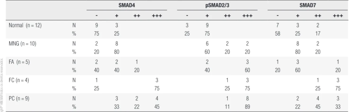
Table 1. Immunohistochemical analysis of SMAD4, pSMAD2/3 and SMAD7
smaD4 psmaD2/3 smaD7
- + ++ +++ - + ++ +++ - + ++ +++
Normal (n = 12) N % 9 75 3 25 3 25 9 75 7 58 3 25 2 17
MNG (n = 10) N % 2 20 8 80 6 60 2 20 2 20 8 80 2 20
FA (n = 5) N % 2 40 2 40 1 20 2 40 3 60 1 20 3 60 1 20
FC (n = 4) N
% 1 25 3 75 1 25 3 75 1 25 3 75
PC (n = 9) N
% 3 33 2 22 4 45 1 11 8 89 2 22 4 45 3 33
Cop
yright
© ABE&M t
odos os dir
eit
os r
eser
vados
.
figure 1. Immunohistochemical staining for pSMAD2/3, SMAD4 and SMAD7 in human thyroid tumors by the peroxidase method. The positivity of immunoreaction is shown by brown staining in representative sections from normal tissue, multinodular goiter (MNG), follicular adenoma, papillary carcinoma and follicular carcinoma. Nuclei were counterstained with hematoxylin, which appear in blue. Bar = 50 µm.
pSMAD2/3 SMAD4 SMAD7
Normal
MNG
Follicular adenona
Papillary carcinoma
Cop
yright
© ABE&M t
odos os dir
eit
os r
eser
vados
.
figure 2. Immunohistochemical analysis of pSMAD2/3 expression in thyroid tumors. (a) Immunohistochemical localization of pSMAD2/3 in multinodular goiter (MNG), follicular adenoma (FA), follicular carcinoma (FC), papillary carcinoma (PC). The cytoplasmic and nuclear positivity of follicular cells is shown by brown staining. The sections were counterstained with hematoxylin and negative nuclei for pSMAD2/3 were stained blue. Bar = 25 µm. (b) Semi-quantitative analysis of pSMAD2/3 immunopositivity. The graph shows the percentage of positive cells in normal tissue (N, n = 12), MNG (n = 10), FA (n = 5), FC (n = 4) and PC (n = 9). * P< 0.05 vs. normal. Black bars in the graph represent the percentage of nuclear positive cells.
150
100
50
0
N MNG FA FC PC
MNG
pSMAD2/3
% positive cells
*
* * *
FA FC PC
a
b
effects by inducing the expression of the CDK inhibi-tors, p15 and p21, and by inhibiting c-MYC expression in several epithelial cell lines responsive to TGF-β or ac-tivin (22,28-30). The loss of TGF-β/activin responsi-veness caused by inactivation of the signaling pathway’s components, such as deletions or mutations in either TGF-β or activin receptors and SMADs, has been iden-tiied in the development and progression of a variety of cancers, conferring a potential tumor suppressive role for the SMADs pathway (2,31,32).
Although the number of pSMAD2/3 positive cells was signiicantly higher in carcinomas (Figure 2B) compared with benign lesion (MNG), the percentage of nuclear positive cells for pSMAD2/3 expression was not different among tumors (Figure 2B). The presence of pSMAD2/3 in the nucleus of neoplastic cells indica-tes that the TGF-β/receptor or activin/receptor com-plex is intact and able to activate downstream mediators of the cascade such as SMADs.
SMAD4 is known as DPC4 (deleted in pancreatic carcinoma locus 4) due to high frequency of deletions in the gene encoding SMAD4 found in pancreatic carci-noma. Inactivating mutations or deletions of this gene in others cancers such as colon carcinomas and, more recently, in thyroid tumors, were also reported (28,33). Differently from what is observed in pancreatic and co-lon carcinomas, in our study, SMAD4 was expressed in most thyroid tumors, displaying high expression mainly in the carcinomas.
Cop
yright
© ABE&M t
odos os dir
eit
os r
eser
vados
.
several carcinomas and is implicated in cancer progres-sion (38-40). In thyroid tumor, we detected increased expression of inhibitory SMAD7 in papillary and folli-cular carcinomas, indicating that SMAD7 may be also involved in thyroid tumorigenesis.
In this study, the presence of nuclear immunopo-sitivity for the stimulatory SMADs, SMAD4 and pS-MAD2/3 indicates that the TGF-β/activin signaling pathway is intact in thyroid tumors. This antiprolife-rative signaling is critical to limit tumor progression. However, the antiproliferative SMADs signal induced by TGF-β/activin proteins could be attenuated by the high expression of an antagonist SMAD, SMAD7, ob-served mostly in thyroid carcinomas.
Acknowledgments: we are grateful to Dr. Alison Colquhoun for cri-tical review of the manuscript. This study was supported by research grants from the Sao Paulo State Research Foundation (Fapesp), Co-ordination for the Improvement of Higher Education Personnel (Ca-pes) and National Counsel of Technological and Scientific Develop-ment (CNPq) and by Fapesp fellowship grants to SEM, APZPF and to KNE, Capes fellowship grants to APZPF and CNPq fellowship grants to SMS. ETK is a CNPq-funded investigator.
Disclosure: no potential conlict of interest relevant to this article was reported.
RefeRences
1. DeLellis RA, Lloyd RV, Heitz PU, Eng C. WHO Classiication of
tu-mours. pathology and genetics of tumours of endocrine organs. Lyon: IARC Press; 2004.
2. Derynck R, Akhurst RJ, Balmain A. TGF-beta signaling in tumor su-ppression and cancer progression. Nat Genet. 2001;29(2):117-29. 3. Massague J, Blain SW, Lo RS. TGFbeta signaling in growth
con-trol, cancer, and heritable disorders. Cell. 2000;103(2):295-309. 4. Derynck R, Feng XH. TGF-beta receptor signaling. Biochim
Bio-phys Acta. 1997;1333(2):F105-50.
5. Moustakas A, Souchelnytskyi S, Heldin CH. Smad regulation in TGF-beta signal transduction. J Cell Sci. 2001;114(Pt 24):4359-69. 6. Pangas SA, Woodruff TK. Activin signal transduction pathways.
Trends Endocrinol Metab. 2000;11(8):309-14.
7. ten Dijke P, Miyazono K, Heldin CH. Signaling inputs converge
on nuclear effectors in TGF-beta signaling. Trends Biochem Sci. 2000;25(2):64-70.
8. Mehra A, Wrana JL. TGF-beta and the Smad signal transduction pathway. Biochem Cell Biol. 2002;80(5):605-22.
9. Shi Y, Massague J. Mechanisms of TGF-beta signaling from cell membrane to the nucleus. Cell. 2003;113(6):685-700.
10. Kimura ET, Matsuo SE, Ricarte-Filho JC. [TGFbeta, activin and SMAD signalling in thyroid cancer]. Arq Bras Endocrinol Metabol. 2007;51(5):683-9.
11. Hayashi H, Abdollah S, Qiu Y, Cai J, Xu YY, Grinnell BW, et al. The MAD-related protein Smad7 associates with the TGFbeta recep-tor and functions as an antagonist of TGFbeta signaling. Cell. 1997;89(7):1165-73.
12. Lebrun JJ, Takabe K, Chen Y, Vale W. Roles of pathway-speciic and inhibitory Smads in activin receptor signaling. Mol Endocri-nol. 1999;13(1):15-23.
13. Roberts AB, Sporn MB. The transforming growth factor-βs. In:
Sporn MB, Roberts AB, editors. Peptide Growth Factors and Their Receptors I. New York: Springer-Verlag; 1991. p. 418-72.
14. Pang XP, Park M, Hershman JM. Transforming growth factor-beta blocks protein kinase-A-mediated iodide transport and protein kinase-C-mediated DNA synthesis in FRTL-5 rat thyroid cells. En-docrinology. 1992;131(1):45-50.
15. Taton M, Lamy F, Roger PP, Dumont JE. General inhibition by transforming growth factor beta 1 of thyrotropin and cAMP res-ponses in human thyroid cells in primary culture. Mol Cell Endo-crinol. 1993;95(1-2):13-21.
16. Franzen A, Piek E, Westermark B, ten Dijke P, Heldin NE. Expres-sion of transforming growth factor-beta1, activin A, and their re-ceptors in thyroid follicle cells: negative regulation of thyrocyte growth and function. Endocrinology. 1999;140(9):4300-10. 17. Gold LI. The role for transforming growth factor-beta (TGF-beta)
in human cancer. Crit Rev Oncog. 1999;10(4):303-60.
18. Schulte KM, Jonas C, Krebs R, Roher HD. Activin A and activin re-ceptors in the human thyroid: a link to the female predominance of goiter? Horm Metab Res. 2000;32(10):390-400.
19. Chen YG, Lui HM, Lin SL, Lee JM, Ying SY. Regulation of cell pro-liferation, apoptosis, and carcinogenesis by activin. Exp Biol Med (Maywood). 2002;227(2):75-87.
20. Kimura ET, Kopp P, Zbaeren J, Asmis LM, Ruchti C, Maciel RM, et al. Expression of transforming growth factor beta1, beta2, and beta3 in multinodular goiters and differentiated thyroid carcino-mas: a comparative study. Thyroid. 1999;9(2):119-25.
21. Matsuo SE, Ebina KN, Kulcsar MA, Friguglietti CU, Kimura ET. Activin betaB expression in rat experimental goiter and human thyroid tumors. Thyroid. 2003;13(3):239-47.
22. Siegel PM, Massague J. Cytostatic and apoptotic actions of TGF-beta in homeostasis and cancer. Nat Rev Cancer. 2003;3(11):807-21. 23. Martins L, Matsuo SE, Ebina KN, Kulcsar MA, Friguglietti CU, Ki-mura ET. Galectin-3 messenger ribonucleic acid and protein are expressed in benign thyroid tumors. J Clin Endocrinol Metab. 2002;87(10):4806-10.
24. Pisarev MA, Thomasz L, Juvenal GJ. Role of transforming gro-wth factor beta in the regulation of thyroid function and grogro-wth. Thyroid. 2009;19(8):881-92.
25. La Rosa S, Uccella S, Marchet S, Capella C, Lloyd RV. Localization of inhibins and activins in normal endocrine cells and endocrine tumors of the gut and pancreas: an immunohistochemical and in situ hybridization study. J Histochem Cytochem. 2004;52(2):217-25. 26. Asmis LM, Kaempf J, Von Gruenigen C, Kimura ET, Wagner HE,
Studer H. Acquired and naturally occurring resistance of thyroid follicular cells to the growth inhibitory action of transforming gro-wth factor-beta 1 (TGF-beta 1). J Endocrinol. 1996;149(3):485-96. 27. Matsuo SE, Leoni SG, Colquhoun A, Kimura ET. Transforming
growth factor-beta1 and activin A generate antiproliferative sig-naling in thyroid cancer cells. J Endocrinol. 2006;190(1):141-50. 28. Ten Dijke P, Goumans MJ, Itoh F, Itoh S. Regulation of cell
prolife-ration by Smad proteins. J Cell Physiol. 2002;191(1):1-16. 29. Ho J, de Guise C, Kim C, Lemay S, Wang XF, Lebrun JJ. Activin
induces hepatocyte cell growth arrest through induction of the cyclin-dependent kinase inhibitor p15INK4B and Sp1. Cell Signal. 2004;16(6):693-701.
30. Burdette JE, Jeruss JS, Kurley SJ, Lee EJ, Woodruff TK. Activin A mediates growth inhibition and cell cycle arrest through Smads in human breast cancer cells. Cancer Res. 2005;65(17):7968-75. 31. Risbridger GP, Schmitt JF, Robertson DM. Activins and inhibins in
endocrine and other tumors. Endocr Rev. 2001;22(6):836-58. 32. Su GH, Bansal R, Murphy KM, Montgomery E, Yeo CJ, Hruban RH,
Cop
yright
© ABE&M t
odos os dir
eit
os r
eser
vados
.
33. Lazzereschi D, Nardi F, Turco A, Ottini L, D’Amico C, Mariani-Costantini R, et al. A complex pattern of mutations and abnor-mal splicing of Smad4 is present in thyroid tumours. Oncogene. 2005;24(34):5344-54.
34. Ishisaki A, Yamato K, Nakao A, Nonaka K, Ohguchi M, ten Dijke P, et al. Smad7 is an activin-inducible inhibitor of activin-induced growth arrest and apoptosis in mouse B cells. J Biol Chem. 1998;273(38):24293-6. 35. Halder SK, Beauchamp RD, Datta PK. Smad7 induces
tumorigeni-city by blocking TGF-beta-induced growth inhibition and apopto-sis. Exp Cell Res. 2005;307(1):231-46.
36. Afrakhte M, Moren A, Jossan S, Itoh S, Sampath K, Westerma-rk B, et al. Induction of inhibitory Smad6 and Smad7 mRNA by TGF-beta family members. Biochem Biophys Res Commun. 1998;249(2):505-11.
37. Itoh S, Landstrom M, Hermansson A, Itoh F, Heldin CH, Heldin NE, et al. Transforming growth factor beta1 induces nuclear export of inhibitory Smad7. J Biol Chem. 1998;273(44):29195-201.
38. Boulay JL, Mild G, Lowy A, Reuter J, Lagrange M, Terracciano L, et al. SMAD7 is a prognostic marker in patients with colorectal cancer. Int J Cancer. 2003;104(4):446-9.
39. Dowdy SC, Mariani A, Reinholz MM, Keeney GL, Spelsberg TC, Podratz KC, et al. Overexpression of the TGF-beta antago-nist Smad7 in endometrial cancer. Gynecol Oncol. 2005;96(2): 368-73.