O
h
r
c
i
r
g
a
in
e
a
s
l
R
e
Özlem Demirpençe1, Mustafa Yıldırım2, Alper Avcı3, Ersin Kılıç4, Havin Bilgetekin5, Hülya Gupse Deniz6, Vildan Kaya7, Hülya Çiçek8 1Department of Biochemistry, Ministry of Health Batman Regional Govermant Hospital, Batman, Turkey, 2Department of Medical Oncology, Medicalpark Gaziantep
Hospital, Gaziantep, Turkey, 3Department of Thoracic Surgery, Ministry of Health Batman Regional Government Hospital, Batman, Turkey, 4 Sağlık Hizmetleri Meslek
Yüksekokulu Tıbbi Lab. Tek. Mardin Artuklu Üniversitesi, Mardin, 5Department of Food Engineering, Rhenish Friedrich-Wilhelms Üniversity Bonn Faculty of Agriculture,
Bonn, Germany, 6Department of Chest Diseases, Ministry of Health Batman Regional Government Hospital, Batman, Turkey, 7Department of Radiation Oncology,
Suleyman Demirel University School of Medicine, Isparta, Turkey, 8Department of Biochemistry, Gaziantep University Hospital, Gaziantep, Turkey Trace Elements in Pleural Efsions
The Role of Trace Elements in the
Malignant-Benign Diferentation of Pleural Efusions
Plevral Efüzyonlarda Malign
Benign Ayrımında Eser Elementlerin Rolü
DOI: 10.4328/JCAM.3366 Received: 05.03.2015 Accepted: 14.04.2015 Printed: 01.05.2016 J Clin Anal Med 2016;7(3): 364-7 Corresponding Author: Mustafa Yıldırım, Department of Medical Oncology, Medicalpark Gaziantep Hospital, 27090, Gaziantep, Turkey.
GSM: +905333948252 F.: +90 3423248860 E-Mail: [email protected] Özet
Amaç: Eser elementlerin bazı kanser türlerinde rol oynadığı öne sürülmekte-dir. Bu çalışmanın amacı plevral sıvıdaki eser elementlerin plevral efüzyonda tanısal yararını incelemektir. Gereç ve Yöntem: Bu çalışmaya, malign ve be-ning plevral efüzyon tanısı olan 38 hasta alındı. Krom, Nikel, Selenyum, Bakır, Kurşun ve Çinko konsantrasyonları örneklerde indüktif eşleşmiş plazma op-tik emisyon spektrometresi ile tespit edildi. Bulgular: Cr, Cu, Ni, Pb, Se ve Zn konsantrasyonları ile malign ve benign efüzyon arasında anlamlı ilişki bulun-madı. Tartışma: Eser elementler, birçok enzim bileşenidir ve bazı kimyasal re-aksiyonlarda katalizör olarak işleve sahiptir. Kanserlerde değişik tip eser ele-mentin eksiklik veya fazlasının ilişkisini gösteren çalışmalar olmuştur. Bizim çalışmamızda, efüzyon ayırıcı tanıda plevra sıvılarında ölçülen eser element-lerin rolü gösterilemedi.
Anahtar Kelimeler
Malign; Plevral Efüzyon; Eser Element
Abstract
Aim: It has been speculated that trace elements may play a role in some type of cancers. The aim of the present study was to examine the diagnos-tic utility of trace elements in pleural luid with pleural efusions. Material and Method: This study consisted of 38 patients diagnosed with malignant and benign pleural efusions. Chrome, nickel, selenium, copper, lead and zinc concentrations in samples were determined by inductively coupled plasma optical emission spectrometry. Results: No signiicant diference was found between malignant and benign efusions with respect to Cr, Cu, Ni, Pb, Se and Zn concentrations in samples. Discussion: Trace elements have func-tion as the component of many enzymes and the catalyst of some chemi-cal reactions. There have been studies demonstrating the association of the deiciency or surplus of trace elements (TEs) with various type of cancers. In our study, the role of TEs measured in the pleural efusions in the diferential diagnosis in the efusion etiology could not be demonstrated.
Keywords
Malignant; Pleural Efusion; Trace Element
Trace Elements in Pleural Efsions
Introduction
Pleural luid is originated from mainly the pleural capillaries of the parietal pleura, lymphatics, intrathoracic blood vessels, pulmonary space and the peritoneal cavity. It is reabsorbed through the lymphatic channels of the parietal pleura [1]. Physiologically, the amount of pulmonary luid should be less than 10 mL in any of the pulmonary cavities. The pleural ef-fusion can be associated with the increase in the production of pleural luid or decrease in its removal, hydrostatic pressure changes, colloidal pressure changes or negative intrathoracic pressure. Because the pulmonary luid accumulation at a clini-cally detectable level may be associated with pleural, pulmonary or non-pulmonary diseases, when the thickness of the pleural luid in the decubitus graphy >10 mm or when the pleural efu-sion is shown by ultrasonography, thoracentesis should be con-sidered for diagnosis. Although the reason of efusion may not be found in all of the pulmonary pathologies, it is determined in 70-80% of the cases. The diferentiation of malignant and benign cases is important for deciding the treatment in pleural efusions [2].
The micro elements for which the daily need is less than 100 mg and which exist in the body luids or tissues at very low concentrations (mg/dL and mg/kg) are deined as the trace ele-ments (TE) and are categorized under micronutrients [3]. The TEs function as the essential cofactor in the physiologi-cal processes. At the same time, some trace elements can be toxic for human health. The TE deiciency or surplus, aside from some chronic diseases such as cardiovascular diseases and dia-betes are also associated with the cancer pathogenesis [4,5]. The environmental exposure of some TEs such as cadmium (Cd), chrome (Cr), nickel C(Ni) and arsenic(As) is classiied under Group 1 of the International Agency for Research on Cancer categories of carcinogen [6]. Again for human beings, lead (Pb, Group 2A) and mercury (Hg) (Group 2B) have been reported as probably suspicious carcinogens [7]. On the other hand, sele-nium (Se) is regarded as an anti-carcinogen TE [8].
In this study, the TE levels of the samples from pleural efusion patients with diferent clinical diagnosis were examined and it was investigated if these levels are diferent in the diferentia-tion between malignant and benign ones.
Material and Method Patient Selection
The patients who were followed for pleural efusion in the Tho-racic Diseases and ThoTho-racic Surgery Clinic of Ministry of Health Batman District Hospital between 2011-2012 were involved in the study. Exclusion criteria were renal or hepatic insuiciency, vascular disease (i.e., peripheral vascular disease, cerebrovas-cular disease), alcohol usage, and intake of supplements con-taining with antioxidants (Cu, Se, Zn or vitamin supplements) or aspirin within 1 week. Subjects taking oral contraceptives, or hormone replacement therapy were also excluded from the study. The patients without suicient samples for analysis were excluded from the study. The TE levels of the samples obtained were measured in the Chemistry Department of Dicle University Faculty of Science and Letters. The patient iles were scanned and the data such as the age, sex, pleural efusion obtainment method were obtained retrospectively.
Obtaining the Samples
The samples to be examined for pleural efusion of the cases were obtained by either thoracentesis, videothoracoscopy or closed thorax drainage methods. The samples remaining ater biochemical and cytological examination were put into 6-mL royal blue top (containing K2EDTA) trace element vacutainer tubes (Becton Dickinson, U.S) and centrifuged at 3000 rpm for 10 minutes. Then, the centrifuged efusion material was put into trace element-free transport tube and the samples were stored at -80 C0. The TE levels were measured from these samples.
TE Measurement
Approximately, 3.0 g of lung liquid were weighted in a beaker and 4.0 mL mixture of HN03:H202 was added and transferred to microwave vessel. It was waited for 20.0 min before micro-wave irridation. The following program was applied to vessel for digestion (Table 1).
Samples were digested in temperature and pressure controlled microwave oven (Berghof MWS-3). Cr, Cu, Ni, Pb, Se and Zn concentrations in samples were determined by Perkin Elmer Optima 2000 DV inductively coupled plasma optical emission spectrometry (ICP-OES) at wavelengths of 267.716, 327.393, 231.604, 220.353, 192.026 and 343.823 nm, respectively. The entire optical system is enclosed and purged with nitrogen.
Statistical Analysis
The SPSS statistical sotware system for Windows (SPSS ver-sion 15.0) was used for the statistical analysis. The variables were investigated using visual (histograms, probability plots) and analytical methods (Kolmogorov-Simirnov/Shapiro-Wilk’s test) to determine whether or not they are normally distributed. Because the Cu and Ni levels were normally distributed, their averages in the benign and malignant patients were evaluated using student t test. Whereas, Cr, Pb, Se, and Zn levels were not normally distributed, they were evaluated via Mann-Whitney U test. A signiicance level of 0.05 (p< 0.05) was used in all tests.
Results
Total 38 patients, of which 28 (73.7%) were male and 10 were female, were involved in the study. The average age of the pa-tients was determined as 61±23 (range 15-92). 23 (60.5%) of the patients with pleural efusion was displaying right-side localisation, 14 (36.8%) of patients let-side and in 1 patient
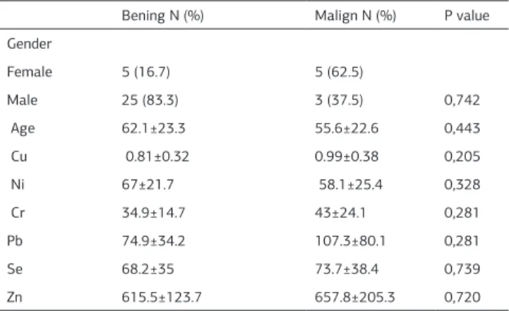
Table 1. According to cytology patients characteristics
Bening N (%) Malign N (%) P value Gender
Female 5 (16.7) 5 (62.5)
Male 25 (83.3) 3 (37.5) 0,742
Age 62.1±23.3 55.6±22.6 0,443
Cu 0.81±0.32 0.99±0.38 0,205
Ni 67±21.7 58.1±25.4 0,328
Cr 34.9±14.7 43±24.1 0,281
Pb 74.9±34.2 107.3±80.1 0,281
Se 68.2±35 73.7±38.4 0,739
Zn 615.5±123.7 657.8±205.3 0,720
Journal of Clinical and Analytical Medicine | 365
Trace Elements in Pleural Efsions
bilateral localisation. In 33 (86.8%) patients, pleural efusions were obtained by thoracentesis, in 4 (10.5%) patients by closed thorax drainage and in 1 (3.2%) patient by videothoracoscopy. The cytology of 30 (78.9%) patients was evaluated as benign and of 8 (21.1%) patients as malignant. A statistically signii-cant relation was determined between the sexes and cytological diagnosis of the patients (P= 0.009). The cytology of 3 (10.7%) male patients was rated as malignant while it was 5 for the female patients. When the patients were grouped by cytology, the average ages of the malignant and benign patients were not diferent (P= 0,443).
In the patients, Cr was determined as 36.6±17 (range 19-99) ng/mL (nanogram /mL) , Cu as 0.85±0.34 (range 0.17-1.63) microgram/mL, Ni as 65.1±22.4 (range 19-101) ng/mL, Pb as 81.7±17 (range 19-99) ng/mL, Se as 64.9±35.2 (range 13-131) ng/mL, and Zn as 624,4±142.3 (range 341-1114) ng/mL . In the patients diagnosed as benign and malignant, the difer-ence with respect to the Cu and Ni levels was analyzed using student t test and no statistically signiicant diference was determined (P= 0.205, P= 0.328, respectively). In the patients diagnosed as benign and malignant, Cr, Pb, Se and Zn levels were analyzed using Mann-Whitney U test and no statistically signiicant inter-group diference was determined (P= 0.281, P= 0.281, P= 0.739, P= 0.720, respectively) (Table 2).
Discussion
In our study, not any correlation between the Cr, Cu, Ni, Pb, Se and Zn levels we measured in the pleural efusions of the pa-tients and the state of being malignant or benign was deter-mined.
Cr exists in all organs of the adults and newborns. The con-centration of Cr is typically high in the lung tissue and has a tendency to increase with age. The increase is associated with the likely inhalation and retention of the water-soluble com-pounds. Cr compounds have been shown to stimulate chromo-somal anomalies and mutations. Cr compounds may cause ge-netic damages such as DNA damage as single-chain fractures, damage in the DNA-protein and DNA-DNA ligaments [9]. In our study, no diference was determined in the Cr levels measured in the pleural efusions of the patients diagnosed as benign and malignant.
In the human body, Cu is the third abundant TE ater Zn and iron (Fe). The normal serum Cu level is 0.6–1.6 microgram/mL [10]. It exists in the structure of many enzymes which take place in many biochemical processes such as antioxidant-prooxidant balance, energy production, metabolism, heme synthesis, iron use and neurotransmitter production [11]. As the cofactor of some antioxidant enzymes eliminating free radicals, Cu is a TE that helps to maintain the cell membrane integrity, slows down the aging process and decreases the cancer risk [12]. Kosova et.al. in their study with benign and malignant thyroid patients have found out that serum Cu level increases in the malignant group and decreases in the benign group [13].
In highly Ni-contaminated environments, exposure causes sev-eral pathological efects. Among these pathologies, skin aller-gies, lung ibrosis and respiratory tract cancers are included. Ni compounds have shown to be carcinogenic for human and animals in many epidemiological studies [14,15]. On the other
hand, in our study it could not be demonstrated that the Ni level is higher in malignant pleural efusions.
The Pb related oxidative stress disturbs the delicate oxidant/ antioxidant balance in the mammals’ cells contributing to Pb in-toxication pathogenesis. In in vivo studies, Pb exposure leads to ROS production and changes the antioxidant defense systems in occupationally exposed workers and in animals [16].
Pb-related oxidative stress has inluences on the cell mem-brane, DNA and the antioxidant defense systems of the cell. In both animal studies and epidemiological studies, Pb exposure has shown to cause oxidative stress in lungs, blood vessels, tes-ticle, sperm, liver, and brain. Oxidative stress also is another mechanism associated with carcinogenesis [17].
Se is a TE that presents at the active center of selenoprotein which has many important duties including the preservation of redox balance [18]. For example, glutathione peroxidase, which protects the body from oxidative damage and from free radi-cal damage, is a selenoenzyme [19]. Se is an essential TE with growth-modulating capacity. Also, in several empirical model systems it was shown to inhibit the growth of the malignant cells [20;21].
In a study from Iran, although not any signiicant diference has been found between the heavy metal accumulation in the samples taken from diferent sites of cancerous tissues of the patients with breast cancer, Se concentration in the tissue has been found maximum as compared to the other elements [22]. Zn takes place in approximately 100 enzyme activities such as polymerase, carbonic anhydrase, superoxide Cu-Zn dismutase. In the genome there exists DNA associated Zn-inger structure. In the human body there is 2-3 g of Zn. Zn deiciency is a com-mon phenomenon in underdeveloped countries. In its deiciency, the immune system, wound healing, the senses of taste and smell and DNA synthesis may be negatively afected [3]. The superoxide dismutase enzyme, of which Zn is the cofactor, plays a key role in the protection of body against free radicals thus inhibiting the initiation and progression of neoplastic phenom-ena [23].
In the study by Martin-Lagos et.al. serum Zn levels in gyneco-logic cancers have been found signiicantly lower than the con-trol group whereas serum Cu levels of cancerous patients were not statistically signiicantly diferent than the control group [24]. There was not a statistically signiicant diference between serum Cu, Zn concentrations of male and female patients and they couldn’t demonstrate any statistically signiicant efect of the patient age on serum Cu and Zn levels. On the other hand in our study, a statistically signiicant relation was determined between the sex and cytological diagnosis of patients.
Pirincci et. al. in their study on the patients with renal cell car-cinoma, have found that serum Pb level is higher in the patient group than the control group whereas Zn level is lower [25]. The role of TEs in various types of cancers has been investi-gated in several studies. Tekşen et.al. have compared the Se, Cu, Zn, and magnesium levels in serum and pleural efusions of malignant patients and non-malignant control group [26]. They have not determined any diference in the result data of malig-nant patients and control group, similar to our study.
In the study of Domej et.al. results similar to our study have been obtained. In that study, no statistically signiicant
difer-| Journal of Clinical and Analytical Medicine 366
Trace Elements in Pleural Efsions
ence has been determined in the serum or pleural luids of the patients with pleural efusion who were diagnosed as benign and malignant [27].
Other than pleural efusion, in a study on the serum and cancer-ous tissues of patients, Zn, Cu, Se, and Fe concentrations have been shown to be high in the malignant tissues. In this study also Cu levels and Cu / Fe and Cu / Zn (in serum and tissue) have been determined at high levels in the advanced malignant tumors [28].
TEs have function as the component of many enzymes and the catalyst of chemical reactions. There have been studies demon-strating the association of the deiciency or surplus of TEs with many type of cancers. In our study, the role of TEs measured in the pleural efusions in the diferential diagnosis in the efu-sion etiology could not be demonstrated. In order to achieve a deinite judgment, we suggest that further studies with greater number of patients should be conducted.
Competing interests
The authors declare that they have no competing interests.
References
1.Villena Garrido V, Ferrer Sancho J, Hernández Blasco L, De Pablo Gafas A, Pérez Rodríguez E, Rodríguez Panadero F et al. Diagnosis and treatment of pleural efu-sion. Arch Bronconeumol 2006;42(7):349-72.
2.Wang XF, Wu YH, Wang MS, Wang YS. CEA, AFP, CA125, CA153 and CA199 in Malignant Pleural Efusions Predict the Cause. Asian Pac J Cancer Prev 2014;15(1):363-8.
3.Fraga CG. Relevance, essentiality and toxicity of trace elements in human health. Mol Aspects Med 2005;26, 235–44.
4.Czerny B, Krupka K, Ożarowski M, Seremak-Mrozikiewicz A. Screening of trace elements in hair of the female population with diferent types of can-cers in Wielkopolska region of Poland. The Scientiic World Journal 2014; DOI: 10.1155/2014/953181.
5.He K . Trace Elements in Nails as Biomarkers in Clinical Research. Eur J Clin Invest 2011;41:98–102.
6.Arita A, Costa M. Epigenetics in metal carcinogenesis: Nickel, Arsenic, Chromium and Cadmium. Metallomics 2009;1(3):222-8.
7.Beyersmann D, Hartwig A. Carcinogenic metal compounds: recent insight into molecular and cel-lular mechanisms. Arch Toxicol 2008;82(8):493-512. 8.Schrauzer GN. Anticarcinogenic efects of selenium. Cell Mol Life Sci 2000;57(13-14):1864-73.
9.Zhitkovich A. Chromium in drinking water: sources, metabolism, and cancer risks. Chem Res Toxicol 2011 Oct 17;24(10):1617-29.
10.Mir MM, Dar NA, Salam I, Malik MA, Lone MM, Yatoo GN et al. Studies on Asso-ciation Be-tween Copper Excess, Zinc Deiciency and TP53 Mutations in Esopha-geal Squamous Cell Carci-noma From Kashmir Valley, India-A High Risk Area. Int J Health Sci (Qassim) 2007;1(1):35-42.
11.Babić Ž, Tariba B, Kovačić J, Pizent A, Varnai VM, Macan J. Relevance of serum copper eleva-tion induced by oral contraceptives: a meta-analysis. Contraception 2013;87(6):790-800.
12.Khanna S, Udas AC, Kumar GK, Suvarna S, Karjodkar FR. Trace elements (cop-per, zinc, sele-nium and molybdenum) as markers in oral sub mucous ibrosis and oral squamous cell carcinoma. J Trace Elem Med Biol 2013;27(4):307-11. 13.Kosova F, Cetin B, Akinci M, Aslan S, Seki A, Pirhan Y et al. Serum copper levels in benign and malignant thyroid diseases. Bratisl Lek Listy 2012;113(12):718-20. 14.Basketter DA, Briatico-Vangosa G, Kaestner W, Lally C, Bontinck WJ. Nickel, cobalt and chromium in consumer products: a role in allergic contact dermatitis? Contact Dermatitis 1993;28(1):15-25.
15.Grimsrud TK, Berge SR, Martinsen JI, Andersen A. Lung cancer incidence among Norwegian nickel-reinery workers 1953-2000. J Environ Monit 2003;5(2):190-7. 16.Monteiro HP, Abdalla DS, Arcuri AS, Bechara EJ. Oxygen toxicity related to ex-posure to lead. Clin Chem 1985;31(10):1673-6.
17.Hsu PC, Guo YL. Antioxidant nutrients and lead toxicity. Toxicology 2002;180(1):33-44.
18.Papp LV, Lu J, Holmgren A, Khanna KK. From selenium to selenoproteins: synthe-sis, identity, and their role in human health. Antioxid Redox Signal 2007;9(7):775-806.
19.El-Bayoumy K. The protective role of selenium on genetic damage and on can-cer. Mutat Res 2001;475(1-2):123-39.
20.Suzuki M, Endo M, Shinohara F, Echigo S, Rikiishi H. Diferential apoptotic re-sponse of human cancer cells to organoselenium compounds. Cancer Chemother Pharmacol 2010;66(3):475-84.
21.Hoeig CS, Renko K, Köhrle J, Birringer M, Schomburg L. Comparison of
difer-ent selenocom-pounds with respect to nutritional value vs. toxicity using liver cells in culture. J Nutr Biochem 2011;22(10):945-55.
22.Mohammadi M, Riyahi Bakhtiari A, Khodabandeh S. Concentration of cd, pb, hg, and se in dif-ferent parts of human breast cancer tissues. J Toxicol 2014; DOI: 10.1155/2014/413870.
23.Johnson MA, Fischer JG, Kays SE. Is copper an antioxidant nutrient? Crit Rev Food Sci Nutr 1992;32(1):1-31.
24.Mártin-Lagos F, Navarro-Alarcón M, Terrés-Martos C, López-G de la Serrana H, López-Martínez MC. Serum copper and zinc concentrations in serum from patients with cancer and cardi-ovascular disease. Sci Total Environ 1997;204(1):27-35. 25.Pirincci N, Gecit I, Gunes M, Kaba M, Tanik S, Yuksel MB et al. Levels of se-rum trace elements in renal cell carcinoma cases. Asian Pac J Cancer Prev 2013;14(1):499-502.
26.Tekşen F, Mungan D, Sayal A, Misurligil Z, Aydin A, Gürbüz L et al. Serum and pleural luid selenium, copper, zinc, and magnesium levels in malignant and non-malignant pleural diseases. Res-piration 1996;63(1):25-7.
27.Domej W, Krachler M, Goessler W, Maier A, Irgolic KJ. Pleural efusions and sera from patients with benign or malignant diseases. Biol Trace Elem Res 2000;78(1-3):13-33.
28.Kuo HW, Chen SF, Wu CC, Chen DR, Lee JH. Serum and tissue trace elements in patients with breast cancer in Taiwan. Biol Trace Elem Res 2002;89(1):1-11.
How to cite this article:
Demirpençe Ö, Yıldırım M, Avcı A, Kılıç E, Bilgetekin H, Deniz HG, Kaya V, Çiçek H. The Role of Trace Elements in the Malignant-Benign Diferentation of Pleural Ef-fusions. J Clin Anal Med 2016;7(3): 364-7.
Journal of Clinical and Analytical Medicine | 367