Contents lists available atScienceDirect
International Journal of Biological Macromolecules
j o u r n a l h o m e p a g e : w w w . e l s e v i e r . c o m / l o c a t e / i j b i o m a c
A novel and efficient and low-cost methodology for purification of
Macrotyloma
axillare
(Leguminosae) seed lectin
M.A. de Santana
a,∗, A.M.C. Santos
a, M.E. Oliveira
b, J.S. de Oliveira
a, E.H. Baba
b,
M.M. Santoro
a, M.H.G. de Andrade
baDepartamento de Bioquímica e Imunologia, Instituto de Ciências Biológicas, Universidade Federal de Minas Gerais, Belo Horizonte, Minas Gerais, MG, Brazil bNúcleo de Pesquisas em Ciências Biológicas, Departamento de Ciências Biológicas, Universidade Federal de Ouro Preto, Ouro Preto, Minas Gerais, MG, Brazil
a r t i c l e
i n f o
Article history: Received 28 May 2008 Accepted 10 July 2008 Available online 25 July 2008
Keywords: Macrotyloma axillare Lectin
GalNac Purification A1 antigen
a b s t r a c t
TheN-acetyl-galactosamine specific lectin fromMacrotyloma axillareseeds (LMA) was purified by precipi-tation and ion exchange chromatography. The LMA 0.2 mol L−1fraction showed hemagglutinating activity
on erythrocytes A1. The results for molecular mass determinations were about 28 kDa. The LMA pH-dependent assays showed best hemagglutinating activity at pH 6.0–8.0; being decreased at acidic/alkaline conditions and by EDTA treatment. LMA is a tetramer at pH 8.2 and a dimer at pH 4.0. Human erythrocytes from ABO system confirmed the A1 specificity for LMA. This new methodology is useful and easy, with low costs, for lectin purification in large amounts.
© 2008 Elsevier B.V. All rights reserved.
1. Introduction
Lectins are a class with several structure-related proteins, which possess considerable specific binding capacity for carbohydrates molecules[1]. They are binding proteins of non-immune origin, with highly ordered three-dimensional structures that can be asso-ciated as dimeric or tetrameric complexes [2]. The leguminous plants, particularly their seeds, are recognized lectins sources[3]. Generally, the lectin basic monomer is described as containing two main-sheets and another one smaller (about to 20–30 kDa). This folding and the features of the carbohydrate-binding site, that consists of a shallow groove on the superimposed-sheets, are very similar in all leguminous lectins[4–6]. Due to the spe-cific binding properties, the lectins can be considered important biotechnological tools with wide applications, such as the ability of agglutinate complex carbohydrates, glycoproteins, erythrocytes, vegetative cells, lymphocytes, fibroblasts, spermatozoids, fungi and bacteria [7,5]. Thus, the lectin in vitro functions are well doc-umented and the molecular basis of this interaction has been studied with a variety of biophysical techniques, including X-ray
∗ Corresponding author at: Departamento de Bioquímica e Imunologia, Instituto de Ciências Biológicas, Universidade Federal de Minas Gerais, Av. Antonio Carlos 6627, Belo Horizonte, MG 31270-901, Brazil. Tel.: +55 31 34092657;
fax: +55 31 34092614.
E-mail address:[email protected](M.A. de Santana).
crystallography, NMR and isothermal titration calorimetry (ITC) [8–10].
The lectin ofMacrotyloma axillare(LMA) was previously isolated by affinity chromatography using antigen A1 + substance H coupled to Sepharose 4BTMresin. LMA hasN-acetylgalactosamine (GalNac)
specificity, similar amino acid composition and the same N-terminal sequence[11]as the lectin isolated fromDolichos biflorus
(DBL)[12,13]. Such as DBL, LMA is specific for A1 human erythro-cytes, being therefore useful for routine blood group identification at blood banks[14,15], in addition to other lectin biotechnology applications that include the GalNac recognition[16,17].
The affinity chromatography techniques generally are the first choice for lectin purification, in accordance to their binding fea-tures[18]but this method can be very expensive and, sometimes, the procedure does not supply enough amounts for industrial appli-cations. The affinity chromatography maintenance is a hardy task when plant crude extracts are loaded into columns because such samples contain pigments, oily components, proteolytic enzymes and other complex substances that could damage the affinity chro-matography system and impair the purification. Considering such problems and the difficulties observed for affinity systems and their importance for lectin purification, the present work proposes a new preparative methodology for the purification ofM. axillareseed lectin, based on thermal and ethanolic precipitation followed by ion exchange chromatography. Such simple and versatile method-ology (accepted patent[19]) yields active lectin amounts applicable for preparative and industrial purposes. Some LMA properties were
studied, such as thermal and pH-dependent stability; molecular mass determination by mass spectrometry (ESI-TOF), SDS-PAGE (denatured form) and by molecular exclusion (native form), since such studies and properties were not fully explored for LMA.
2. Materials and methods
2.1. Reagents and materials
TheM. axillareseeds (“Java” cultivar) were purchased from Mat-sudaSeeds, Brazil. Q-SepharoseTMion exchange and SephacrylTM
S-200 HR 16/60 size exclusion column were purchased from Amersham Pharmacia Biotech (Uppsala, Sweden). Molecular mass standards were obtained from Sigma–Aldrich (St. Louis, MO, USA). All other reagents were of analytical grade or of high quality and purchased from reputed commercial suppliers.
2.2. Lectin extraction from M. axillare seeds—crude extract
M. axillareseeds were powdered with semi-industrial grinder and the powder obtained was stored at−20◦C. For lectin extrac-tion, the crude extract was prepared with 80 g of seed powder and sufficient volume of 0.15 mol L−1NaCl solution with 5 mmol L−1of
MnCl2 and 5.0 mmol L−1 of CaCl2 to complete a final suspension
about 20% (w/v) (400 mL), and incubated at 4◦C for 24 h. After incu-bation, the suspension was filtered and the retained precipitate was washed with the same solution described above until adding up the volume to 400 mL. Then, theM. axillarecrude extract obtained was centrifuged at 4800×gby 30 min at 4◦C for the removal of insoluble fibers. The supernatant was submitted to the next step.
2.3. Thermal precipitation of the crude extract
TheM. axillare20% (w/v) crude extract was submitted to ther-mal precipitation at 85◦C in water bath for 30 min and was soon transferred to a cold ice bath. After this cooling, the thermally treated crude extract was submitted to centrifugation at 4800×g
for 30 min at 4◦C. The precipitate material was discarded and the supernatant was submitted to the next precipitation step.
2.4. Precipitation by cold ethanol
Commercial ethanol was cooled to−20◦C previously to perform the precipitation step. The thermally treated crude extract was sub-mitted to precipitation at 4◦C by addition of increasing ethanol volumes from 20 to 60% (v/v), regarding the initial crude extract volume and the ethanol volume added. The precipitate obtained in the range of 20–60% (v/v) of ethanol was submitted to centrifu-gation at 4800×gfor 30 min at 4◦C. The pellet was resuspended at 0.01 mol L−1of NaHCO
3, pH 8.3, drop by drop. The redissolved
fraction was submitted to centrifugation to discard denatured and insoluble proteins. The supernatant was freeze-dried and stored until the next ion exchange chromatography step.
2.5. Ion exchange chromatography of the fraction precipitated with 20–60% ethanol
The 20–60% ethanol precipitated fraction (500–800 mg aliquots) was dissolved in 10 mL of 0.01 mol L−1of NH
4HCO3, pH 8.3 buffer
and loaded on a HiloadTM 16/10 Q-SepharoseTManion exchange
chromatography column (10.0 cm×1.6 cm) coupled to a Fast Per-formance Liquid Chromatography System (FPLCTMPharmaciaTM)
equilibrated with the same buffer. The elution was performed by stepwise NaCl gradients 0.10; 0.20; 0.30; 0.40; 0.50 and 1.0 mol L−1,
all of them in 0.01 mol L−1 of NH
4HCO3 buffer, pH 8.3. The
chro-matographic flow rate was 3.0 mL min−1, the eluate was monitored
at 280 nm and the hemagglutinating activity was determined (described in Section 2.6). The fractions with A1 hemaggluti-nating activity were collected and submitted to dialysis against 0.01 mmol L−1NH
4HCO3 buffer, pH 8.3. After an exhaustive
dial-ysis procedure (10,000 dilution factor), the active fractions were freeze dried and stored at−20◦C.
2.6. Protein assays, hemagglutination activity (HA) determination and SDS-PAGE analysis
The protein determinations were carried out by the method described by Lowry et al.[20]using bovine serum albumin (BSA) as a standard. The hemagglutination activity (HA) was measured by twofold serial dilutions of the samples on microtitrer plates (96×96 wells) using 2.5% (v/v) of A1 human erythrocytes suspen-sion (treated or not treated with trypsin) in 0.15 mol L−1of NaCl.
The lectin samples were incubated with erythrocytes suspension for 60 min at room temperature and the hemagglutination activity units (HAU) were expressed by the reciprocal of the highest dilu-tion with visible positive hemagglutinadilu-tion. The specific activity (HAU mg−1) was defined as the ratio between HAU mL−1and the
protein concentration mg mL−1. The total activity was calculated
by the product between the total lectin mass (mg) and the specific activity. The purification factor was determined by the ratio of the initial specific activity to the final specific activity.
The samples from each purification step were analyzed on 12.5% SDS-PAGE (10 cm×14 cm plate), in accordance to the methodology previously described[21]. The gels were revealed, by silver[22]or Coomassie Blue staining[23]for purification evaluation and rela-tive molecular mass determination, respecrela-tively. For glycoprotein gel staining, the Schiff periodic acid (PAS) method was used[24].
2.7. Molecular mass determination
The lectin molecular mass was determined by electrospray mass spectrometry (ESI Q-TOF MicroTM, Micromass, UK) in the positive
ion mode. Mass spectrometer calibrations were conducted by using sodium iodide in the 100–3000m/zrange. Lectin samples from the ion exchange final step (20–25g) was solubilized in 50L of 50% (v/v) acetonitrile in water solution plus 0.2% (v/v) formic acid and applied to the mass spectrometer by a syringe pump system at 10L min−1flow rate. The capillary voltage was 3.5 kV and cone
voltage was 60 V. The spectrum data obtained was the result from 30 scans (2.5 s) combined. Original data (m/z) were treated (base-line subtraction, smoothing and centering) and analyzed by the Mass LynxTM4.0 software.
2.8. Determination of the relative mass of the native protein by size exclusion chromatography
The LMA native molecular mass determination was performed by size exclusion chromatography using a SephacrylTM HR
S-200 column (1.6 cm×60 cm) coupled to a FPLCTMSystem at flow
rate of 1.0 mL min−1. Lectin sample solutions of 500L (about
1.5–3.0 mg mL−1) were loaded in the chromatography system at pH
4.0 (0.05 mol L−1acetate buffer) and pH 8.3 (0.01 mol L−1NaH 2CO3)
Table 1
The purification board of results for one purification lot of the crude extract ofMacrotyloma axillareseeds
Step no. Step Total protein (mg) Total activitya(HAUb) Specific activity (HAU) mg−1
Purification factorc Yield (%)
1 Crude extract (CE) 6760 26,062 3.8 1.0 100.0
2 Thermal precipitation (85◦C, 30 min) 4360 25,367 5.8 1.5 97.3
3 Ethanol precipitation (20–60%) 1040 24,960 24 6.2 95.8
4 Ion exchange first fractiond 841 19,511 23.2 6.1 74.9
4 Ion exchange second fractiond 85 3,400 40.0 10.4 13.0
4 Ion exchange third fractiond 13 978 75.3 19.6 3.8
All purification lots (n= 4) performed presented similar results, although small differences were found because of the variability into hemagglutinating activity determination. aThe total activity was obtained by multiplication between the HAU and the LMA total mass for each step.
bThe hemagglutinating activity unit (HAU) was calculated using the reciprocal of the highest title with visible hemagglutination on A1 human erythrocytes. cThe purification factor is defined as the ratio between the initial total activity and the total activity of each step.
dThe first, second and third fraction from the ion exchange chromatography were obtained with 0.2, 0.3 and 0.4 mol L−1NaCl, in this order.
molecular mass and aggregation states determination for LMA at pH 4.0 and 8.0.
2.9. Heat and pH stability for LMA
The heat stability of the LMA was determined by incubation of aliquots of lectin solution (1.3–1.4 mg mL−1) at different
temper-atures (70, 75, 80, 85 and 90◦C) for 10, 20, 30, and 90 min and the remaining hemagglutinating activity (HA) was determined. The results were expressed by relative percentage to a control sam-ple (not thermally treated). For pH stability, 5.0 mL of purified active LMA (2.5–2.8 mg mL−1) were dialyzed for 24 h against buffers
with different pH: pH 2.0–3.0 (0.1 mol L−1glycine buffer); pH 5–6
(0.1 mol L−1sodium acetate buffer); pH 7.0–9.0 (0.1 mol L−1of
Tris-buffer); pH 10.0 and 12.0 (0.1 mol L−1glycine buffer); a 0.15-mol L−1
NaCl solution was employed as control and reference of 100% of activity. After dialysis, the pH of all LMA samples was adjusted to 7.4 by another dialysis procedure for 24 h against 0.1 mol L−1of
ammonium acetate buffer at pH 7.4. The hemaglutination assay was conducted at pH 7.4 and the results were expressed as percentage of residual activity relative to the control.
2.10. Influence of EDTA, Ca(II) and Mn(II) on LMA activity
The LMA purified (1 mg mL−1) was incubated for 10 h against
50 mmol L−1EDTA solution at 4◦C with continuous shaking. After the test, the samples treated with EDTA were dialyzed exhaustively (dilution factor of 10,000) against 0.15 mol L−1of NaCl at 4◦C, and the hemagglutinating activity was assessed before and after the addition of 50 mmol L−1of CaCl
2and MnCl2. As a HA control sample,
one aliquot of the same LMA solution was submitted to dialysis pro-cedure against aqueous solution of NaCl 0.15 mol L−1, 5 mmol L−1
CaCl2and MnCl2(no EDTA treatment, only dialysis effect control).
2.11. Hemagglutinating activity of LMA on the O, A, B and AB human erythrocytes
The LMA specificity on the ABO system (human blood groups A1, A2, B and O) was assessed by using erythrocytes, treated or not with trypsin, from voluntary healthy donors. The HA experi-ment with native cells (not treated with trypsin) was performed as mentioned before. For the erythrocyte trypsin treatment, a 10% (v/v) erythrocyte suspension in 0.15 mol L−1of NaCl was prepared
with 0.1 % (w/v) of commercial trypsin (Merck®
). The tryptic sus-pension was incubated at 37◦C for 70 min with occasional stirring. The trypsin-treated erythrocytes (TTE) was washed three times with 0.15 mol L−1of NaCl solution by centrifugation at 1100×gfor
15 min, each time, followed by suspension at 0.15 mol L−1of NaCl.
The final sediment of TTE was used for 2.5 % (v/v) erythrocytes
suspension (0.15 mol L−1of NaCl) for HA determination of the LMA
purified samples.
3. Results
3.1. First steps of partial purification (crude extract, thermal precipitation of the crude extract and precipitation by cold ethanol)
The comparative results of partial purification of lectins from crude extract, thermal precipitation and ethanol precipitation are shown inTable 1. These procedures were considered satisfactory for the next step of purification.
3.2. Ion exchange chromatography of the 20–60% ethanol precipitated fraction
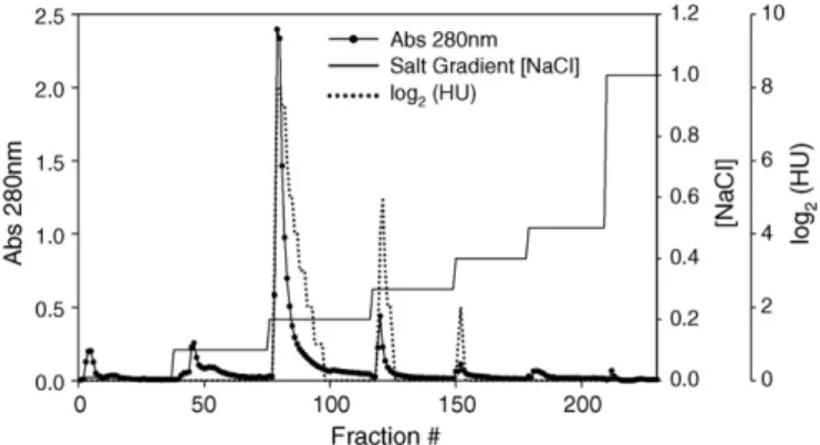
The resultant profile of purification by ion exchange chromatog-raphy (Fig. 1) showed four main peaks with satisfactory resolution among them. More data about this purification step and it efficiency are presented inTable 1.
3.3. Protein assay, hemagglutination activity determination and SDS-PAGE analysis
LMA peak at elution NaCl concentration of 0.2 mol L−1 was
chosen for allhemagglutination activitytests and characterization assays, because it comprises about 70% of the LMA total activity (Fig. 1) and the largest mass percentage, if compared to the other fractions (Table 1). The SDS-PAGE analyses for all fractions have shown the same double-band profiles (Fig. 2), with an average molecular mass value of 28 kDa.
Fig. 2.SDS-PAGE (12.5%) analyses. Lane 1, molecular mass standards (high mass of InvitrogenTM, silver stained); lane 2, crude extract; lane 3, thermally treated crude extract; lane 4, sample after ethanol precipitation at 20–60%; lanes 5–7, LMA 0.2, 0.3 and 0.4 NaCl mol L−1fractions respectively, lanes 2–7 were stained by Silver Staining methodology and Coomassie Blue lanes 8–10 correspond to LMA eluted with 0.2, 0.3 and 0.4 mol L−1NaCl and were stained by the PAS methodology, indicating that the LMA is a glycoprotein.
3.4. Molecular mass determination by size exclusion chromatography and mass spectrometry
Molecular mass of LMA determined by ESI-MS showed single Gaussians curves that, after being processed, give rise to molecular masses from 27 to 28 kDa (Table 2). By size exclusion chromatogra-phy some molecular masses values from 55 to 110 kDa (Table 3) were found depending on the experimental conditions (e.g., pH variation).
Table 2
Molecular mass obtained by ESI-MS (Q-TOF MicroTM, Micromass, UK) for LMA and comparison with results previously published for DBL
LMAa DBLb
“Truncated” monomer (processed) 27,137.8±3.5 27,139
27,186.3±1.7 27,188
27,297.0±8.7 27,349
Intact monomer (not processed) 28,269.8±1.7 28,273
28,316.0±1.3 28,314
28,432
aThe molecular mass data for LMA are represented by average±1 standard devi-ation (n= 5).
bExperimental data obtained from Ref.[29].
Table 3
Relative molecular mass determination for LMA at pH 4.0, 6.0 and 8.0 on SephacrylTM S-200 HR 16/60 column coupled in FPLC system
Condition Molecular mass (kDa)
pH 4.0 54±1
pH 6.0 106±5
pH 8.0 116±3
Flow rate was 1.0 mL min−1and monitored by UV absorbance at 280 nm. The linear
fitting obtained from molecular mass standards was defined by linear regression yielding the following equation:y=−0.0515x+ 5.4978 (R2= 0.9915). The data are
expressed as the average of three independent experiments and one standard devi-ation.
Fig. 3.Thermal stability of LMA lectin at different temperatures (from 60 to 90◦C)
as a function incubation time. The hemoaglutination activity (HA) was expressed as a percentage function of the control HA (not thermally treated).
3.5. Heat and pH stability for LMA
Results for thermal stability of LMA as a function of the incu-bation time are shown inFig. 3. It can be noticed that after 10 min incubation none differences are noted for all temperatures tested, but prolonged incubation led to significant differences between the samples in the assay InFig. 4the residual activity dependence as a function of pH is presented.
3.6. Influence of EDTA, calcium(II) and manganese(II) on LMA activity
The LMA hemagglutination activity on EDTA treatment and in the presence Ca(II) and Mn(II) metal ions are shown inTable 4. The LMA activity is null before EDTA treatment and the hemaglutina-tion activity recovery is uncompleted after addihemaglutina-tion of any divalent metal ions.
The specificity study towards human ABO erythrocytes (Fig. 5) demonstrated that LMA has hemagglutinating activity only for A1 erythrocytes, as it was also verified for DBL[11,12].
4. Discussion
TheM. axillareseed lectin (LMA) was isolated firstly by Haylett and Swart[11]. The purification procedure adopted was affinity
Table 4
EDTA treatment effect at LMA hemaglutination activity on erythrocytes A1
Treatment HAUa
Controlb 272.0±18.5
EDTA 50 mmol−1(pH 7.0) 0
CaCl250 mmol−1(pH 6.8)c 31.0±5.0
MnCl250 mmol−1(pH 6.8)c 0
CaCl2and MnCl250 mmol−1(pH 6.8)c 56.0±9.0
aThe hemaglutination activity units are expressed as the average of four repeats and the standard deviation for each test.
bAqueous solution of 0.15 mol L−1NaCl plus CaCl
2and MnCl2. cAqueous solution of 0.15 mol L−1NaCl.
chromatography using the antigen A1 + substance H coupled to Sepharose 6B (PharmaciaTM). Such procedure was the same
tech-nique used by Etzler and Kabat[12]to purify theD. bifloruslectin (DBL).
The affinity chromatography procedures are frequently used for lectin purification because of the relative facility to bind (covalently link) carbohydrates to insoluble resins employed for this technique. Thus, fast and efficient procedures can be performed for lectins purification in only one step. The isolation by affinity is possible in this case because the lectins do not suffer any modification after binding to carbohydrates[18].
However, the affinity chromatography processes are very expen-sive methods because they need specific materials with high purification degree that elevate substantially the process costs. Besides, the affinity column maintenance can be a difficult task when crude extracts from plant are loaded into the columns. Such samples contain, frequently, many pigments and oily components that can impregnate the resin and damage the affinity columns.
The LMA has the same specific properties of DBL, in this way the procedure based on the precipitation by ammonium sulfate and molecular exclusion chromatography in Sephadex G100TM
followed by ion exchange in Carboxymethyl Cellulose[25]could be a simple alternative method for preparative purification of LMA. However, such process comprises three purification steps with difficult intermediate procedures, requiring desalinization and concentration of large volumes of samples.
The new development strategy of purification resulted in a fast, simple and low-cost procedure that could obtain large amounts of the purified lectin (Table 1). The methodology consists of two
pre-Fig. 5. LMA specificity study on native and trypsin-treated erythrocytes for the ABO antigen system.
cipitation steps (by temperature and by ethanol,Table 1) followed by ion exchange chromatography in Q-SepharoseTMpH 8.3 (Fig. 1).
According to the proposed methodology, the LMA purification yields an average around 0.6% (w/w) (lectin/seed powder), while Haylett and Swart[11]isolated LMA by affinity chromatography, obtaining a 0.9% (w/w) yield of LMA. However, taking into account the readiness to obtain LMA by the new methodology proposed, this apparent yield deficit can be balanced by the methodology easiness and the low cost per purification cycle performed.
The inclusion of the thermal treatment as a purification step turned the crude extract (CE) protein profile simpler and increased the lectin specific activity, because the total mass pro-tein was decreased. Subsequently, the ethanol precipitation step contributed for additional enrichment, as can be seen in the purifi-cation table (Table 1) and SDS-PAGE analysis (Fig. 2). The choice of commercial ethanol for protein precipitation (range of 20–60%) allowed some experimental advantages, such as the recovering of about 95% hemagglutinating activity and the dialysis suppres-sion because ethanol can be removed by evaporation at reduced pressure. Furthermore, the first precipitation steps inactivate pro-teinases (trypsin-like) of the crude extract, thus preventing a possible LMA degradation.
As can be verified in Fig. 2, the protein profile from this step revealed just the LMA bands (relative mass =Mr= 28±1 kDa). The ion exchange chromatography provides the final purification step, yielding three active LMA fractions named LMA 0.2, 0.3 and 0.4 mol L−1elution salt concentration (Fig. 1).
Among them, LMA 0.2 mol L−1 was chosen for all tests and
characterization assays, because it comprises about 70% of the LMA total activity and the largest mass percentage, if compared to the other fractions. Besides, the LMA 0.3 and 0.4 mol L−1revealed
some pigments that, possibly, interfered with the protein assay and with the hemmagglutinating activity determination. More-over, such latter fractions showed not reproducible data when they were submitted to different blood group assays using trypsin-treated erythrocytes (data not shown) while the LMA 0.2 mol L−1
had shown the expected results. On the other hand, the SDS-PAGE analyses for all fractions have shown the same double-band pro-files (Mr= 28±1 kDa,Fig. 2), which indicates to be the same LMA but with different charge density, considering ion exchange profile. Thus, only LMA 0.2 mol L−1was used for all assays and from now,
it will be referred just as LMA.
The total LMA activity recovery from four repetitions of this purification methodology demonstrated similar results, andTable 1 is a representative and reproducible example of the purification. The LMA hemagglutinating activity remained unaffected at tem-peratures up to 50◦C for 90 min. This finding is in agreement with the high values of the denaturation temperature observed for LMA at differential scanning calorimeter (DSC) experiments (melting temperature-Tm, about 100◦C at pH 6.2, data not shown). This
results show that LMA has high thermal stability. The thermally treated crude extract did not suffer significant HA decrease on heating up to 85◦C for 30 min (Fig. 3). It is important to point out that the temperature test around 85◦C is 15◦C lower than theTm obtained by DSC experiments (100◦C), therefore the LMA at 85◦C is 100% of native state. Moreover, the SDS-PAGE of this purification step revealed good refinements at the protein profile (Fig. 2) with increased specific activity (Table 1).
at acid pH range (<5.5), as well as at basic pH range (>8.0). On LMA purification using affinity chromatography, Haylett and Swart[11] proposed the decreasing of pH up to 5.0 as an alternative to perform LMA elution, because to removeD. bifloruslectin (DBL) from the affinity column[12]. GalNac is useful in analytical scale but more expensive and not applicable for preparative chromatography. In the present study, it was verified that the pH decreasing affects the HA when LMA is incubated for long periods (24 h), thus the purifi-cation procedures for LMA must be made near neutral pH (6.0–8.0) for total HA maintenance.
The LMA activity shows to be dependent of divalent metal ions (Table 4). The EDTA treatment completely removed the LMA hemagglutinating activity in all the experiments performed (n= 4). The HA assessed before the treatment with CaCl2 and MnCl2 at
50 mmol L−1 (dilution factor of 10,000), both together or
sepa-rated, revealed that the total activity is not recovered, remaining about 20% of the total activity if compared with the control samples (n= 4). Interestingly, the Ca(II) addition alone induced a recovering of about 11% of LMA activity as compared to the control, which is similar to that previously observed with the DBL activity[26]. Such results suggest that the LMA requires both ions for optimal hemag-glutinating activity. As discussed previously[4,27], the Ca(II) and Mn(II) metal ions, although not directly involved, are necessary for carbohydrate binding to leguminous lectins (metaloproteins). Indeed, the metal ion removal process forErythrina speciosalectin [28], a Gal/GalNac lectin, led to a complete loss of hemagglutinat-ing activity, which is in agreement with our LMA results, but the HA recovery was complete when Ca(II) and Mn(II)+were added to
E. speciosalectin. Such different results could be related to LMA incapacity to recover the proper topological carbohydrate bind-ing site, thus the complete activity could not be measured. In fact, Haylett and Swart[11]showed that the LMA has other divalent metal ion species, such as Mg(II) and Zn(II) present at different pro-portions, however not structurally identified. However the addition of these metal ions (at 5 mmol L−1) did not cause a full recovering of
LMA activity after the EDTA treatment, even with Ca(II) and Mn(II) present. Additionally, it must be pointed out that long dialysis pro-cedures (>36 h) decreased the LMA activity by up to 10%. Thus, the LMA HA recovery could not be determined with real accuracy, even regarding control samples that had gone through the same dialysis procedure.
The molecular mass of the DBL monomers, determined by ESI-TOF, showed two distinct mass groups with mass difference about 1129, on average[29]. Such difference is due to the processing that takes place on the monomeric C-terminal and this cleavage is nec-essary for the proper DBL assembly. This processing removes about 10–11 amino acids from the C-terminus (an␣-helix segment)[29]. The LMA molecular mass determination by ESI-TOF showed the same profile as that determined earlier for DBL[31](Table 2).
The mass spectra data from LMA were grouped in the same way as performed for the DBL data, i.e., the molecular mass data were separated according to their dimensions considering 50 as a min-imal relative mass difference. In this way, LMA can be considered identical to DBL. In fact, Haylett[11]demonstrated 100% of homol-ogy between the first fifty amino acid that comprise the N-terminal sequence of the two lectins. Thus, our mass data reinforce a possible identity between DBL and LMA.
The LMA relative molecular mass estimated by size exclusion chromatography (native state) revealed that the LMA has a pH-dependent oligomeric behavior. At acid pH (4.0) the molecular mass is close to a dimeric form (about 55 kDa), while at neutral and neu-tral and slightly alkaline conditions pH (6.0–8.0) the mass is close to a tetrameric form (about 110 kDa), as it could be seen inTable 3. These behaviors share similarity with other leguminous lectins reported earlier[30,31], which showed that the lectin monomeric
subunits can assume different aggregation forms depending on pH. The tetramer form at a slightly alkaline condition is in agreement with earlier results for LMA[11], when the relative mass about 104 kDa at pH 7.4 was found through size exclusion chromatog-raphy on SephadexTMG200 (40.0 cm×1.4 cm) column.
The specificity study towards human ABO erythrocytes (Fig. 5) demonstrated that LMA has hemagglutinating activity only for A1 erythrocytes, as it was also verified for DBL[11,12]. However, when the erythrocyte suspension treated with trypsin was used, it was observed a significant titre increase for HA, but with A2 subgroup positive. This result was expected because the GalNac antigen is the determinant carbohydrate for A group. The only difference is in the antigen quantities and qualities, but the higher titre observed for A1 subgroup defines the A1 specificity for LMA[14].
5. Conclusions
The proposed purification methodology is easy to perform, avoiding the use of the expensive affinity chromatography. Large amounts of LMA can be purified by this new methodology and the yields are comparable with earlier methodology values reported for LMA isolation. Thus, the industrial process for LMA purification can be adopted using the methodology described herein, with eco-nomic advantages, because the equipments and reagents used are widespread at biotechnology factories. Due to the direct industrial applicability of the LMA purification, provided that the LMA has the same GalNac specificity as does DBL (used for A1 blood type determination at blood bank) a patent of the process was submit-ted and accepsubmit-ted at National Institute of Intellectual Property from Brazil in 2004. LMA has a significant thermal stability (up to 85◦C for 30 min), what could enable the thermal treatment as the first step of purification, since most of the total activity was recovered with a substantial increment of the specific activity. As verified for other legume lectins, the LMA is Ca(II), Mn(II) and pH dependent for hemagglutinating activity, with full activity in the pH range from 6.0 to 8.0. In this pH range, the LMA tetramer prevails at neutral to slightly alkaline range (>6.0), while the dimeric form prevails in the acid pH range (<4.0).
Acknowledgements
The authors would like to thank the CAPES and CNPq by fel-lowship and the research supporting and the Núcleo de Estrutura e Func¸ão de Biomoléculas (UFMG) for mass spectrometry analyses (FAPEMIG EDT 24000) and Rodrigo Silva Reston and Dr. Marcelo P. Bemquerer for discussions and manuscript revision.
References
[1] S. Elgavish, B. Shaanan, Trends Biochem. Sci. 22 (1997) 462–467. [2] N. Sharon, Trends Biochem. Sci. 18 (1993) 221–226.
[3] C.R. Carlini, M.F. Grossi-de-Sa, Toxicon 40 (2002) 1515–1539.
[4] R. Loris, T. Hamelryck, J. Bouckaert, L. Wyns, Biochim. Biophys. Acta. 1383 (1998) 9–36.
[5] M. Vijayan, N. Chandra, Curr. Opin. Struct. Biol. 9 (1999) 707–714. [6] N. Sharon, H. Lis, J. Agric. Food Chem. 50 (2002) 6586–6591.
[7] R.S. Singh, A.K. Tiwary, J.F. Kennedy, Critic. Rev. Biotechnol. 19 (1999) 145–178. [8] R. Loris, Biochim. Biophys. Acta-Gen. Subjects 1572 (2002) 198–208. [9] N. Sharon, Cell. Mol. Life Sci. 62 (2005) 1057–1062.
[10] T.K. Dam, C.F. Brewer, Chem. Rev. 102 (2002) 387–429. [11] T. Haylett, L.S. Swart, South Afr. J. Chem. 35 (1982) 33–36. [12] M.E. Etzler, E.A. Kabat, Biochemistry 9 (1970) 869–876. [13] M.E. Etzler, C.F. Talbot, P.R. Ziaya, FEBS Lett. 82 (1977) 39–41.
[14] J.B. Henry, Clinical Diagnosis and Management by Laboratory Methods, John Bernard Henry, Philadelphia, 1996.
[15] N. Sharon, H. Lis, Glycobiology 14 (2004) 53R–62R. [16] H. Lis, N. Sharon, Annu. Rev. Biochem. 55 (1986) 35–67.
[19] M.H.Guerra de Andrade, E.H.Baba, and M.A.Santana. Purification and isolation of lectin ofMacrotyloma axillareby obtaining lectin ofMacrotyloma axillare through isolation of lectin by thermally treating brute extract of leguminous plant seeds, UNIVERSIDADE FEDERAL OURO PRETO [BR200401517-A], 2004. [20] O.H. Lowry, N.J. Rosebrough, A.L. Farr, R.J. Randall, J. Biol. Chem. 193 (1951)
265–275.
[21] U.K. Laemmli, Nature 227 (1970) 680–694.
[22] W. Wray, T. Boulikas, V.P. Wray, R. Hancock, Anal. Biochem. 118 (1981) 197– 203.
[23] P.J. Wirth, A. Romano, J. Chromatogr. A 698 (1995) 123–143.
[24] G. Fairbank, T.L. Steck, D.F.H. Wallach, Biochemistry 10 (1971) 2606–2613. [25] J. Font, A.M. Leseney, R. Bourrill, Biochim. Biophys. Acta 243 (1971) 434–446.
[26] C.A.K. Borrebaeck, B. Lonnerdal, M.E. Etzler, Biochemistry 20 (1981) 4119–4122. [27] T.W. Hamelryck, R. Loris, J. Bouckaert, M.H. Dao-Thi, G. Strecker, A. Imberty, E.
Fernandez, L. Wyns, M.E. Etzler, J. Mol. Biol. 286 (1999) 1161–1177.
[28] E.H.E. Konozy, E.S. Bernardes, C. Rosa, V. Faca, L.J. Greene, R.J. Ward, Arch. Biochem. Biophys. 410 (2003) 222–229.
[29] N.M. Young, D.C. Watson, M. Yaguchi, R. Adar, R. Arango, E. Rodriguez-Arango, N. Sharon, P.K. Blay, P. Thibault, J. Biol. Chem. 270 (1995) 2563–2570. [30] K.J. Lightwahl, B.L. Schwartz, R.D. Smith, J. Am. Chem. Soc. 116 (1994)
5271–5278.