Vol.48, n. 5 : pp. 705-716, September 2005
ISSN 1516-8913 Printed in Brazil BRAZILIAN ARCHIVES OF BIOLOGY AND TECHNOLOGY
A N I N T E R N A T I O N A L J O U R N A L
Isolation and Partial Characterization of a
D-Galactose-Binding Lectin from the Latex of
Synadenium carinatum
Maria Aparecida Souza
*, Francielle Amâncio-Pereira, Cristina Ribeiro Barros Cardoso,
Adriano Gomes da Silva, Edmar Gomes Silva, Lívia Resende Andrade, Janethe Deolina
Oliveira Pena, Henrique Lanza and Sandra Regina Afonso-Cardoso
Laboratório de Imunologia; Instituto de Ciências Biomédicas; Universidade Federal de Uberlândia; Av. Pará, 1720; Bloco 4C; Campus Umuarama; 38400-902; Uberlândia - MG - Brasil
ABSTRACT
A lectin from the latex of Synadenium carinatum was purified by affinity chromatography on immobilized-D-galactose-agarose and shown to be a potent agglutinin of human erythrocytes. The haemagglutination of human red cells was inhibited by 3.0 mM N-acetyl-D-galactopyranoside, 6.3 mM methyl-β-D-galactopyranoside, 50 mM methyl-α-D-galactopyranoside and 50 mM D-fucose but not by L-fucose, demonstrating an anomeric and a conformational specificity. According to SDS-PAGE analysis, the lectin appeared to be a glycoprotein composed of two polypeptide chains of ca. 28 and 30 kDa, but size exclusion chromatography (Sephadex G-100) and native PAGE revealed a protein of apparent molecular weight 120 - 130 kDa made up of 28 and 30 kDa subunits. The lectin was stable in the range pH 6 - 9, and 4 - 56oC. The N-terminal sequence of the 30 kDa subunit contained the conserved consensus sequence GPN observed in other D-galactose-binding lectins found in latex of members of the Euphorbiaceae.
Key words: D-Galactose-binding, lectin, latex, Synadenium carinatum
*
Author for correspondence
INTRODUCTION
Lectins are a complex and heterogeneous group of proteins with diverse molecular structures, biochemical properties and carbohydrate-binding specificities (Licastro et al., 1993). They are widely distributed in nature, particularly in the plant kingdom where they can be found in seeds, leaves, bark, bulbs, rhizomes, roots, cotyledons and tubers (Cavada et al., 1998; Witisuwannakul et al., 1998; Yagi et al., 2002; Oliveira et al., 2002; Konozy et al., 2002, 2003). Seeds of leguminous species often contain large amounts of lectins that are similar to
those present in other tissues of the same plant, including the latex.
thus involved in diverse mechanisms such as endocytosis, intracellular translocation of glycoproteins, cellular regulation, migration and adhesion, phagocytosis and the binding of micro-organisms to host cells (Sharon and Lis, 1993). Lectin specificity is usually defined according to the mono- or oligo-saccharide that is able to inhibit the agglutinating activity induced by the lectin. Many galactose-specific lectins have been isolated from plants. A particularly rich source of such lectins is the latex of members of the Euphorbiaceae, for example, Hura crepitans L., Euphorbia characias L. (Barbiere et al., 1983), E. marginata (Stirpe et al., 1993), E. neriifolia (Seshagirirao and Prasad, 1995), E. milii (Dias-Baruffi et al., 2000), Hevea brasiliensis (Gidrol et al 1994; Witisuwannakul et al., 1998; Rojas et al., 2001), Synadenium grantii (Premaratna et al., 1981), and S. cupulare (Jager et al., 1996).
The plant Synadenium carinatum (Euphorbiaceae) is common in ornamental gardens in Brazil, and an aqueous preparation of the latex has been used in popular medicine to treat a number of diseases. In the present study, we describe the isolation and partial characterization of a D-galactose-binding lectin present in the aqueous extract of the latex of this species.
MATERIALS AND METHODS
Extraction of latex protein
Plants of S. carinatum were authenticated by Dr Glein Monteiro Araujo (Instituto de Biologia, Universidade Federal de Uberlândia, MG, Brazil) and a voucher specimen is located in the herbarium of this institution. Proteins were extracted from fresh latex by gentle shaking with deionised water, in the proportion 1:5, for 48 h at 4oC. The mixture was centrifuged (3500 x g, 30 min, 4º C; Eppendorf centrifuge) and filtered through nitrocellulose membranes (0.45 µm pore size; Merck, Göttingen, Germany) to yield a crude extract. Protein concentration was determined according to the procedure of Lowry et al. (1951), and the extract was stored at -20°C until required for assay.
Haemagglutination assay
Fresh human, mouse, rabbit, and horse erythrocytes were used in the initial haemagglutination assay. For all subsequent assays, human erythrocytes of type A Rh+ (A+) were employed. Following the approval of the Ethical Committee of the Universidade Federal de Uberlândia, (approval number 064/2001), erythrocytes were collected from healthy volunteers and separated from the platelet-rich plasma and buffy coat by differential centrifugation (500 x g, 15 min, 25oC). Red cells were washed in 0.15 M NaCl and then re-centrifuged (1000 x g, 10 min, 25oC). The haemagglutination titre was assayed by preparing two-fold serial dilutions of the crude extract (1:2 to 1:1024) in V-well microtitre plates (50 µl) and adding 25 µl of a fresh erythrocyte suspension (2%) in 0.15 M NaCl. After 1 h at room temperature, when the erythrocytes had fully sedimented, each well was examined for agglutination. The haemagglutination titre was defined as the reciprocal of the largest dilution that was able to induce visible erythrocyte agglutination.
Assay of inhibition of haemagglutination
Gel filtration chromatography
A sample of crude extract (20 mg) was applied at room temperature to a column (85 x 2.5 cm i.d.) containing 450 ml of Sephadex G-200 (Amersham Pharmacia Biotech, Uppsala, Sweden) in 0.02 M Tris buffer at pH 7.2 (TBS). The column was eluted with TBS at a flow rate of 0.285 ml/min and fractions were collected and monitored spectrophotometrically at 280 nm. Fractions associated with each peak were pooled, concentrated, dialysed against water using Amicon YM 10kDa membrane (Millipore Corp., Belford, MA, USA) and then analysed by SDS-PAGE. In order to determine the molecular weight of the lectin, size exclusion chromatography on a column (26 x 0.9 cm i.d.) of Sephadex G-100 (Amersham Pharmacia Biotech) was carried out. Purified lectin (2 mg) obtained by affinity chromatography (see below) was applied to the gel, which had previously been equilibrated with TBS at room temperature, and eluted at a flow rate of 0.16 ml/min: the collected fractions were monitored spectrophotometrically at 280 nm.
Affinity chromatography
Galactose-binding lectin was purified on immobilised D-galactose-agarose (Pearce, Rockford, IL, USA) equilibrated with 0.05 M borate buffer at pH 7.2 (BBS). The lectin was eluted with 0.4 M D-galactose in BBS (BBS-D-Gal): the effluent was pooled, concentrated and dialysed against TBS.
Cation exchange chromatography
The purified lectin (2 mg/ml) was subjected to cation exchange chromatography on a CM-Sepharose CL6B (Amersham Pharmacia Biotech) column equilibrated with 0.01 M sodium acetate (pH 5.0). After elution of the non-adsorbed proteins, a linear gradient of NaCl (0 to 2 M) in sodium acetate buffer was applied and the effluent monitored spectrophotometrically at 280 nm: fractions collected were dialysed against TBS.
Polyacrylamide gel electrophoresis
Sodium dodecyl sulphate - polyacrylamide gel electrophoresis (SDS-PAGE) was carried out under denaturing conditions using 12 or 15% homogeneous polyacrylamide gels and the discontinuous Tris-glycine system of Laemmli
(1970). In addition, 8% native PAGE was performed using Tris-glycine alkaline (pH 8.3) buffer (Davis, 1964). Proteins were visualised by silver staining (Heukeshoven and Dernick, 1988), and periodic acid-Schiff (PAS) staining reagent (Glossmann and Neville, 1971) was used to detect glycosidic linkages in glycoproteins. The molecular markers employed were phosphorylase B (97 kDa), bovine serum albumin (66 kDa) ovalbumin (43 kDa) carbonic anhydrase (29 kDa) trypsin inhibitor (18 kDa), lysozyme (14 kDa), aprotinin (6.3 kDa), and insulin (b) chain (3.4 kDa), all from Amersham Pharmacia Biotech.
Red blood cells overlay assay
Following separation by 15% SDS-PAGE, proteins on the gel were electro-transferred onto Immobilon-NC (Millipore) nitrocellulose transfer membranes using a semi-dry system (Amersham Pharmacia Biotech) operating at 0.8 mA/cm2 for 2h. The membrane was incubated in TBS containing 1% Triton X-100 for 1 h at room temperature, washed three times with TBS and subsequently incubated with TBS containing 1% bovine serum albumin (TBS-BSA) for 1 h at room temperature. The membrane was then incubated with a human erythrocyte suspension (2%) in TBS-BSA for 1 h at room temperature with gentle shaking. After washing, the membrane was fixed for 10 min in 3% buffered formalin in TBS.
Effect of temperature and pH on haemagglutination
The influence of temperature on the haemagglutinating activity of the lectin was determined by incubation of aliquots of the purified protein at 4, 25, 37, 56 and 95oC for 30 min prior to assay. The pH sensitivity of the lectin was established by incubating aliquots of the purified protein for 1 h in buffers at pH values 3-12: the haemagglutinating activity of the lectin was then measured after adjusting the pH of the assay solution to 7.0
Analytical isoelectric focusing (IEF)
isoelectric-focusing protein calibration kit (Pharmalyte 3-9, Pharmacia).
Sequencing N-terminal amino acids
A sample of the affinity-purified lectin was run on a 15% SDS-PAGE under denaturing, but not reducing, conditions and transferred to a PVDF membrane (Bio-Rad, Hercules, CA, USA). The blot was stained with Coomassie Brilliant Blue and the 30 kDa band was submitted to N-terminal sequencing by the Edman degradation method in an automatic sequencer model ABI 477A (Amersham Pharmacia Biotech, Freiburg, Germany). Sequence homologies were searched within an on-line protein database (http://srs.ebi.ac.uk).
Cytotoxicity assay
In order to determine if the crude latex extract or the isolated lectin exhibited cytotoxic activity, an assay was performed using J774.A1 cells and peritoneal murine macrophages. Peritoneal exudate cells (PEC) from BALB/c mice, previously inoculated with 3% sodium thioglycolate medium, were harvest with RPMI-1640 medium. PEC were seeded at 4 x 10⁶ cells/well into 24 well tissue culture plates and incubated in 5% CO2 for 2 h at 37ºC. Non-adherent cells were removed by washing vigorously with RPMI-1640 medium. Adherent cells were incubated in 1 ml of RPMI-1640 medium supplemented with 10% foetal bovine serum (Cultilab, Campinas, Brazil), 100 U/ml
penicillin, 100 µg/ml streptomycin and 2 mM L-glutamine (Sigma), in the presence or absence of Escherichia coli lipopolysaccharide (Sigma) (10 µg/ml), or crude latex extract or purified lectin (1, 2, 5, 10, 20, 30, 50, 100 or 200 µg/ml). After incubation for 24, 48 and 72 h in 5% CO2 in a humidified chamber at 37oC, cells were collected and their viabilities determined by Trypan Blue exclusion (Sigma).
RESULTS
Analysis of the crude extract
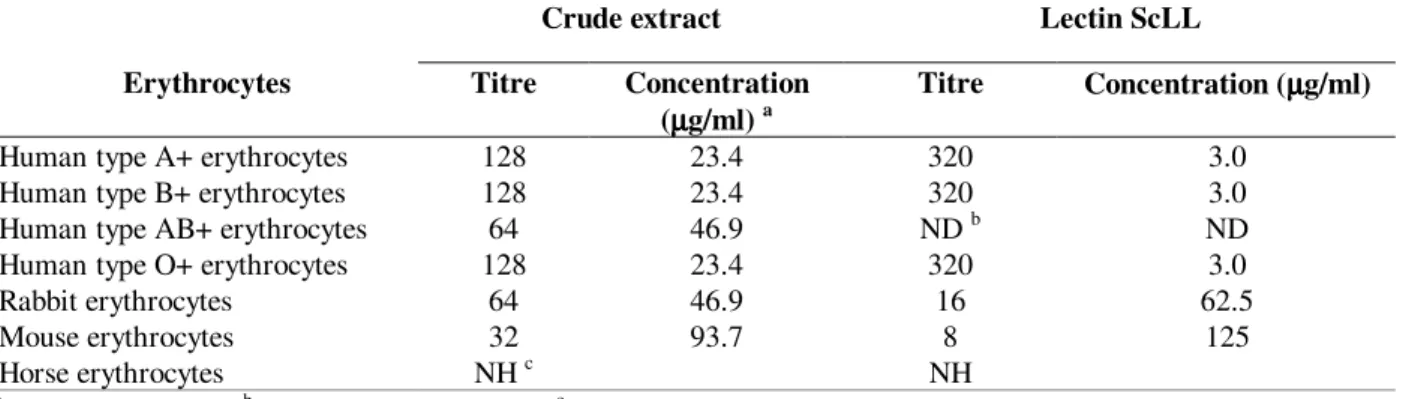
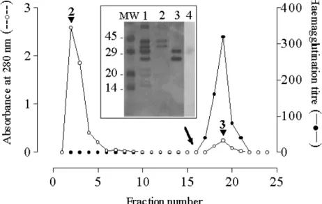
The crude extract of the latex of S. carinatum agglutinated all human blood groups and erythrocytes from rabbit and mouse, but not from horse (Table 1), revealing the presence of a lectin that was subsequently named ScLL. The crude extract was separated into five fractions on Sephadex G-200 (Fig. 1) each of which was subjected to SDS-PAGE and assayed for haemagglutinating activity. The protein profile revealed on the gel (Fig. 1 insert; lanes 1-5) was compatible with that exhibited by gel filtration, but only the fraction associated with the third peak showed haemagglutinating activity (Fig. 1; blocked triangles) against human A+ erythrocytes with a titre of 16.
Table 1 - Haemagglutination assay
Crude extract Lectin ScLL
Erythrocytes Titre Concentration
(µµµµg/ml) a
Titre Concentration (µµµµg/ml)
Human type A+ erythrocytes 128 23.4 320 3.0
Human type B+ erythrocytes 128 23.4 320 3.0
Human type AB+ erythrocytes 64 46.9 ND b ND
Human type O+ erythrocytes 128 23.4 320 3.0
Rabbit erythrocytes 64 46.9 16 62.5
Mouse erythrocytes 32 93.7 8 125
Horse erythrocytes NH c NH
aµ
g/ml of the protein;b ND - assay not performed; cNH - no haemagglutination observed
Assay of inhibition of haemagglutination
The level of inhibition by various carbohydrates of the haemagglutinating activity against human A+ erythrocytes was assessed in order to determine the carbohydrate-affinity of the lectin. It was observed
Figure 1 - Gel filtration (Sephadex G-200) chromatogram of an aqueous extract of the latex of
Synadenium carinatum and the haemagglutination titres of the components. The eluted fractions were collected (fraction size - 2 ml; flow rate - 0.5 ml/min) and monitored spectrophotometrically at 280 nm (----); the asterisk corresponds to the void volume of the column. For each peak, fractions with the highest absorbance values were analysed for haemagglutinating activity ( ✁✂✄☎✆✝✞ ✟✆✠✡✟☎☛
☞ ✟✡☎✆
electrophoretic profiles (12% SDS-PAGE; proteins visualised by silver stain) of the five eluted peaks: lanes 1-5 correspond to peaks 1-5, respectively, in the chromatogram, whilst lane 6 corresponds to the original crude aqueous extract.
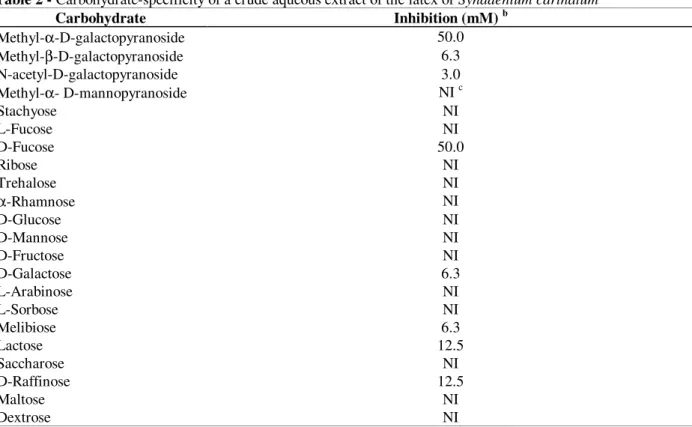
Table 2 - Carbohydrate-specificity of a crude aqueous extract of the latex of Synadenium carinatuma
Carbohydrate Inhibition (mM) b
Methyl-α-D-galactopyranoside 50.0
Methyl-β-D-galactopyranoside 6.3
N-acetyl-D-galactopyranoside 3.0
Methyl-α- D-mannopyranoside NI c
Stachyose NI
L-Fucose NI
D-Fucose 50.0
Ribose NI
Trehalose NI
α-Rhamnose NI
D-Glucose NI
D-Mannose NI
D-Fructose NI
D-Galactose 6.3
L-Arabinose NI
L-Sorbose NI
Melibiose 6.3
Lactose 12.5
Saccharose NI
D-Raffinose 12.5
Maltose NI
Dextrose NI
a
The haemagglutination assay was performed using human type A+ erythrocytes b Minimum concentration of carbohydrate required completely to inhibit the haemagglutinating activity (4 units) of the crude extract
c
Thus activity was completely eliminated by N-acetyl-D-galacto-pyranoside and methyl-β -D-galacto-pyranoside at 3.0 and 6.3 mM, respectively, and by methyl-α-D-galacto-pyranoside and D-fucose at a somewhat higher concentration (50.0 mM). No inhibition by L-fucose was observed even at 100 mM.
Affinity chromatography analysis
The crude extract was applied to an immobilized D- galactose-agarose column and the electrophoretic profiles and the haemagglutinating activities of the fractions were determined. The protein fraction that bound to D-galactose (labelled
3 in Fig. 2) presented a high haemagglutination titre (320) and, according to SDS-PAGE, was composed of two subunits, one with a molecular weight of 28 kDa and another of 30 kDa (Fig. 2 insert; lane 3). Staining with PAS reagent (Fig. 2 insert, lane 4) indicated that the 30 kDa polypeptide was glycosylated. The protein fraction labelled 2 in
Fig. 2 (corresponding to lane 2 in the insert) did not bind to D-galactose and no agglutination activity was observed.
Overlay analysis
In order to examine which of the two subunits possessed haemagglutinating activity, a blood cell overlay assay was performed. Samples of the crude extract of the latex were separated by SDS-PAGE, electro-blotted onto a nitro-cellulose sheet and incubated with human A+ erythrocytes. Erythrocyte binding was observed to be associated only with the 30 kDa band (Fig. 3, lane 1). In addition, when the nitrocellulose membrane was pre-incubated with D-galactose in order to inhibit haemagglutinating activity, no erythrocyte binding could be observed (Fig. 3, lane 2).
Figure 2 - D-galactose-affinity chromatogram of the crude aqueous extract of the latex of Synadenium carinatum. A sample (2 mg) was applied to 3 ml of immobilised D-galactose- agarose, previously equilibrated with BBS, and eluted initially with BBS followed by BBS-D-Gal (changeover indicated by an arrow): eluted fractions were collected (fraction size - 2 ml; flow rate - 1 ml/min), monitored spectrophotometrically at 280 nm (--o--) and assayed for haemagglutinating activity (-- ✁✁
✁✂✂ ✆✄☎✟ ✆✄✝✆✆✆✆✞ ✟ ✄✞✞ ✠ ✡☛✠✠✆ ✟ ☛
☛✞✞ ✡☛ ✡☎✆ ✆✆ ☞
✡✝☛✞ ☛✌ ✞☛✞ ✁
✝☛ ☞
✞✞ ✄✞✞ ✝☛ ☞
✞✞
Figure 3 - Erythrocytes overlay. Lane 1 shows the result of electro-transferring the gel after 15% SDS-PAGE separation of the original crude aqueous extract of the latex of Synadenium carinatum onto a nitrocellulose membrane and then incubating with human type A+ erythrocytes (see Materials and Methods). Lane 2 shows exactly the same experiment conducted using a nitrocellulose membrane which had been pre-incubated with D-galactose.
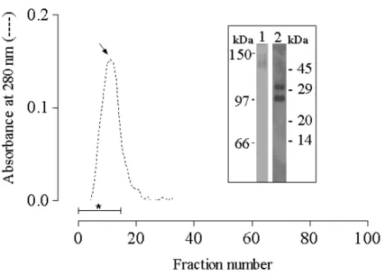
Determination of the native molecular weight of the lectin
Following size exclusion chromatography over Sephadex G-100, the lectin fraction was obtained as a single peak (arrowed in Fig. 4) within the void volume (indicated by an asterisk) signifying that the lectin possessed a molecular mass >100 kDa. This fraction showed a single band with an apparent molecular weight of 120-130 kDa when analysed by native PAGE (Fig. 4 insert, lane 1). In contrast, SDS-PAGE of the fraction showed two bands with
apparent molecular weights of 28 and 30 kDa (Fig. 4 insert, lane 2).
Ion exchange chromatography and pI
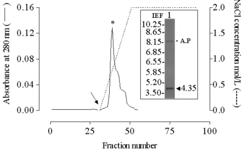
The determined pI value of 4.4 (Fig. 5 insert) was in agreement with the observation that when the lectin was buffered with 0.05 M sodium acetate (pH 5.0) and loaded onto a CM-cellulose column, it was fully adsorbed and eluted as a single peak with 0.77 M NaCl (Fig. 5).
Figure 5 - Cation exchange chromatogram of ScLL and determination of pI. An aliquot (2 ml) of ScLL solution (1 mg/ml) was applied to a CM-Sepharose DEAE column (20 ml) that had been pre-equilibrated with 0.01M sodium acetate buffer (pH 5.0). The column was eluted with the same buffer to elute the non-bound proteins, and then (changeover indicated by an arrow) with a continuous gradient of NaCl in this buffer () to elute the bound proteins. The eluted fractions were collected (fraction size -1.5 ml; flow rate - 0.562 ml/min) and monitored spectrophotometrically at 280 nm (_____). The insert shows the isoelectric focusing (IEF) gel of the fraction indicated by an asterisk with the arrowhead indicating the pI of ScLL and the label A.P indicating the loading point.
Effect of pH and temperature
Thermal denaturation experiments revealed that the lectin remained stable below 56oC for more 30 min with no loss of haemagglutinating activity.At 95oC, ScLL lost its activity completely within 30 min (Fig. 6A). Whilst the lectin retained its haemagglutinating activity within the pH range 6.0-9.0 (Fig. 6B), it was sensitive to very acidic (pH 3.0) and to very basic (pH 12.0) conditions, under which the activity was completely lost.
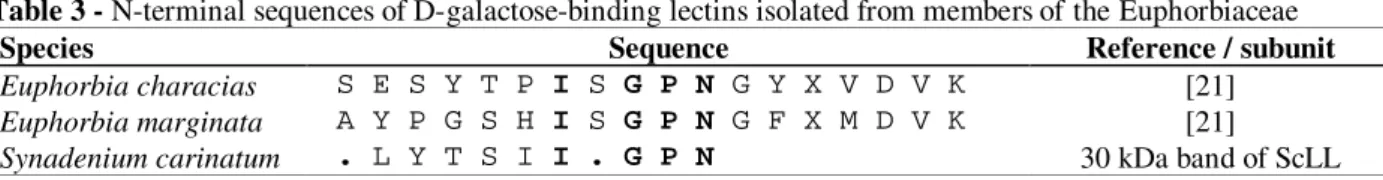
N-terminal sequence
The nine amino acid N-terminal sequence of the 30 kDa band of ScLL was determined using the Edman degradation method (Table 3). The GPN consensus sequence was homologous to two other lectins derived from Euphorbiaceae species, both of which showed affinity for D-galactose.
Analysis of cytotoxic activity
µg/ml were observed with J774 cells or with peritoneal murine macrophages within 24 to 72 h
(data not shown).
Table 3 - N-terminal sequences of D-galactose-binding lectins isolated from members of the Euphorbiaceae
Species Sequence Reference / subunit
Euphorbia characias S E S Y T P I S G P N G Y X V D V K [21]
Euphorbia marginata A Y P G S H I S G P N G F X M D V K [21]
Synadenium carinatum . L Y T S I I . G P N 30 kDa band of ScLL
Figure 6 - Effects of pH (A) and temperature (B) on the haemagglutinating activity of ScLL. The percentage activity was calculated assuming that the maximum measured activity was 100%.
DISCUSSION
Although the latex of S. carinatum has been used extensively in the treatment of various diseases, it is actually very toxic to humans. For this reason an analysis of the components of the latex is central to the elucidation of the risks associated with the ingestion of this material. It has recently been
a water solution of the latex of this plant has been used in popular medicine, and (ii) an aqueous solution of the latex is stable and the lectin properties are not lost.
Purified ScLL exhibited strong agglutination of different species of erythrocytes, however, the titre against human type A+ erythrocytes was 20- and 40-fold higher than against rabbit or mouse red blood cells, respectively. In contrast, no agglutination of horse erythrocytes was observed. These findings could be explained by differences in glycosylation of the surface proteins of red blood cells in the species tested.
It is interesting to note that treatment with SDS did not influence the binding of the lectin to red blood cells. The agglutinating activity was, however, completely inhibited by D-galactose, as has been reported for other lectins derived from the Euphorbiaceae (Stirpe et al., 1993). Although a number of D-Gal-binding lectins exhibited preferential haemagglutinating activity against human A, B and O blood groups (Datta et al., 1988; Witisuwannakul et al., 1998; Machuka et al., 1999), ScLL showed no such specificity.
Methyl-α- and methyl-β-D-galacto-pyranoside inhibited haemagglutinating activity of ScLL to a different extent suggesting that the lectin differentiated between α- and β-galactose. Moreover, α-rhamnose, L-arabinose and L-fucose did not inhibit agglutination whilst D-fucose did, suggesting that the configuration of the anomeric galactose residue had a significant effecton binding to ScLL. The higher binding capability of β -D-galactopyranoside compared with free D-galactose indicated a preference of the lectin for the β-D-pyranose form, which was predominantly in the 4C1 chain conformation (Yeasmin et al., 2001; Kenoth et al., 2003). The lectin PSA isolated from Polyporus squamosus also exhibited an anomeric preference for the beta configuration (Mo et al., 2000).
Staining with PAS reagent, which detected glycosidic linkages, revealed that ScLL was a glycoprotein and the intact molecular weight of the lectin was determined as ca. 120-130 kDa by size exclusion gel filtration on Sephadex G-100 and native PAGE. These results clearly indicated that the intact lectin was a tetrameric glycoprotein with a quaternary structure based on four heterogeneous subunits of 28 and 30 kDa, as revealed by
SDS-PAGE. Most lectins have molecular weights in the range 26-400 kDa and consisted of 2-18 homogeneous or heterogeneous subunits (Castagna et al., 1996; Machuka et al., 1999; Yeasmin et al., 2001). Galactose-specific lectins isolated from other plants have been reported to be dimeric or tetrameric proteins (Calvete et al., 1998; Machuka et al., 1999; Campana et al., 2002; Jung et al., 2003).
The 30 kDa subunit of ScLL showed N-terminal homology with D-galactose-binding lectins from E. marginata and E. characias that have been reported as strong mitogenics for human T lymphocytes (Stirpe et al., 1993). The three lectins exhibited a three amino acid (GPN) domain in common with the D-galactose-binding lectin from Maclura pomifera (Osage orange) (Young et al., 1989). The acidic pI value of ScLL was typical of many galactose-specific lectins, for example, those from Erythrina species (Bhattacharyya et al., 1986; Konozy et al., 2002, 2003), Maclura pomifera, Sophora japonica (Hankins et al., 1988), and Luetzelburgia auriculata (Oliveira et al., 2002).
Although plant latex is often cytotoxic, neither the crude aqueous extract nor the purified lectin from the latex of S. carinatum showed any cytotoxic activity against J774 cells or murine peritoneal macrophages. It seemed that aqueous extraction was efficient for the separation of non-toxic proteins from the potentially toxic components present in the latex of S. carinatum.
The isolation and characterisation of the lectin described in the present work could permit further advances to be made in the clarification of the biological effects of lectins on human cells and on the activation of the immune system .
ACKNOWLEDGEMENTS
RESUMO
No presente trabalho, foi purificada por cromatografia de afinidade em D-galactose imobilizada em agarose, uma lectina do latex de Synadenium carinatum (ScLL). Essa lectina é uma potente aglutinina para eritrócitos humanos, cuja atividade hemaglutinante foi inibida com 3,0 mM de N acetil-D-galactopiranosidio, 6,3 mM de metil--D-galactopiranosidio ou 50 mM metil-✁
-D-pironosidio ou D-fucose, porém, nenhuma inibição foi evidenciada por L-fucose, revelando uma especificidade anomérica e conformacional da lectina. A análise por SDS-PAGE dessa lectina pareceu ser uma glicoproteína composta por duas cadeias polipeptídicas de aproximadamente 28 e 30 kDa, porém, em cromatografia de exclusão por tamanho sobre Sephadex G100 e em gel nativo apresentou um peso molecular aparente de 120-130 kDa, a qual mostrou ser composta de uma mistura de subunidades de peptídeos de 28 e 30 kDa. Essa lectina manteve-se estável em pH de 6 a 9 e temperatura de 4 a 56ºC. A seqüência N-terninal contem uma região conservada GPN a qual também é observada em outras lectinas de látex de outras Euphorbiaceas ligante de D-galactose.
REFERENCES
Barbiere, L.; Falasca, A.; Franceshi, C.; Licastro, F.; Rossi, C. A. and Stirpe, F. (1983), Purification and propeties of two lectins from latex of the euphorbiaceous plants Hura crepitans L. (sand-box tree) and Euphorbia characias L. (Mediterranean spurge). Biochem. J., 215, 433-439.
Bhattacharyya, L.; Ghosh, A. and Sen, A. (1986) A comparative study on lectins from four Erythrina
species. Phytochemstry, 25, 2117-2122.
Calvete, J. J.; Santos, C. F.; Mann, K.; Grangeiro, T. B.; Nimtz, M.; Urbanke, C. and Sousa-Cavada, B. (1998), Amino acid sequence, glycan structure, and proteolytic processing of the lectin of Votairea macrocarpa seeds. FEBS Letters, 425, 286-292. Campana, P. T.; Moraes, D. I.; Monteiro-Moreira, A.
C. O. and Beltramini, L. M. (2002), Unfolding and refolding studies of frutalin, a tetrameric D-galactose binding lectin. Eur. J. Biochem., 269, 753-758.
Castagna, L.; Zarzur, J.; Filipetti, M. and Landa, C. (1996), Isolation and partial characterization of N-acetyl-D-galactosamine-binding lectins from
Epiphragmophora trenquelleonis snail. J. Biochem.,
119, 372-377.
Cavada, B. S.; Santos, C. F.; Grangeiro, T. B.; Nunes, E. P.; Sales, P. V. P.; Ramos, R. L.; De Souza, F. A. M.; Crisostomo, C. V. and Calvete, J. J. (1998), Purification and characterization of a lectin from seeds of Vatairea macrocarpa Duke. Phytochemistry,
49, 675-680.
Datta, P. K.; Basu, P. S. and Datta, T. K. (1988), Purification of human erythrocytes specific lectins from rice bean, Phaseolus calcaratus syn, by high-performance liquid chromatography. J. Chromatogr.,
431, 37-44.
Davis, B. J. (1964), Disc electrophoresis II, methods and application to human serum proteins. Ann. N. Y. Acad. Sci., 121, 404-427.
Dias-Baruffi, M.; Sakamoto, M.; Rosseto, S.; Vozari-Hampe, M. M. and Roque-Barreira, M. C. (2000), Neutrophil migration and aggregation induced by euphorbin, a lectin from the latex of Euphorbia milii, var. milii.Inflamm. Res., 49, 732-736.
Dodd, R. B. and Drickamer, K. (2001), Lectin-like proteins in model organisms: implications for evolution of carbohydrate-binding activity.
Glycobiology, 11, 71R-79R.
Gidrol, X.; Chrestin, H.; Tan, H. L. and Kush, A. (1994), Hevein, a lectin-like protein from Hevea brasiliensis (rubber tree) involved in the coagulation of latex. J. Biol. Chem., 269, 9278-9283.
Glossmann, H. and Neville Jr., D. M. (1971) Glycoproteins of cell surfaces. Comparative study of three different cell surfaces of the rat. J. Biol. Chem.,
246, 6339-6346.
Hankins, C. N.; Kindinger, J. I. and Shannon, L. M. (1988), The lectins of Sophora japonica. 2. Purification, properties, and N-terminal amino acid sequences of 5 lectins from bark. Plant Physiol.,
86, 67-80.
Heukeshoven, J. and Dernick, R. (1988), Improved silver staining procedure for fast staining in PhasSystem development unit. I. Staining of sodium dodecyl sulphate gels. Electrophoresis, 9, 28-32. Jager, A.; Hutchings, A. and Van Staden, J. (1996),
Screening of Zulu medicinal plants for prostaglandin-synthesis inhibitors. J. Ethnopharmacol., 52, 95-100.
Kenoth, R.; Komath, S. S. and Swamy, M. J. (2003), Physicochemical and saccharide-binding studies on the galactose specific seed lectin from Trichosanthes cucumerina. Arch Biochem. Biophys, 413, 131-138. Konozy, E. H. E.; Bernardes, E. S.; Rosa, C.; Faca, V.;
Greene, L. J. and Ward, R. J. (2003), Isolation, purification and physicochemical characterization of a D-galactose-binding lectin from seeds of Erythrina speciosa. Arch. Biochem. Biophys., 410, 222-229. Konozy, E. H. E.; Mulay, R.; Faca, V.; Ward, R. J.;
Greene, L. J.; Roque-Barreira, M. C.; Sabharwal, S. and Bhide, S. V. (2002), Purification, some properties of a D-galactose-binding leaf lectin from
Erythrina indica and further characterization of seed lectin. Biochimie, 84, 1035-1043.
Laemmili, U. K. (1970) Cleavage of structural proteins during the assembly of the head of bacteriophage T4.
Nature, 227, 680-685.
Liscastro, F.; Davis, L. J. and Morini, M. C. (1993), Lectins and superantigens: membrane interactions of these with T lymphocytes affect immune responses.
Int. J. Biochem., 25, 845-852.
Lowry, O. H.; Rosebrough, A. L.; Farr, A. L. and Randall, R. J. (1951) Protein measurement with the folin-phenol reagent. J. Biol. Chem., 193, 165-275. Machuka, J. S.; Okeola, O. G.; Van Damme, E. J. M.;
Chrispeels, M. J.; Van Leuven, F. and Peumans, W. J. (1999), Isolation, and partial characterisation of galactose-specific lectins from African yam beans,
Sphenostyles stenocarpa Harms. Phytochemistry, 51, 721-728.
Mo, H.; Winter, H. C. and Goldstein, I. J. (2000), Purification and characterization of a Neu5Acα 2-6Galβ1-4Glc/GlcNac-specific lectin from the fruiting body of the polypore mushroom Polyporus saquamosus. J.Biol. Chem., 275, 10623-10629. Oliveira, J. T. A.; Melo, V. M. M.; Câmara, M. F. L.;
Vasconcelos, I. M.; Beltramini, L. M.; Machado, O. L. T.; Gomes, V. M.; Pereira, S. P.; Fernandes, C. F.; Nunes, E. P.; Capistrano, G. G. G. and Monteiro-Moreira, A. C. O. (2002), Purification and physicochemical characterization of a cotyledonary lectin from Luetzelburgia auriculata. Phytochemstry,
61, 301-310.
Premaratna, A.; Shadaksharaswamy, M. and Nanjappa, S. (1981) Isolation, purification and properties of a lectin from the latex of Synadenium grantii Hook f. Indian J. Biochem Biophys, 18, 32-35.
Rojas, E.; Llinas, P.; Rodriguea-Romero, A.; Hernandez, C.; Linares, M.; Zanteno, E. and Lascurain, R. (2001), Havein, an Allergenic lectin from rubber latex, activates human neutrophils oxidadtive burst. Glycoconj. J., 18, 339-345.
Seshagirirao, K. and Prasad, M. N. (1995), Purification and partial characterization of a lectin from
uphorbia neriifolia latex. Biochem. Mol. Biol. Int.,
35, 1199-1204.
Sharon, N. and Lis, H. (1993), Carbhydrates in cell recognition. Sci. Am., 268, 82-89.
Souza, C. P.; Modesto, M. M. and Alves, T. M. A. (2000) Estudo químico da caramboleira e da leiterinha. FESB O jovem e a ciência no futuro -Abstract 15.P.7. Disp. in: [http://www.fesbe.org.br/ra/ fesbe2000/ojcf.html. Stirpe, F.; Licastro. F.; Morini, M. C.; Parente, A.;
Savino, G.; Abbondanza, A.; Bolognesi, A.; Falasca, A. I. and Rossi, C. A. (1993), Purification and partial characterization of mitogenic lectin from the latex of Euphorbia marginata. Biochem. Biophys. Acta, 1158, 33-39.
Van Damme, E. J. M.; Peumans, W. J.; Barre, A. and Rougé, P. (1998), Plant lectin: A composite of several distinct families of structurally and evolutionary related proteins with diverse biological roles. Cri. Rev. Plant Sci., 17, 575-692.
Witisuwannakul, R.; Witisuwannakul, D. and Sakulborirug, C. (1998), A lectin from bark of the rubber tree (Hevea brasiliensis). Phytochemstry, 47, 183-187.
Yagi, F.; Iwaya, T.; Haraguch, T. and Goldstein, I. J. (2002), The lectin from leaves of japanese cycad,
Cycas revoluta Thunb. (gymnosperm) is a member of the jacalin-related family. Eur. J. Biochem, 269, 4335-4341.
Yeasmin, T.; Tang, M. A. K.; Razzaque, A. and Absar, N. (2001), Purification and characterization of galactose specific lectins from Mulberry seeds (Morus sp.). Eur. J. Biochem., 268, 6005-6010. Young, N. M.; Johnston, R. A.; Szabo, A. G. and
Watson, D. C. (1989), Homology of the D-galacotse-specific lectins from Artocarpus integrifolia and
Maclura pomifera and the role of an unusual small polypeptide subunit. Arch. Biochem. Biophys, 270, 596-603.