The antioxidant and anti-inflammatory properties of lycopene in mice lungs exposed
to cigarette smoke
Keila Karine Duarte Campos
a, Glaucy Rodrigues Araújo
b, Thais Lourenço Martins
a,
Ana Carla Balthar Bandeira
b, Guilherme de Paula Costa
c, André Talvani
c, Camila Carrião Machado Garcia
d,
Laser Antônio Machado Oliveira
e, Daniela Caldeira Costa
b, Frank Silva Bezerra
a,⁎
aDepartamento de Ciências Biológicas, Laboratório de Fisiopatologia Experimental, Universidade Federal de Ouro Preto, Minas Gerais, Brazil bDepartamento de Ciências Biológicas, Laboratório de Bioquímica Metabólica, Universidade Federal de Ouro Preto, Minas Gerais, Brazil cDepartamento de Ciências Biológicas, Laboratório de Imunobiologia da Inflamação, Universidade Federal de Ouro Preto, Minas Gerais, Brazil dDepartamento de Ciências Biológicas, Laboratório de Bioquímica e Biologia Molecular, Universidade Federal de Ouro Preto, Minas Gerais, Brazil eDepartamento de Ciências Biológicas, Laboratório de Biomateriais e Patologia Experimental, Universidade Federal de Ouro Preto, Minas Gerais, Brazil
Received 7 November 2016; received in revised form 9 May 2017; accepted 6 June 2017
Abstract
Lycopene is a carotenoid with known antioxidant and anti-inflammatory properties. We aimed to evaluate thein vitroandin vivoeffects of lycopene on reducing the redox imbalance and inflammation induced by cigarette smoke (CS). For thein vitrostudy, J774A.1 (macrophages) cells were incubated in the presence of 0.5, 1.0, 2.0, 4.0, 8.0, 10.0 and 25μM of lycopene for 3, 6 and 24 h or in the presence of 0.1%, 0.25%, 0.5%, 0.625%, 1.25%, 2.25%, 5% and 10% cigarette smoke extract (CSE) for 3, 6 and 24 h to assess cell viability and measurement of intracellular reactive oxygen species (ROS). For thein vivostudy, 40 mice were divided into 5 groups: a control exposed to ambient air (CG), a vehicle-control group that received 200μl of sunflower oil by orogastric gavage, a group exposed to CS and two groups administered lycopene (diluted in sunflower oil) at doses of either 25 or 50 mg/kg/day prior to exposure to CS (LY25+CS and LY50+CS). The total treatment time lasted 5 days. A cell viability decrease was observed at 10- and 25-μM concentrations of lycopene in 3, 6 and 24 h compared with CG. There was an increase of ROS production in 24 h in CS compared with CG. Lycopene concentrations of 1μM and 2μM were able to reduce the production of ROS in 24 h compared with CS. In the bronchoalveolar lavage fluid, the total number of leukocytes increased in the CS group compared with the control groups (CG). Administration with lycopene at the highest dose suppressed this CS-induced increase in leukocytes. Lipid peroxidation and DNA damage increased in the CS group compared with that in the controls, and this increase was suppressed by lycopene at the highest dose. In contrast, superoxide dismutase activity decreased in the CS group compared with that in the controls. Catalase activity also increased in the CS group compared with that in both control groups, and this increase was suppressed in LY25+CS and LY50+CS. There was an increase in the levels of tumor necrosis factor-α, interferon-γand interleukin-10 after exposure to CS, and these effects were suppressed by both doses of lycopene. These data elucidate the role of lycopene as an antioxidant and anti-inflammatory agent in these two models of short-term exposure to CS.
© 2017 Elsevier Inc. All rights reserved.
Keywords:Lycopene; Cigarette smoke; Redox imbalance; Inflammation; Mice
1. Background
Chronic obstructive pulmonary disease (COPD) is a major cause of
morbidity and mortality worldwide[1,2]. COPD is characterized by a
slowly progressive and largely irreversible airflow limitation due
to chronic bronchitis and/or emphysema, associated with an abnormal
inflammatory response of the lungs [1,3–5]. The inflammatory
response is mediated by increased expression of inflammatory
mediators, such as cytokines, chemokines, adhesion molecules and inflammatory enzymes[6]. Although it is well established that genetic
and environmental factors contribute to the development of COPD[7],
smokers are a higher risk of this disease[2,8,9].
Cigarette smoke (CS) is a complex mixture of more than 6000 chemicals[10]. Exposure to CS can lead to the production of reactive
oxygen species (ROS) via NADPH oxidase, which catalyzes the
reduction of oxygen to a superoxide anion (O2•–)[11,12], leading to
the activation of the inflammatory cascade in the epithelium of the
upper and lower airways[13]. Oxidizing species interact directly with cell membranes and tissues, causing damage that results in the induction of inflammation, and this in turn causes a further release of oxidizing species leading to an overall imbalance in the redox state[4]. An imbalance between oxidants and antioxidants in favor of the oxidants, leading to a disruption of redox signaling and control and/or
molecular damage[14]. The increase in redox imbalance can be due to
oxidants present in CS and pollutants in ambient air, but they can also
be generated by activated inflammatory cells, such as macrophages
and neutrophils[15–18]. However, the increase in intracellular ROS can activate various redox-sensitive signaling pathways[12].
ScienceDirect
Journal of Nutritional Biochemistry 48 (2017) 9–20
⁎ Corresponding author. Tel.: +55 31 35591672; fax: +55 31 35591352. E-mail address:[email protected](F.S. Bezerra).
During human evolution, endogenous defenses were gradually developed to maintain the balance between oxidants and antioxidants. This primary antioxidant defense comprises three important enzymes that prevent the formation and promote the neutralization of free radicals: glutathione peroxidase (GPx) catalyzes the conversion of hydrogen peroxide (H2O2) and organic peroxides to H2O while at the same time
converting glutathione (GSH) into glutathione disulfide (GSSG)[16,19]. GSH is the predominant nonprotein thiol in the cells and is a key player in the maintenance of the cellular redox status. GSH exists primarily in two redox forms,i.e., reduced GSH and GSSG (the oxidized form). The latter represents about 1% of the total GSH pool[20]. The GSH/GSSG ratio can serve as a good indicator of the cellular redox state[21]. Catalase (CAT), which converts hydrogen peroxide into water and molecular oxygen, and superoxide dismutase (SOD), which converts the superoxide anion into hydrogen peroxide[16]. Moreover, exogenous antioxidant defenses, such as vitamins and some minerals, also play key roles[22,23].
NF-E2 p45-related factor 2 (Nrf2) is a transcription factor that plays an important role in endogenous antioxidation processes. Nrf2 regulates the expression of antioxidant genes under both physiological and oxidative stress conditions. Nrf2 induction occurs in the cell during oxidative stress caused by various environmental factors, such as CS exposure[24,25]. These external stimuli induce the generation of ROS, which in turn triggers Nrf2 activation and its translocation into the nucleus[26]. Upon translocation into the nucleus and binding to an antioxidant response element (ARE), Nrf2 regulates the expression of antioxidant proteins[25].
The antioxidant lycopene, a naturally occurring carotenoid found in tomatoes and tomato-plant extracts, exhibits potent free-radical-scavenging activity. Structurally, it is a highly unsaturated hydrocar-bon open chain, composed of 11 linearly conjugated double hydrocar-bonds and 2 nonconjugated double bonds, and is responsible for the red color of
many fruits and vegetables, such as tomatoes[27,28]. Lycopene is
found in nature in the all-transform; however, thermal processing, as well as intestinal digestion, of raw tomato products facilitates itscis -isomerization. Recent studies suggest that a diet rich in lycopene can
reduce the risk of serious diseases, such as cancer [29] and
cardiovascular disease, because this compound acts to suppress
singlet oxygen, being 2 times more effective thanβ-carotene and 10
times more potent thanα-tocopherol[28,30]. The anti-inflammatory effects of lycopene observed in most studies can be attributed mainly to its ability to modulate signaling pathways responsible for the
induction of inflammatory mediators as well as to activate the
expression of antioxidant genes[31]. In this latter regard, lycopene induces the nuclear translocation of Nrf2. One possible mechanism by which it is believed to achieve this involves the direct interaction of lycopene with Keap1 cysteine residues, which would trigger the release of Nrf2 from the complex. Lycopene-generated metabolites may also activate various kinases, which can also induce the release and nuclear translocation of Nrf2[32].
Knowing that lycopene has the characteristics of an antioxidant and that it may be useful to prevent many diseases through its effects on inflammation and redox imbalance[31,33], in this study, we aimed
to investigate the antioxidant and anti-inflammatory effects of
lycopene on lung induced by CS. We used anin vitromodel (J774A.1
macrophage cell line) to examine the effects of lycopene and a CS extract (CSE) on cell cytotoxicity as well as intracellular ROS production. In addition, we used a murine model of short-term exposure to CS to evaluate the inhibitory effects of lycopene on redox
imbalance and inflammation.
2. Materials
2.1. Cell culture
The J774A.1 macrophage cell line was a kind gift from the cell culture laboratory at the Pharmacy School, Federal University of Ouro
Preto (UFOP). J774A.1 cells were cultured in 75-cm2growth bottles
(Sardesdt AG & Co., Germany) containing J774A.1 culture medium
[Dulbecco's modified Eagle's medium (DMEM), Sigma] supplemented
with 10% (v/v) fetal bovine serum (FBS) (Invitrogen Co. Ltd., Carlsbad, CA, USA), 1% (v/v) penicillin, 3.7% (v/v) sodium bicarbonate, 1% (v/v)
glutamine (200 mM) and 2.5 ml of HEPES (1 M) (final concentration
200 g/ml). Growth bottles were incubated at 37°C in a humidified
atmosphere of 5% carbon dioxide (CO2). The medium was replaced
every 2–3 days depending on cell confluency. Cells were passaged
upon reaching 100% confluency by aspirating the medium and
detaching the cells from the monolayer by scraping. Cell number was determined using exclusion of a Trypan blue solution (0.3%) in a Neubauer chamber.
2.2. CSE preparation
Commercial Marlboro full-flavorfiltered cigarettes were obtained,
and a CSE was prepared as previously described [34]. Briefly, the
smoke from one cigarette was bubbled into 10 ml of DMEM
supplemented with 1% FBS with a modified syringe-driven apparatus.
The resulting suspension was adjusted to pH 7.4 and thenfiltered
through a 0.22-μm Milliporefilter. CSE was freshly prepared for each experiment and diluted with culture media supplemented with 1% FBS immediately before use. Control medium was prepared by bubbling air through 10 ml of culture media supplemented with 1% FBS, and the pH was adjusted to 7.4 and sterilefiltered as described above.
2.3. 3-(4,5-Dimethylthiazol-2-yl)-2,5-diphenyltetrazolium bromide test (MTT) cell viability assay
The MTT was performed as described previously[35]. J774A.1 cells
(5.0×10⁴/well) were plated into wells of a 96-well plate containing
J774A.1 culture medium (see above), and cells were cultured for 24 h to allow adherence. Following this, J774A.1 cells were incubated in absence (untreated) or presence of either lycopene (0.5, 1, 2, 4, 8, 10
and 25 μm, # Sigma L9879) or CSE (0.1%, 0.25%, 0.5%, 0.625%,
1.25%, 2.25%, 5% and 10%) for 3, 6 or 24 h to assess cell viability.
After cell treatment, the supernatant was removed, and 200μl of MTT
(5.0 mg/ml) was added and incubated for 1 h at 37°C in humidified air supplemented with 5% CO2. The MTT solution was removed, and 100μl
of dimethyl sulfoxide (DMSO) was added to each well. The absorbance was read at 570 nm (Biotek EL 808). Cell viability was assessed relative to vehicle-treated control cells. The control cells were considered 100% viable. Lycopene was dissolved in tetrahydrofuran (THF) containing 0.025% butylated hydroxytoluene (BHT) to prevent the formation of peroxides. Stock solutions of lycopene were prepared immediately before each experiment. From the stored solutions, aliquots of lycopene were rapidly added to the culture medium to obtain the
final concentrations indicated. Vehicle-treated control cells were
treated with the THF/BHT solvent. The amount of THF added to the cells was not more than 0.5% (v/v).
2.4. Measurement of intracellular ROS
Intracellular ROS generation was detected using the probe 5-(and-6)-carboxy-2′,7′-dichlorodihydrofluorescein diacetate (carboxy-H2DCFDA)
as a nonfluorescent cell-permeable compound. After penetration of the cell by carboxy-H2DCFDA, intracellular nonspecific esterases generate the
molecule carboxy-DCFH. In the presence of nonspecific ROS (produced
during oxidative stress), the reducedfluorescein compound is oxidized and emits a brightfluorescent signal. J774A.1 cells (2.5×10⁴cells/well) were
seeded into the wells of a 96-well plate containing DMEM supplemented
with 10% FBS and cultured for 45 min at 37°C in humidified air
supplemented with 5% CO2. Following this, the supernatant was removed,
(dissolved in THF) for 6 h at 37°C in humidified air supplemented with 5% CO2. After 6 h, the cells were washed with Hanks' balanced salt solution
(HBSS) and exposed to media containing 0.5% CSE and incubated in the dark
with carboxy-H2DCFDA for 3 and 24 h at 37°C in humidified air
supplemented with 5% CO2. After incubation (3 and 24 h), cells were
washed with HBSS, medium without red phenol was added, and
fluorescence intensity was determined using 485-nm excitation and
535-nm emission using an automated plate reader model VICTOR X3,
software PerkinElmer 2030 workstation and workout 2.5. Meanfl
uores-cence intensity was reported as percentage (%) increase over control.
2.5. In Vivo model of CS exposure
Male C57BL/6 mice (8 weeks old) were housed under controlled conditions in standard laboratory cages (Laboratory of Experimental Nutrition, Department of Food, School of Nutrition, UFOP, Brazil) and given free access to water and food. Allin vivoexperimental protocols at the UFOP were approved by the ethics committee. Forty mice (C57BL/6) were divided into 5 groups: a control group exposed to
ambient air (CG), a group that received 200 μl of sunflower oil
(sunflower seed oil fromHelianthus annus, Sigma Aldrich LTDA)via
orogastric gavage (OG) [36], a group exposed to CS (total of 12
cigarettes/day; see protocol below) and groups administered lyco-pene 25 or 50 mg/kg/day (BASF, LycoVit Dispersion 10%, Germany)
diluted in sunflower oil[37]plus CS (LY25+CS and LY50+CS) for 5
days. The administration of lycopene was performedviaOG at doses of
25 and 50 mg/kg/day 12 h before exposure to CS[27,28,36,37].
2.6. CS exposure protocol
In a smoking chamber, mice were exposed to 12 commercial
full-flavorfiltered Virginia cigarettes per day (tar, 10 mg; nicotine, 0.9 mg; and carbon monoxide, 10 mg) spread over 3 times a day (morning, afternoon and evening) for a total of 5 days as described previously
[38]. The total number of 12 cigarettes had been already established in the literature causing emphysema in a long-term exposure to cigarette smoke[39].
Briefly, mice in the CS and LY+CS groups were placed in the
inhalation chamber (40 cm long, 30 cm wide and 25 cm high) inside an exhaustion chapel. A cigarette was coupled to a plastic 60-ml syringe so that puffs could be drawn in and subsequently expelled into the exposure chamber. One liter of smoke from one cigarette was aspirated with the syringe (20 puffs of 50 ml) and was immediately injected into the chamber. The CS and LY+CS groups were maintained in this smoke-filled atmosphere (±3% CS concentration) for 6 min. The cover was then removed from the inhalation chamber, and by turning on the exhaust fan in the chamber of the chapel, the smoke was
evacuated within 1 min[40]. Animals exposed to ambient air under
the same conditions were considered the control group (CG;n=8).
Twenty-four hours after their last exposure to CS, animals were euthanatized by cervical dislocation[17,41].
2.7. Assessment and analysis of bronchoalveolar lavage fluid (BALF)
Immediately after euthanasia, the chest of each animal was opened to collect the BALF. The left lung was clamped, and the right lung and the trachea were cannulated and perfused with 1.5 ml of saline solution. Samples were kept on ice until the end of the procedure to avoid cell lysis. Total mononuclear and polymorphonuclear cells were stained with Trypan blue and counted in a Neubauer chamber (Sigma-Aldrich, MA, USA). Differential cell counts were performed on cytospin preparations (Shandon, Waltham, MA, USA) stained with a fast panoptic coloration kit (Laborclin, Pinhais, Paraná)[42].
2.8. Tissue processing and homogenization
After BALF collection, the right ventricle of each mouse was perfused with saline to remove blood from the lungs. The right lung was clamped such that only the left lung could be perfused with 4% buffered formalin (pH 7.2) at a pressure of 25 cm H2O for 2 minviathe
trachea. The left lung was then removed and immersed in afixative
solution for 48 h[43]. The material was then processed as follows: a tap water bath rinse for 30 min, 70% and 90% alcohol bath rinses for 1 h
each, two bath rinses in 100% ethanol for 1 h each and finally
embedding in paraffin. Serial 5-μm sagittal sections of the left lung were stained with hematoxylin and eosin for histologic analyses. After removal of the left lung for histological analyses, the right lung was immediately removed and stored on crushed ice in labeled tubes. The lungs were subsequently homogenized in 1 ml potassium phosphate buffer (pH 7.5) and centrifuged at 1500×gfor 10 min. The supernatant
was collected, and the final volume of all samples was adjusted
to 1.5 ml with phosphate buffer. The samples were stored in a freezer (−80°C) for biochemical analyses.
2.9. Morphometric and stereological analyses
All morphometric analyses were performed at the Multiuser Laboratory of the Research Center for Biological Sciences of the UFOP. Twenty random images obtained from the lung histology slides were digitized using the Leica Application Suite software in associa-tion with a Leica DM5000b optical microscope and a CM300 digital microcamera. All images were scanned using a ×40 objective. The analysis of the volume density values of alveolar air space (Vv[a]) and
volume densities of alveolar septa (Vv[sa]) was performed on a test
system that consists of 16 points and a known test area in which the boundary line was considered forbidden in order to avoid an overestimation of the number of structures. The test system was connected to a monitor attached to a microscope. The number of points (PP) that touched the Vv[a] andVv[sa] values was assessed
according to the total number of test points (Pt) in the system by using
the following equation: Vv¼PP
PT [44]. To obtain uniform and
proportional lung samples, we analyzed 18 randomfields in a cycloid
test system attached to the monitor screen. The reference volume was estimated by point counting using the test point system. A total area of
1.94 mm2from each of the hematoxylin-and-eosin-stained slides was
analyzed to determine the values for volume density of alveolar air space and volume density of alveolar septa[45,46].
2.10. Nitrite content
The nitrite content in lung homogenates was determined using a
method based on the Griess reaction [47]. The lung homogenate
(100μl) was mixed with 100μl of Griess reagent (1% sulfanilamide in 5% phosphoric acid and 0.1% naphthylenediamide dihydrochloride in water) and incubated at room temperature for 10 min. The absorbance was then measured with a plate reader at 550 nm (PerkinElmer Microplate Reader model 0602, Life Sciences). Nitrite concentrations in the samples were determined from a standard curve that was generated using different concentrations of sodium nitrite.
2.11. Antioxidant defense and oxidative stress biomarkers in lung homogenates
The lung homogenates were used to determine CAT activity. CAT activity was measured using the Aebi method[48]. H2O2decomposition
was calculated using a molar absorption coefficient of 39.4 M−1cm−1. Results are expressed as activity per milligram of protein. One unit of CAT is equivalent to the hydrolysis of 1μmol of H2O2per min. The initial
spectrophotometrically at 240 nm, over a 5-min time frame. SOD activity was assayed using the spectrophotometric method of Marklund
and Marklund [49], which is an improved pyrogallol autoxidation
inhibition assay. SOD reacts with O2•–, and this reaction slows down the
rate of formation of o-hydroxy-o-benzoquinone and other polymer
products. One unit of SOD is defined as the amount of enzyme that
reduces the rate of autoxidation of pyrogallol by 50%. The total glutathione content in homogenates was determined using an assay adapted from a commercial kit (CS0260, Sigma, St. Louis, MO, USA). The kit uses a kinetic method to measure the total levels of glutathione (GSH+GSSG). In this assay, the reduction of 5,5′ -dithiobis-(2-nitro-benzoic acid to 5-thio-2-nitro-dithiobis-(2-nitro-benzoic acid was assessed spectrophoto-metrically at 570 nm[50]. Subtracting the concentration of GSSG of the total glutathione concentration value provides the value of the concentration of GSH, and the GSH/GSSG ratio was obtained by dividing the concentration of GSH by the GSSG results. GPx activity was measured by monitoring the oxidation of NADPH at 340 nm in the
presence of H2O2 [51]. Total protein content in each sample was
determined using the method of Bradford[52].
As a measure of lipid peroxidation, we used the formation of thiobarbituric acid reactive substances (TBARS), a process that occurs as a result of heating in the presence of acid, as previously described by Draper et al.[46]. Specific details of the method used for measurement
of the TBARS level were as described by Buege and Aust[53]. For
analysis of DNA damage, approximately 50 mg of the lung was
homogenized, and an aliquot of homogenate (100μl) was mixed in
0.5% low-melting point agarose in phosphate-buffered saline (PBS) at 37°C. The mixture was placed onto a glass microscope slide precoated with a layer of 1.5% normal melting-point agarose in PBS. The slides were immersed in a cold lysis solution (2.5 M NaCl, 100 mM EDTA, 10
mM Tris–HCl, 10% DMSO, 1% Triton X-100, Sigma-Aldrich) overnight
at 4°C. The slides were then placed in an electrophoresis tank immersed in ice, and DNA was unwound by incubating with an alkaline solution (300 mM NaOH and 1 mM EDTA, pH 13.0) for 20 min. Electrophoresis was then performed at 300 mA, 25 V for 20 min in the same alkaline solution. The slides were neutralized with 0.4 M
Tris–HCl buffer (pH 7.4), fixed in 100% ethanol and stained with
ethidium bromide (20 μg/ml, Sigma-Aldrich). The DNA comets
were analyzed using an Axioplan microscope (Carl Zeiss, Germany),
with a ×20 objective, influorescent mode (510–560-nmfilter and a
barrier of 590 nm). One hundred comets on each slide were scored according to their tail length. Comets were classified from 1 to 4, and the damage index was calculated using the formula:
index = (tnumber of comets Class 1×0)+(number of comets Class
2×1)+(number of comets Class 3×2)+(number of comets class 4×3)/
number of comets analyzed[54].
2.12. Enzyme-linked immunosorbent assay (ELISA) for inflammatory mediators
Lung homogenates were used to determine the concentrations of the inflammatory mediators tumor necrosis factor (TNF)-α, interferon (IFN)-γand interleukin (IL)-10. After euthanasia, 100 mg of lung from each animal was homogenized in cold phosphate buffer, and the supernatants were stored to evaluate the previously mentioned
soluble inflammation mediators. For the analysis, the samples were
thawed and excess proteins were removed by acid/salt precipitation,
as previously described[55]. Briefly, equal volumes of homogenate
and 1.2% trifluoracetic acid/1.35 M NaCl were mixed and incubated at
room temperature for 10 min followed by centrifugation for 5 min at 10,000 rpm. The salt content of the supernatant was adjusted to be 0.14 M sodium chloride and 0.01 M sodium phosphate at a pH of 7.4 prior to determination of the concentrations of TNF-α, IFN-γand IL-10 using commercially available ELISA kits (BioSource International, Inc.,
CA, USA) according to the manufacturer's guidelines. All samples were measured in duplicate[56,57].
2.13. Immunostaining and confocal microscopy
The lungs were collected, washed in PBS and cryofixed in a solution of 80% methanol and 20% dimethyl sulfoxide at−80°C. After 5–7 days
of freeze substitution, samples were embedded in Paraplast.
Five-micrometer-thick sections were mounted on slides, deparaffinized,
rehydrated and then incubated in blocking solution (1% bovine serum albumin and 0.1% Tween-20 in PBS) at room temperature for 1 h. Sections were incubated overnight at 4°C with rabbit anti-human Nrf2
(1:100; Abcam, Cambridge, MA, USA). After four tofive rinses in PBS,
donkey anti-rabbit IgG conjugated with AlexaFluor 488 (1:500; Jackson Immuno Research Laboratories, West Grove, PA, USA) was
added for 1 h in the dark at room temperature[58]. Following washes
with PBS, sections were mounted and viewed with a laser scanning confocal microscope (Zeiss LSM780, Center for Research in Biological Sciences, NUPEB/UFOP). Optimal confocal settings (aperture, gain and laser power) were determined at the beginning of each imaging session and then held constant during sample analysis. Nuclei were
labeled with 4′6-diamidino-2-phenylindole dihydrochloride
(Molec-ular Probes, Carlsbad, CA, USA). The number of Nrf2-positive nuclei in lung parenchyma was then determined. For each analysis, 15 random
microscopicfields were analyzed (area: 65,536μm2each), and the
number of Nrf2-positive nuclei (expressed as Nrf2/μm2) was counted
using Zen 2011 software (blue edition). Nrf2-positive nuclei were
quantified in the lung parenchyma. For each group, 15 microscopic
fields (15 randomfields of 65,536μm2 each) were analyzed. The
number of Nrf2-positive nuclei (Nrf2/μm2) was counted using
Software Zen 2011 blue edition.
2.14. Statistical analysis
Data with a normal distribution were assessed through univariate analysis of variance [either one-way analysis of variance (ANOVA) or two-way ANOVA] followed by the Bonferroni posttest. Data were expressed as mean±standard error of mean. We used the Kruskal–Wallis test followed by Dunn's posttest for discrete data and expressed them as median, minimum and maximum values. In both cases, differences were
considered significant when theP value wasb.05. All analyses were
performed with GraphPad Prism version 5.03 (GraphPad Software, San Diego, CA, USA) for Windows 7.
3. Results
3.1. Cellular viability assessment
We used both in vitro and in vivo models to examine the
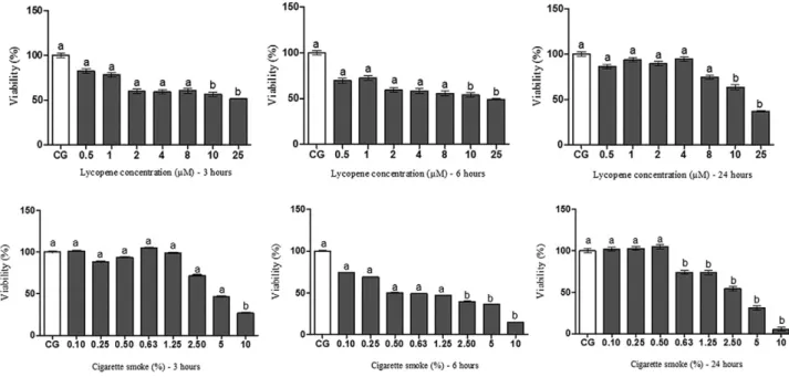
antioxidant effect of lycopene. Macrophage cells were exposed to different concentrations of lycopene and CSE for varying amounts of time, and the MTT assay was performed to assess cellular cytotoxicity by measuring the activity of mitochondrial dehydrogenase enzymes. A decrease in cell viability was observed after 3-, 6- and 24-h exposure to
10 and 25μM lycopene when compared to that in the CG (untreated
cells) (Fig. 1). In cells exposed to CSE, cell viability decreased after 3-h exposure to 10% CSE. Cell viability decreased at lower CSE levels as the time of exposure increased (2.50%, 5% and 10% CSE decreased cell viability after 6-h exposure, whereas 0.63%, 1.25%, 2.50%, 5% and 10%
CSE decreased cell viability after 24-h exposure) (Fig. 1). Thus,
lycopene concentrations (0.5, 1 and 2μM) were noncytotoxic, and the
3.2. Quantitative analysis of ROS production in J774A.1 cells
To evaluate the possible antioxidant effect of lycopene through a ROS mechanism, we measured ROS production in cells treated with
either lycopene or CSE. There was no significant difference in ROS
production following 3-h exposure to CSE (Fig. 2). There was,
however, a significant increase in ROS production when the cells
were exposed to CSE and in the cells pretreated with lycopene at 0.5
μM and then exposed to CSE for 24 h in relation to CG (Fig. 2). When the cells were pretreated with lycopene at 0.5μM and then exposed to CSE for 24 h, there was a reduction in ROS production when compared to CSE. However, when cells were pretreated with lycopene at 1 and 2
μM and then exposed to CSE for 24 h, we observed a reduction of the
production of ROS in 24 h compared with CSE and 0.5μM+CSE (Fig. 2).
3.3. Cellular influx in BALF
Having observed an antioxidant effect of lycopene in vitro, we
evaluated whether administration of lycopenein vivowould be able to
decrease the influx of inflammatory cells into lungs following 5 days of exposure to CS. For this, we determined the amount of total leukocytes
present in BALF of administered mice (Table 1). Total leukocyte
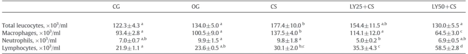
number increased in the CS group compared to that in the CG and OG groups. There was a decrease in the number of total leukocytes in the LY50+CS group compared to that in the CS group. The number of macrophages also increased in the CS group compared to that in the CG and OG groups and decreased in the LY25+CS group compared to that in the CS group. There was a decrease in the number of macrophages in the LY50+CS group compared with that in all other groups. The number of neutrophils also decreased in the LY25+CS group compared with that in the OG and CS groups. The number of lymphocytes increased in the CS group compared with that in the CG group and in the LY25+CS group compared to that in the CG and OG groups. The number of lymphocytes in the LY50+CS group increased compared to that in all other groups (Table 1).
3.4. Morphometric evaluations of the lung parenchyma
We performed morphometric assessments of the lung parenchyma by using stereology to evaluate whether administration of lycopene reduces lung damage caused by oxidants present in CS. Campos et al. reported that mice exposed to a total of six cigarettes/day for 5
consecutive days showed no significant difference in either the
volume density of alveolar air space (Vva) or the volume densities of alveolar septa (Vvsa)[17]. In contrast, when the animals used in this study were exposed to 12 cigarettes/day for 5 consecutive days, we observed changes in lung parenchyma following morphometric analysis. We observed an increase in the Vva in the CS group compared to that in the CG and OG control groups, whereas compared to that in the CS group alone, there was a decrease in Vva in both of the CS groups also treated with lycopene (LY25+CS and LY50+CS). The Vvsa decreased in the CS compared with that in the CG and OG control
Fig. 1. Cellular viability assessment by MTT test at different concentrations of lycopene (0.5, 1, 2, 4, 8, 10 and 25μM) and CSE (0.10%, 0.25%, 0.50%, 0.63%, 1.25%, 2.50%, 5% and 10%) at different times (3, 6 and 24 h). Bars with a common superscript letter do not differ. Bars without a common superscript letter differ. Data were expressed as mean±standard error of the mean and were analyzed by Kruskal–Wallis followed by Dunns posttest (Pb.05). This test was performed in sextuplicate from three independent experiments.
groups, and it increased in the LY25+CS group compared with that in the CS group (Fig. 3A and B).
3.5. Evaluation of the translocation of Nrf2
Studies using immunostaining and other methods have shown that activation of transcription of genes containing an ARE by various agents is preceded by nuclear translocation of the transcription factor
Nrf2[59]. Thus, using immunofluorescence, we investigated whether
administration of lycopene results in the translocation of Nrf2 from the cytosol into the nucleus in pulmonary tissue. Translocation of Nrf2 from the cytoplasm into the nucleus was predominant in the CS group compared to CG, OG, LY25+CS and LY50+CS groups (Fig. 4A and B). The administration with lycopene (LY25+CS and LY50+CS) promoted a
reduction of this parameter compared to the CS group (Fig. 4A and B). We did not observe any nonspecific labeling in negative controls (NC) (Fig. 4A, panel A). In contrast, the merged image shows the nuclear localization of the Nrf2 protein (bar=50μm) visualized using an anti-Nrf2 antibody and an Alexa-Fluor-488-conjugated secondary antibody. Arrows indicate the location of the cell nucleus (Fig. 4A, panels B–F).
3.6. Effects of treatment with lycopene on antioxidant enzyme activities (SOD, CAT and GPx), GSH/GSSG ratio, TBARS, DNA comet tail and nitrite in lung parenchyma
In our previous study, we showed that short-term exposure to CS
associated with the influx of inflammatory cells led to an oxidant/
antioxidant imbalance [17]. In this context, we analyzed whether
Table 1
Effects of treatment with lycopene on cellular influx in BALF from the CG, OG, CS and LY+CS groups
CG OG CS LY25+CS LY50+CS
Total leucocytes, ×103/ml 122.3±4.3a 134.0±5.0a 177.4±10.0b 154.4±11.5a,b 130.0±5.5a
Macrophages, ×103/ml 93.4±2.8a 100.5±9.0a 137.5±4.0b 114.1±12.0a 64.5±3.0c
Neutrophils, ×103/ml 7.0±0.7a,b 9.9±1.5a 9.8±1.8a 5.0±0.2b 6.9±0.5a,b
Lymphocytes, ×103/ml 21.9±1.1a 23.6±0.5a,b 30.1±2.0b,c 35.3±4.3c 58.5±2.8d
For comparison among groups, one-way ANOVA followed by the Bonferroni's posttest was performed (Pb.05,n=8). Values without a common superscript letter indicate statistical differences.
administration of lycopene reduced this redox imbalance. In order to do this, we evaluated the activities of SOD, CAT, GPx and the GSH/GSSG ratio in lung homogenates and measured the formation of TBARS as an
index of lipid peroxidation and assessed DNA damage (Table 2).
SOD activity decreased in the CS group relative to the CG and OG. However, we found no statistical differences in SOD activity in groups treated with lycopene and exposed to CS compared to the CS group. CAT activity was higher in the CS group compared to the GC and OG control groups. In contrast, animals administered lycopene and exposed to CS (LY25+CS and LY50+CS) had considerably lower CAT activity relative to the CS group. GPx activity increased in the CS group compared to the CG and OG control groups. We also observed an increase in GPx activity in the LY25+CS group compared to that in the CG, OG and CS groups. GPx activity was also lower in the LY50+CS
group than that in the LY25+CS group (Table 2). The GSH/GSSG ratio
was lower in the CS group than that in the CG group. Administration of lycopene increased the GSH/GSSG ratio in the LY50+CS group
compared to the CS group (Table 2). There was an increase in the
TBARS concentration in the CS group compared to the CG group. In contrast, administration of lycopene reduced TBARS concentrations in
the LY50+CS group compared to the CS group alone. Lung DNA damage was assessed using a comet tail assay. Increased DNA damage was observed in the CS group compared to that in the CG group. We observed a decrease in DNA damage in the LY50+CS group relative to the CS group. Finally, there were no
significant differences in lung nitrite content in any of the mice
treatment groups (Table 2).
3.7. Evaluation of the levels of inflammatory cytokines in lung parenchymal
It is known that acute exposure to CS leads to an inflammatory
response[60]. Therefore, we evaluated the possible effects of lycopene administration on CS-induced increases in the levels of TNF-α, IFN-γ
and IL-10 (Fig. 5). As expected, we observed that there was an increase in the levels of these cytokines in the CS group compared with the control CG and OG groups. In contrast, lycopene led to a decrease in the levels of these cytokines in both the LY25+CS and LY50+CS groups compared with the CS group alone (Fig. 5).
4. Discussion
Our present data substantiate that lycopene is a potent antioxidant agent bothin vitroandin vivo. Using both types of models, we assessed lycopene's antioxidant and anti-inflammatory activities in the tested dosage. We observed a decrease in cellular cytotoxicity and ROS production in cultured cellsin vitro, while,in vivo, we observed several
beneficial effects of lycopene with regard to redox imbalance and
inflammation in animals that had been exposed to CS for a short term (5 days). The experiments were performed using consistent physio-logical lycopene concentrations, which can be applied in human diet rich in foods containing, as tomatoes[30].
Due to the beneficial effects of lycopene, numerous studies in
different organs have been carried out with the purpose of clarifying the pathophysiological processes that involve certain pathologies. According to Rao and Shen, a consumption between 5 mg and 10 mg of lycopene per day is sufficient to obtain the benefits of this nutrient
[61]. For Rao and Agarwal, the average consumption of this
antioxidant should be 35 mg/day. It should be noted that these dosages are suggested for the healthy population[62]. Li et al. reported that daily supplementation for 8 weeks with tomato juice containing 32.5 mg of lycopene in a healthy young person showed antiobesity, antioxidation and anti-inflammatory effects[63].
4.1. Effect of lycopene through the cellular cytotoxicity and ROS production
In our study, we observed a decrease in cell viability in cells treated for 3, 6 and 24 h with different concentrations of lycopene or CSE compared to that in the control cells. Lower concentrations of lycopene (1 and 2μM) did not affect cell viability. In relation to ROS production, we showed that these low lycopene concentrations (1 and
2μM) were able to reduce ROS production in cells caused by 24-h
exposure to 0.5% CSE. Our data corroborate the results of a previous
study by Palozza et al.[34]that measured ROS production as a means
to quantify the level of cellular oxidative stress generated by oxysterols. In addition, in this study, lycopene at a concentration of 2
μM strongly reduced ROS production induced by oxysterol. In another
study, Lian et al.[64]observed that treatment with apo-10′-lycopenoic acid at a concentration of 3μM for 24 h decreased cellular ROS levels by about 40%[64].
4.2. Effects of administration with lycopene on cellular influx in BALF
Various studies have reported that repeated exposure to CS may
induce prolonged airway inflammation associated with cellular
infiltration of macrophages and neutrophils[17,65]. Differential cell counts in BALF have been used to support the diagnosis of lung
diseases [66]. Our findings concerning total leukocytes in the
pulmonary parenchyma corroborate the results of a previous study by Campos et al.[17]who reported a gradual increase in the number of
inflammatory cells in pulmonary tissue beginning on the second day
and persisting until the last day of CSE. Bao et al.[67]reported an
increase in the number of inflammatory cells in pulmonary tissue in
mice exposed to CS for 4 days. In this latter study, animals exposed to CS and administered an apple polyphenol showed a decrease in the influx of total leukocytes. Macrophages play an important role in host defense against noxious substances and are involved in various disease processes, including autoimmune diseases, infections and inflammatory disorders[68]. We observed that exposure to CS led to a
high influx of macrophages into BALF, whereas preadministration of
lycopene decreased the amount of macrophage infiltration in response
to CS. Preadministration of lycopene also decreased the amount of neutrophil infiltration in response to CSE. Bao et al.[67]reported a similar result in animals exposed to CS where administration of an apple-derived polyphenol decreased the amount of macrophages and neutrophils in BALF following CSE. In regard to lymphocyte subpop-ulations, an increase in lymphocytes was observed following CSE, and we observed that lycopene increased the number of lymphocytes compared to that in all other treatment groups. In a previously
Table 2
Effects of treatment with lycopene on antioxidant enzyme activities (SOD, CAT and GPx), GSH/GSSG ratio, TBARS, DNA comet tail and nitrite in lung samples from the CG, OG, CS and LY+CS groups
CG OG CS LY25+CS LY50+CS
SOD (U/mg prot) 32.00±3.00a 26.50±4.00a 17.00±2.00b 22.00±1.60a,b 22.00±1.50a,b
CAT (U/mg/prot) 0.45±0.06a 0.40±0.05a 1.06±0.03b 0.70±0.10a 0.55±0.07a
GPx (mM/min/mg prot-1) ×10−5 0.06±0.01a 0.06±0.01a 0.14±0.01b 0.20±0.01c 0.09±0.01a,b
GSH/GSSG (nmol/ml) 0.50±0.08a 0.40±0.06a,b 0.30±0.07b 0.45±0.01a,b 1.00±0.20a
TBARS (nM/mg ptn) 4.95±0.50a 5.90±0.90a,b 8.00±0.01b 5.45±0.40a,b 4.90±0.40a
DNA (% nuclei with comet tail) 1.30(1.8/0.7)a 1.60(1.9/1.0)a,b 1.83(2.6/1.4)b 1.40(1.6/1.0)a,b 1.08(1.5/1.0)a
Nitrite (μmol/mg ptn) 25.80±1.90a 26.00±2.30a 32.00±2.10a 31.00±2.40a 30.00±3.20a
For comparison among groups, one-way ANOVA followed by the Bonferroni's posttest was performed (Pb.05,n=7). Values without a common superscript letter indicate statistical differences.
published study by our group, we showed that CD4+ and CD8+ T-lymphocytes are present in BALF from mice exposed for a short time to CS, and an increase in T-cell recruitment was observed following the third day of CSE[17]. The recruitment of lymphocytes in response to CS is possibly a result of pulmonary aggression caused by toxic compounds present in CS[17,69]. We hypothesize that the marked elevation in the number of lymphocytes observed in mice preadministered with
lycopene prior to exposure to CS (i.e., the LY50+CS group) compared
to that in the CS group alone might occur owing to lycopene's ability to activate the adaptive immune response since that response is important
to maintain an adequate defense against microorganisms[70].
4.3. Administration of lycopene was effective in reversing morphological parameters
Proteases and ROS are inherent by-products of activated
leuko-cytes and contribute to the inflammatory response and parenchyma
destruction[71]. The results of our study show for thefirst time that short-term exposure to CS can cause morphological changes in the lung parenchyma and that administration of lycopene was effective in reversing these changes. We propose that this result was due to the antioxidant action of lycopene possibly acting directly against oxidants present in CS, thereby reducing oxidative damage in the
lung parenchyma and thus preventing initiation of the inflammatory
process. Murta et al.[19]related that Fischer rats exposed to 5% of formaldehyde had reduced Vva of the alveolar septa in the lung parenchyma compared to that in controls.
4.4. Lycopene decrease translocation of Nrf2 protein into the nucleus
Activation of the Nrf2 signaling pathway can induce the expression of endogenous protective genes in various tissues and cells, including the transcription of target genes involved in redox homeostasis[72]. Overall, the results of the present study showed that lycopene did not induce translocation of Nrf2 into the nucleus. In our experimental model, lycopene acts directly as a powerful antioxidant, whereby it scavenges the reactive species, thereby decreasing their levels. Activation of antioxidant genes through Nrf2 nuclear translocation does not appear to be necessary for its antioxidant effects. This hypothesis is supported by the observation that exposure to CS increases translocation of Nrf2 into the nucleus and that this translocation is decreased in mice administered lycopene prior to
exposure to CS. Ours results show a significant reduction in Nrf2
expression along with a concomitant reduction in the activities of the enzymes CAT and GPx in mice administered lycopene and then exposed to CS, reinforcing the data showing decreased translocation of Nrf2 in mice administered lycopene prior to CSE. In the case of SOD activity, although CS did decrease the levels of SOD, we did not observe any subsequent differences between lycopene-administered and untreated mice that were exposed to CS. Our data corroborate the data of a previous study by Teasdale et al.[72], who studied the effect of CS extract on Nrf2-responsive genes in human coronary artery endothelial cells. They observed that the human coronary artery endothelial cells responded to the noxious components in CS extract
by activating the Nrf2 pathway. The present study is the first to
describe the effects of lycopene administration and CSE on Nrf2 expression in mice. However, there are several studies conducted in different cell types that have examined the effect of other antioxidant
compounds on Nrf2 nuclear translocation[64,73].
4.5. Effects of administration with lycopene on the activities of antioxidant enzymes and GSH/GSSG ratio
An imbalance between oxidants and antioxidants in favor of the oxidants leads to a disruption of redox signaling and control and/or
molecular damage, an increase in CAT activity and SOD activity, and
depletion of reduced GSH[74,75]. The superoxide anion is generally
rapidly scavenged, whereas hydrogen peroxide is much more stable and may cross the cell membrane. According to Lushchak et al., high reactive species levels can inhibit the activity of antioxidant enzymes
[76]. We believe that the lack of effect of lycopene on SOD levels in mice exposed to CS can be explained as follows: lycopene directly reduces the production of the superoxide anion by NADPH oxidase
[11]; in the presence of such low superoxide anion levels, the lycopene can now neutralize reactive species present without the need for activation of SOD. Hadad et al. reported that in macrophages exposed
to lipopolysaccharide and treated with lycopene (1μM), there was a
significant inhibition of p47phox, and consequently, there was a
decrease in superoxide anion production[68]. Previous work from our
group has also reported a decrease in SOD activity in lung
homogenates in the groups exposed to CS in the third andfifth days
of exposure compared to the SOD activity from CG[17]. Despite H2O2
metabolism, we analyzed CAT activity[16]. This enzyme shows high
activity in mice exposed to CS, and treatment with lycopene reestablishes the levels.
In regard to CAT and GPx, our research group[17]has previously
showed an increase in their activities following exposure to CS, presumably with them acting in a protective role. We suggest that the reduction in CAT and GPx activities we noted may be due in consequence of the scavenger lycopene property. This could arise due to the protective effect of lycopene acting as an antioxidant and singlet oxygen quencher, thereby inhibiting the destructive effects of
reactive species[77]. We believe that the increase in GPx activity
observed in the group of mice that were administered lycopene (25 mg) prior to exposure to CS (i.e., the LY25+CS group) it has a display effect to CS. In this case, the concentration of lycopene may not have been sufficiently high enough to achieve a compensatory effect as was seen in mice administered a higher dose of lycopene (i.e., the LY50+CS group). Our results are consistent with previous studies that showed a reduction in the activities of both SOD and CAT in the brains of mice
administered lycopene and monosodium glutamate[78]. Our data on
the GSH/GSSG ratio in lung homogenates suggest that lycopene prevented either GSH reduction or GSSG production. A recent study has indicated that there is an increase in the GSH/GSSG ratio in a test of all statins compared to CSE on lungs from mice exposed to cigarette
smoke [75]. Srinivasan et al. showed that γ-irradiation causes a
significant decrease in the levels of GSH compared with those in
normal lymphocytes[79]. Pretreatment ofγ-irradiated lymphocytes
with lycopene caused a significant increase in the levels of GSH
compared to γ-irradiated lymphocytes alone. The decreased GSH
levels inγ-irradiated lymphocytes may be due to enhanced
produc-tion of ROS[79].
4.6. Effects of administration with lycopene on the lipid peroxidation and DNA damage
Exposure to CS is associated with an increased level of malondial-dehyde, a product of the lipid oxidation that can be measured in body
fluids by using the TBARS method[46]. Our data corroborate other
reported data in which short-term CSE has been shown to be
associated with acute lung inflammation and oxidatively generated
damage[17,80]. Moreover, when animals were administered
lyco-pene (LY50+CS) and exposed to CS, there was a decrease in
malondialdehyde levels[60]. Other researchers have reported that,
in vitro,lycopene protects mammalian cells against lipid peroxidation
induced by a ferric nitrilotriacetate [32,81]. With regard to DNA
damage, our data are in accordance with those of Muzandu et al. who reported that in Chinese hamster lungfibroblasts, lycopene can inhibit
DNA damage induced by peroxynitrite [82]. Similar results were
lycopene protected lymphocytes fromγ-radiation-induced damage
by inhibiting the peroxidation of membrane lipids and DNA strand break formation[77].
4.7. Lycopene administration on nitrite levels
Exposure to CS is also associated with increased expression of inducible nitric oxide synthase, which consequently increases the levels of nitric oxide (NO). NO can then react with the superoxide anion to form peroxynitrite, a toxic reactive species[75,83]. In our study, nitrite levels, which are an indirect measure of NO levels, were not significantly changed by lycopene administration or by exposure to CS.
4.8. Lycopene decreased levels of inflammatory cytokines
An important proinflammatory cytokine is TNF, which triggers a
positive feedback loop during inflammation by activating NF-kB. As a
consequence of this, the increase in TNF transcription and its subsequent release amplify the inflammatory process[31]. In contrast, some cytokines display anti-inflammatory activity (e.g., IL-4, IL-10 and
IL-13). Monocytes/macrophages are, in part, overstimulated by IFN-γ
and become the most important sources for the release of other inflammatory mediators[57]. As stated earlier, we observed an increase in the number of macrophages in BALF, which would explain the increases in the levels of IFN-γand TNF. The reduction in macrophages observed in lycopene-administered mice that had been exposed to CS was mirrored by similar reductions in the levels of TNF, IFN-γand IL-10. We believe that lycopene promotes a down-regulation of cytokines.
This hypothesis was suggested by Palozza et al.[34]who reported an
inhibition of cytokine production by lycopene operating though a redox mechanism. This hypothesis is supported by previous observations that
lycopene possesses redox properties bothin vitroandin vivoin many
biological systems[84,85]. Moreover, a down-regulation of cytokine
production by lycopene or tomato products has been reported in studies using cell culture as well as human subjects[86]. Lycopene has also been
found to decrease inflammatory cytokine and chemokine expression by
inhibiting TNF-mediated activation of the NF-κB signaling pathway
bothin vitroandin vivo[87,88]. We believe that the increase in IL-10 levels noted following lycopene treatment indicates the stimulation of a negative feedback mechanism. This hypothesis is supported by Hazewindus et al. who reported that tomato ketchup mitigates the early phases of inflammation through two distinct mechanisms,i.e., by
inhibiting the proinflammatory positive feedback loop and by
stimu-lating the anti-inflammatory feedback loop[31]. Palozza et al.[34]
suggest that the potential ability of lycopene to influence cytokine levels may be, at least in part, explained by the fact that it is a lipophilic compound able to closely associate with, or integrate into, the cell membrane, where surface molecules regulate the primary immune response and where the carotenoid could modulate reactive species production, the activity of redox sensitive kinases and the activity of transcription factors, such as NF-κB.
In conclusion, in these two models of short-term CSE, the results clearly show a role for lycopene as an antioxidant agent both through a
decrease in intracellular reactive species production in vitro and
in vivothrough the neutralization of reactive species, the restoration of the GSH/GSSG ratio, decrease in oxidatively generated damage and
decrease in proinflammatory cytokines through decreased cell influx
as well as direct suppression of cytokine production. Overall, the consumption of lycopene in the diet might contribute to the prevention of and therapy for treatment of patients with COPD.
Conflict of interest
On behalf of all authors, the corresponding author states that there is no conflict of interest.
Sponsorship
This work was supported by the Conselho Nacional de
Desenvolvimento Científico e Tecnológico (#461495/2014-7), the
Coordenação de Aperfeiçoamento de Pessoal de Nível Superior (CAPES) and the UFOP.
Acknowledgments
This work was supported by the Higher Education Personnel Improvement Coordination (CAPES) and the UFOP. We thank the Laboratory of Experimental Nutrition (LABNEX-UFOP) and the Laboratory of Metabolic Biochemistry (LBM-UFOP).
AT is in credit with CNPq for the fellowship of research productivity.
References
[1]Phillips B, Veljkovic E, Peck MJ, Buettner A, Elamin A, Guedj E, et al. A 7-month cigarette smoke inhalation study in C57BL/6 mice demonstrates reduced lung inflammation and emphysema following smoking cessation or aerosol exposure from a prototypic modified risk tobacco product. Food Chem Toxicol 2015;80: 328–45.
[2]Tabata C, Tabata R, Takahashi Y, Nakamura K, Nakano T. Thalidomide prevents cigarette smoke extract-induced lung damage in mice. Int Immunopharmacol 2015;25:511–7.
[3]Lee JS. Heterogeneity of lung mononuclear phagocytes in chronic obstructive pulmonary disease. J Innate Immun 2012;4:489–97.
[4]Lee IT, Yang CM. Role of NADPH oxidase/ROS in pro-inflammatory mediators-induced airway and pulmonary diseases. Biochem Pharmacol 2012.
[5]Shaykhiev R, Crystal RG. Innate immunity and chronic obstructive pulmonary disease: a mini-review. Gerontology 2013;59(6):481–9.
[6]Barnes J. Immunology of asthma and chronic obstructive pulmonary disease. Nature 2008;8:10.
[7]Wood AM, Stockley RA. The genetics of chronic obstructive pulmonary disease. Respir Res 2006;7:130.
[8]Eisner MD, Balmes J, Katz PP, Trupin L, Yelin EH, Blanc PD. Lifetime environmental tobacco smoke exposure and the risk of chronic obstructive pulmonary disease. Environ Health 2005;4:7.
[9]Kearley J, Silver JS, Sanden C, Liu Z, Berlin AA, White N, et al. Cigarette smoke silences innate lymphoid cell function and facilitates an exacerbated type I interleukin-33-dependent response to infection. Immunity 2015;42: 566–79.
[10]Dalrymple A, Ordonez P, Thorne D, Dillon D, Meredith C. An improved method for the isolation of rat alveolar type II lung cells: use in the comet assay to determine DNA damage induced by cigarette smoke. Regul Toxicol Pharmacol 2015;72: 141–9.
[11]Asano H, Horinouchi T, Mai Y, Sawada O, Fujii S, Nishiya T, et al. Nicotine- and tar-free cigarette smoke induces cell damage through reactive oxygen species newly generated by PKC-dependent activation of NADPH oxidase. J Pharmacol Sci 2012; 118:275–87.
[12]Wu YL, Lin AH, Chen CH, Huang WC, Wang HY, Liu MH, et al. Glucosamine attenuates cigarette smoke-induced lung inflammation by inhibiting ROS-sensitive inflammatory signaling. Free Radic Biol Med 2014;69:208–18. [13]Rueff-Barroso CR, Trajano ETL, Alves JN, Paiva RO, Lanzetti M, Pires KMP, et al.
Organ-related cigarette smoke-induced oxidative stress is strain-dependent. Med Sci Monit 2010;16:BR218–26.
[14]Sies H. Oxidative stress: a concept in redox biology and medicine. Redox Biol 2015;4:180–3.
[15]Boutten A, Goven D, Boczkowski J, Bonay M. Oxidative stress targets in pulmonary emphysema: focus on the Nrf2 pathway. Expert Opin Ther Targets 2010;14: 329–46.
[16]de Oliveira TH, Campos KK, Soares NP, Pena KB, Lima WG, Bezerra FS. Influence of sexual dimorphism on pulmonary inflammatory response in adult mice exposed to chloroform. Int J Toxicol 2015;34:250–7.
[17]Campos KKD, Manso RG, Goncalves EG, Silva ME, Lima WG, Menezes CAS, et al. Temporal analysis of oxidative effects on the pulmonary inflammatory response in mice exposed to cigarette smoke. Cell Immunol 2013.
[18]Lanzetti M, da Costa CA, Nesi RT, Barroso MV, Martins V, Victoni T, et al. Oxidative stress and nitrosative stress are involved in different stages of proteolytic pulmonary emphysema. Free Radic Biol Med 2012;53:1993–2001.
[19]Murta GL, Campos KK, Bandeira AC, Diniz MF, de Paula Costa G, Costa DC, et al. Oxidative effects on lung inflammatory response in rats exposed to different concentrations of formaldehyde. Environ Pollut 2016;211:206–13.
[20] Biswas SK, Rahman I. Environmental toxicity, redox signaling and lung inflammation: the role of glutathione. Mol Asp Med 2009;30:60–76.
[22]Carocho M, Ferreira IC. A review on antioxidants, prooxidants and related controversy: natural and synthetic compounds, screening and analysis method-ologies and future perspectives. Food Chem Toxicol 2013;51:15–25.
[23]Rahman K. Studies on free radicals, antioxidants, and co-factors. Clin Interv Aging 2007;2:219–36.
[24]Itoh K, Mimura J, Yamamoto M. Discovery of the negative regulator of Nrf2, Keap1: a historical overview. Antioxid Redox Signal 2010;13:1665–78. [25]Koo YC, Pyo MC, Nam MH, Hong CO, Yang SY, Lee KW. Chebulic acid prevents
hepatic fibrosis induced by advanced glycation end-products in LX-2 cell by modulating Nrf2 translocation via ERK pathway. Toxicol in Vitro 2016;34:8–15. [26]Verma AK, Yadav A, Dewangan J, Singh SV, Mishra M, Singh PK, et al. Isoniazid
prevents Nrf2 translocation by inhibiting ERK1 phosphorylation and induces oxidative stress and apoptosis. Redox Biol 2015;6:80–92.
[27]Palozza P, Simone R, Catalano A, Russo M, Bohm V. Lycopene modulation of molecular targets affected by smoking exposure. Curr cancer drug targets, vol. 12; 2012 640–57.
[28]Palozza P, Parrone N, Catalano A, Simone R. Tomato lycopene and inflammatory cascade: basic interactions and clinical implications. Curr Med Chem 2010;17: 2547–63.
[29]Singh SP, Razani-Boroujerdi S, Pena-Philippides JC, Langley RJ, Mishra NC, Sopori ML. Early postnatal exposure to cigarette smoke impairs the antigen-specific T-cell responses in the spleen. Toxicol Lett 2006;167:231–7.
[30]Rao AV. Lycopene, tomatoes, and the prevention of coronary heart disease. Exp Biol Med (Maywood) 2002;227:908–13.
[31]Hazewindus M, Haenen GR, Weseler AR, Bast A. Protection against chemotaxis in the anti-inflammatory effect of bioactives from tomato ketchup. PLoS One 2014;9: e114387.
[32]Trejo-Solis C, Pedraza-Chaverri J, Torres-Ramos M, Jimenez-Farfan D, Cruz Salgado A, Serrano-Garcia N, et al. Multiple molecular and cellular mechanisms of action of lycopene in cancer inhibition. Evid Based Complement Alternat Med 2013;2013: 705121.
[33]Simone RE, Russo M, Catalano A, Monego G, Froehlich K, Boehm V, et al. Lycopene inhibits NF-kB-mediated IL-8 expression and changes redox and PPARgamma signalling in cigarette smoke-stimulated macrophages. PLoS One 2011;6:e19652. [34]Palozza P, Simone R, Catalano A, Monego G, Barini A, Mele MC, et al. Lycopene prevention of oxysterol-induced proinflammatory cytokine cascade in human macrophages: inhibition of NF-κB nuclear binding and increase in PPARγ expression. J Nutr Biochem 2011;22:259–68.
[35]Mosmann T. Rapid colorimetric assay for cellular growth and survival: application to proliferation and cytotoxicity assays. J Immunol Methods 1983;65:55–63. [36]Venkateswaran V, Klotz LH, Ramani M, Sugar LM, Jacob LE, Nam RK, et al. A
combination of micronutrients is beneficial in reducing the incidence of prostate cancer and increasing survival in the lady transgenic model. Cancer Prev Res (Phila) 2009;2:473–83.
[37]Polívková Z,Šmerák P, Demová H, Houška M. Antimutagenic effects of lycopene and tomato purée. J Med Food 2010;13:1443–50.
[38]Kennedy-Feitosa E, Okuro RT, Pinho Ribeiro V, Lanzetti M, Barroso MV, Zin WA, et al. Eucalyptol attenuates cigarette smoke-induced acute lung inflammation and oxidative stress in the mouse. Pulm Pharmacol Ther 2016;41:11–8.
[39]Bezerra FS, Valenca SS, Pires KMP, Lanzetti M, Pimenta WA, Schmidt AC, et al. Long-term exposure to cigarette smoke impairs lung function and increases HMGB-1 expression in mice. Respir Physiol Neurobiol 2011;177:120–6. [40]Campos KK, Dourado VA, Diniz MF, Bezerra FS, Lima WG. Exposure to cigarette
smoke during pregnancy causes redox imbalance and histological damage in lung tissue of neonatal mice. Exp Lung Res 2014;40:164–71.
[41]Diniz MF, Dourado VA, Pedrosa ML, Bezerra FS, Lima WG. Cigarette smoke causes changes in liver and spleen of mice newborn exposed during pregnancy. J Cytol Histol 2013;4:1–5.
[42]Kinnula VL, Crapo JD. Superoxide dismutases in the lung and human lung diseases. Am J Respir Crit Care Med 2003;167:1600–19.
[43]Nagato AC, Bezerra FS, Lanzetti M, Lopes AA, Silva MA, Porto LC, et al. Time course of inflammation, oxidative stress and tissue damage induced by hyperoxia in mouse lungs. Int J Exp Pathol 2012;93:269–78.
[44]Pires KMP, Aguila MB, Mandarim-de-Lacerda CA. Early renal structure alteration in rat offspring from dams fed low protein diet. Life Sci 2006;79:2128–34. [45]Bannister JV, Calabrese L. Assays for superoxide dismutase. Methods Biochem
Anal 1987;32:279–312.
[46]Draper HH, Squires EJ, Mahmoodi H, Wu J, Agarwal S, Hadley M. A comparative evaluation of thiobarbituric acid methods for the determination of malondialde-hyde in biological materials. Free Radic Biol Med 1993;15:353–63.
[47]Fox JB, Doerr RC, Lakritz L. Interaction between sample preparation techniques and three methods of nitrite determination. J Assoc Off Anal Chem 1982;65:690–5. [48]Aebi H. Catalase in vitro. Methods Enzymol 1984;105:121–6.
[49]Marklund S, Marklund G. Involvement of the superoxide anion radical in the autoxidation of pyrogallol and a convenient assay for superoxide dismutase. Eur J Biochem 1974;47:469–74.
[50]Griffith OW. Determination of glutathione and glutathione disulfide using glutathione reductase and 2-vinylpyridine. Anal Biochem 1980;106:207–12. [51]Flohé L, Günzler WA. Assays of glutathione peroxidase. Methods Enzymol 1984;
105:114–21.
[52]Bradford MM. A rapid and sensitive method for the quantitation of microgram quantities of protein utilizing the principle of protein-dye binding. Anal Biochem 1976;72:248–54.
[53]Buege JA, Aust SD. Microsomal lipid peroxidation. Methods Enzymol 1978;52: 302–10.
[54]Cortat B, Garcia CC, Quinet A, Schuch AP, de Lima-Bessa KM, Menck CF. The relative roles of DNA damage induced by UVA irradiation in human cells. Photochem Photobiol Sci 2013;12:1483–95.
[55]Lula JF, Rocha MO, Nunes Mdo C, Ribeiro AL, Teixeira MM, Bahia MT, et al. Plasma concentrations of tumour necrosis factor-alpha, tumour necrosis factor-related apoptosis-inducing ligand, and FasLigand/CD95L in patients with Chagas cardiomyopathy correlate with left ventricular dysfunction. Eur J Heart Fail 2009;11:825–31.
[56]Silva RR, Shrestha-Bajracharya D, Almeida-Leite CM, Leite R, Bahia MT, Talvani A. Short-term therapy with simvastatin reduces inflammatory mediators and heart inflammation during the acute phase of experimental Chagas disease. Mem Inst Oswaldo Cruz 2012;107:513–21.
[57]Martins RF, Martinelli PM, Guedes PM, da Cruz Padua B, Dos Santos FM, Silva ME, et al. Protein deficiency alters CX3CL1 and endothelin-1 in experimental Trypa-nosoma cruzi-induced cardiomyopathy. Tropical Med Int Health 2013;18:466–76. [58]Gava E, de Castro CH, Ferreira AJ, Colleta H, Melo MB, Alenina N, et al. Angiotensin-(1–7) receptor mas is an essential modulator of extracellular matrix protein expression in the heart. Regul Pept 2012;175:30–42.
[59]Huang HC, Nguyen T, Pickett CB. Regulation of the antioxidant response element by protein kinase C-mediated phosphorylation of NF-E2-related factor 2. Proc Natl Acad Sci U S A 2000;97:12475–80.
[60]Lopes AA, Ferreira TS, Nesi RT, Lanzetti M, Pires KM, Silva AM, et al. Antioxidant action of propolis on mouse lungs exposed to short-term cigarette smoke. Bioorg Med Chem 2013;21:7570–7.
[61]Rao AV, Shen H. Effect of low dose lycopene intake on lycopene bioavailability and oxidative stress. Nutr Res 2002;22:7.
[62]Rao AV, Agarwal S. Role of antioxidant lycopene in cancer and heart disease. J Am Coll Nutr 2000;22:7.
[63]Li YF, Chang YY, Huang HC, Wu YC, Yang MD. Tomato juice supplementation in young women reduces inflammatory adipokine levels independently of body fat reduction. Nutrition 2015;31:6.
[64]Lian F, Wang XD. Enzymatic metabolites of lycopene induce Nrf2-mediated expression of phase II detoxifying/antioxidant enzymes in human bronchial epithelial cells. Int J Cancer 2008;123:1262–8.
[65]Castro P, Machado AL, Reis LC, Valenca S, Porto LC, Walker C, et al. Inhibition of interleukin-1 beta reduces mouse lung inflammation induced by exposure to cigarette smoke. Eur J Pharmacol 2004;498:279–86.
[66]Campos KK, Leal SF, Costa DC, Lima WG, Bezerra FS. Long-term exposure to ultrasonically nebulized distilled water and saline causes cellular influx and oxidative stress in lung tissue of rats. Exp Lung Res 2015;41:546–53.
[67]Bao MJ, Shen J, Jia YL, Li FF, Ma WJ, Shen HJ, et al. Apple polyphenol protects against cigarette smoke-induced acute lung injury. Nutrition 2013;29:235–43. [68]Hadad N, Levy R. The synergistic anti-inflammatory effects of lycopene, lutein,
beta-carotene, and carnosic acid combinations via redox-based inhibition of NF-kappaB signaling. Free Radic Biol Med 2012;53:1381–91.
[69]Hodge S, Matthews G, Mukaro V, Ahern J, Shivam A, Hodge G, et al. Cigarette smoke-induced changes to alveolar macrophage phenotype and function are improved by treatment with procysteine. Am J Respir Cell Mol Biol 2011;44:673–81. [70]Lederer JA, Rodrick ML, Mannick JA. The effects of injury on the adaptive immune
response. Shock 1999;11:153–9.
[71]Rahman I, Adcock IM. Oxidative stress and redox regulation of lung inflammation in COPD. Eur Respir J 2006;28:219–42.
[72]Teasdale JE, Newby AC, Timpson NJ, Munafo MR, White SJ. Cigarette smoke but not electronic cigarette aerosol activates a stress response in human coronary artery endothelial cells in culture. Drug Alcohol Depend 2016;163:256–60. [73]Shie PH, Huang SS, Deng JS, Huang GJ.Spiranthes sinensissuppresses production of
pro-inflammatory mediators by down-regulating the NF-kappaB signaling pathway and up-regulating HO-1/Nrf2 anti-oxidant protein. Am J Chin Med 2015;43:969–89.
[74]Valenca SS, Pimenta WA, Rueff-Barroso CR, Ferreira TS, Resende AC, Moura RS, et al. Involvement of nitric oxide in acute lung inflammation induced by cigarette smoke in the mouse. Nitric Oxide 2009;20:175–81.
[75]Ferreira TS, Lanzetti M, Barroso MV, Rueff-Barroso CR, Benjamim CF, de Brito-Gitirana L, et al. Oxidative stress and inflammation are differentially affected by atorvastatin, pravastatin, rosuvastatin, and simvastatin on lungs from mice exposed to cigarette smoke. Inflammation 2014;37:1355–65.
[76]Lushchak VI. Free radicals, reactive oxygen species, oxidative stresses and their classifications. Ukr Biochem J 2015;87:11–8.
[77]Srinivasan M, Devipriya N, Kalpana KB, Menon VP. Lycopene: an antioxidant and radioprotector against gamma-radiation-induced cellular damages in cultured human lymphocytes. Toxicology 2009;262:43–9.
[78]Sadek K, Abouzed T, Nasr S. Lycopene modulates cholinergic dysfunction, Bcl-2/Bax balance, and antioxidant enzymes gene transcripts in monosodium glutamate (E621) induced neurotoxicity in a rat model. Can J Physiol Pharmacol 2016;94:394–401.
[79]Prasad NR, Menon VP, Vasudev V, Pugalendi KV. Radioprotective effect of sesamol on gamma-radiation induced DNA damage, lipid peroxidation and antioxidants levels in cultured human lymphocytes. Toxicology 2005;209: 225–35.
[80]Lanzetti M, Bezerra FS, Romana-Souza B, Brando-Lima AC, Koatz VLG, Porto LC, et al. Mate tea reduced acute lung inflammation in mice exposed to cigarette smoke. Nutrition 2008;24:375–81.
[82]Muzandu K, Ishizuka M, Sakamoto KQ, Shaban Z, El Bohi K, Kazusaka A, et al. Effect of lycopene and beta-carotene on peroxynitrite-mediated cellular modifications. Toxicol Appl Pharmacol 2006;215:330–40.
[83]Bar-Shai M, Hasnis E, Wiener-Megnazi Z, Reznick AZ. The role of reactive nitrogen species and cigarette smoke in activation of transcription factor NF-kappaB and implication to inflammatory processes. J Physiol Pharmacol 2006;57(Suppl. 4):39–44. [84]Sies H, Stahl W. Vitamins E and C, beta-carotene, and other carotenoids as
antioxidants. Am J Clin Nutr 1995;62:1315s–21s.
[85]Rousseau EJ, Davison AJ, Dunn B. Protection by beta-carotene and related compounds against oxygen-mediated cytotoxicity and genotoxicity: implications for carcinogenesis and anticarcinogenesis. Free Radic Biol Med 1992;13:407–33.
[86]Radhakrishnan G, Suzuki R, Maeda H, Yamamoto M, Hirose N, Gopalrao RK, et al. Inhibition of neointimal hyperplasia development by MCI-186 is correlated with downregulation of nuclear factor-kappaB pathway. Circ J 2008; 72:800–6.
[87]Gouranton E, Thabuis C, Riollet C, Malezet-Desmoulins C, El Yazidi C, Amiot MJ, et al. Lycopene inhibits proinflammatory cytokine and chemokine expression in adipose tissue. J Nutr Biochem 2011;22:642–8.