ORIGINAL
RES
EAR
CH
Correspondence to: Ana Carolina Brandt de Macedo – Rua Aristides Pereira da Cruz, 21, casa 57 – CEP: 80330-290 – Curitiba (PR), Brasil – E-mail: [email protected] Presentation: May 2013 – Approved for publication: Feb. 2014 – Financing source: UFPR, UniBrasil and National Council for the Development of Science and Technology (CNPq,
ABSTRACT | The aim of this study was to evaluate the
acute effects of stretching after gastrocnemius contusion in rats. Thirty-three male Wistar rats were selected (8 weeks, 219±35 g) and divided into 4 groups: Control (CG, n=3) – intact; Lesion (LG, n=10); Stretching (SG, n=10): Lesion and Stretching (LSG, n=10). The right gastrocnemius (RG) was submitted to contusion. Stretching on RG was performed manually, with 4 repetitions of 30 seconds each day, for 5 consecutive days, beginning 72 hour after contusion. One week later, rats were weighed and both paws were removed for investigation of muscle length, serial sarcomere number and sarcomere length. The final body weight increased in all groups. The muscle weight and length, as well as the se-rial sarcomere number (SSN) of LG, were higher than SG. However, the SSN of LSG was higher than SG. The sarco-mere length of SG was the highest among all groups. It was concluded that the contusion and stretching did not affect body weight gain. The stretching induced sarcomerogene-sis in injured muscle, but did not modify the healthy muscle.
Keywords | Muscle, Skeletal/injuries; Muscle Stretching Exercises/methods..
RESUMO | O objetivo do estudo foi avaliar os efeitos
agudos do alongamento após contusão do gastrocnê-mio de ratos. Foram selecionados 33 ratos Wistar machos (8 semanas, 219±35 g), que foram divididos em 4 gru-pos: Controle (GC, n=3) – intacto; Lesão (GL, n=10); Alongamento (GA, n=10); Lesão e Alongamento (GLA,
Acute effects of gastrocnemius
muscle stretching after contusion in rats
Efeitos agudos do alongamento muscular do gastrocnêmio após contusão em ratos
Efectos agudos del estiramiento muscular del gastrocnemio tras contusión en ratones
Ana Carolina Brandt de Macedo1,2, Julye Leiko Ywazaki1, Jaqueline Pacheco2,Sibelly Gonçalves2, Anna Raquel Silveira Gomes1
Study conducted at the Faculdades Integradas do Brasil (UniBrasil) – Curitiba (PR), Brazil.
1Postgraduate Program in Physical Education of Universidade Federal do Paraná (UFPR) – Curitiba (PR), Brazil. 2Physical Therapy course at UniBrasil – Curitiba (PR), Brazil.
n=10). O gastrocnêmio direito (GD) foi submetido à con-tusão. O alongamento do GD foi realizado manualmente, 4 repetições de 30 segundos, durante 5 dias, iniciado 72 horas após a lesão. Após uma semana, os ratos foram pe-sados, e os músculos de ambas as patas foram retirados para análise do peso e comprimento muscular, número e comprimento dos sarcômeros. O peso corporal final au-mentou em todos os grupos. O peso, comprimento mus-cular e número de sarcômeros em série (NSS) do GL fo-ram maiores que o GA. Porém, o NSS do GLA foi superior ao GA. O comprimento dos sarcômeros do GA foi maior que os demais grupos. Conclui-se que a contusão e o alongamento não interferiram no ganho de peso corporal. O alongamento induziu sarcomerogênese em músculos lesados, porém não modificou o músculo hígido.
Descritores | Músculo Esquelético/lesões; Exercícios de Alongamento Muscular/métodos.
INTRODUCTION
Contusion is a lesion caused by direct trauma on the skeletal muscle, tangentially or directly, and represents 60% of sports injuries1. It can be described as mild, moderate and severe, and it presents signs and symp-toms such as pain, edema, limited movement range, muscle strength, function and gait, proportionally to the severity of the injury2.
After an acute muscle lesion, rest has been rec-ommended, as well as cryotherapy/compression/ elevation and early mobilization2,3. Frequently, an-ti-inlammatory medicines are prescribed4-6, as well as actogevin shots, manipulations and exercises. However, systematic reviews point to reduced scien-tiic evidence about the treatment7,8.
Among the several recommended exercises to treat muscle contusion, muscle stretching is usually used for rehabilitation and the practice of sports1,7. However, the efects of this exercise on the process of muscle regeneration are not clear9,10,11. According to Kannus et al.12, free movements and mobilization on the treadmill improve the orientation of collagen ibers and the atrophy caused by immobilization. Other studies with rats reported that the passive static muscle stretching may reduce muscle atrophy13, increase the cross-section area of muscle ibers and the serial sarcomere number (SSN)14.
herefore, the objective of this study was to assess the acute efects of muscle stretching after the contu-sion of the gastrocnemius muscle in rats.
METHODOLOGY
Animals and experimental groups
After the approval of the Ethics Committee (CEEA) (491/2010), 33 young albino rats were selected (8 to 9 weeks old), with initial body weight of 129±35 g.
he animals were groups and kept in standard plas-tic cages, under controlled environmental conditions (lighting: bright/dark 12 hour cycle), with free access to water and pelleted ration.
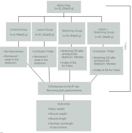
Animals were divided into four groups: Control Group (CG, n=3): the gastrocnemius muscle was not submitted to contusion and received no treat-ment; Lesion Group (LG, n=10): the right gastroc-nemius muscle (RGM) was injured, as described by Minamoto et al.15; the Stretching Group (SG, n=10): RGM was not submitted to contusion, however, 72 hours after the animal arrived to the bioterium, the passive stretching of RGM began, once a day, 5 days in a role. Four repetitions of stretching were performed, and each of them lasted 30 seconds, with 30 second intervals between each repetition16 — it was repeated 4 times17; he Lesion and Stretching Group (LSG, n=10): RGM was submitted to injury, and after 72 hours, stretching began according to the protocol de-scribed earlier. All of the rats in all of the groups were submitted to orthotanasia on the 8th day (Figure 1).
Protocol to cause muscle contusion
Animals were anesthetized with intraperitoneal Ketamine (95 mg/kg) and Xylazine (12 mg/kg) and kept in ventral decubitus position. he right paw was manually immobilized, with knee extension and dorsal lexion at 90º from the tibial tarsal joint. After animals were anesthetized, the contusion was in accordance with the protocol by Minamoto et al.15.
Protocol for gastrocnemius muscle stretching
In order to stretch the RGM, animals were manually immobilized, and the maximum dorsilexion was per-formed at the right paw, for 30 seconds, with 30-sec-ond intervals between each repetition16, which was repeated 4 times17. Stretching began 72 hours after the lesion, and was conducted daily, once a day, for 5 consecutive days.
ratas fueron pesadas y se eliminaron los músculos de las patas para el análisis de peso y longitud muscular, número y longitud de
los sarcómeros. El peso corporal final aumentó en todos los gru-pos. El peso, la longitud muscular y el número de sarcómeros en serie (NSS) del GL fueron superiores al GE. Sin embargo, el NSS del
GLE fue superior al GE. La longitud del sarcómero del GE fue más
alta que la de los otros grupos. Se concluye que la contusión y el estiramiento no afectaron a la ganancia de peso corporal. El esti-ramiento generó sarcomerogenesis en músculos lesionados, pero no modificó el músculo sano.
Orthotanasia of animals and muscle removal
Twenty-four hours after the end of the experi-ment, animals received intraperitoneal anesthe-sia (Ketamine, 95 mg/kg and Xylazine, 12 mg/kg) to remove the gastrocnemius muscles bilaterally. Afterwards, animals were submitted to orthotanasia with anesthetic overdose.
During dissection, muscles were periodically dripped with saline solution (NaCl 0.9%). Afterwards, the muscle was weighed with a precision digital scale, and after that, the length of the muscle was measured by a digital pachymeter. h en, the gastrocnemius muscle was immersed into a glutaraldehyde solution (2.5%) for 3 hours, and then it was transferred into a nitric acid solution (30%) for 48 hours, being after-wards stored in glycerol (50%).
Estimation of serial
sarcomeres and lenght of sarcomeres
For the confection of histological blades, i ve muscle i -bers were isolated from the venter of each gastrocnemius
muscle, right and left sides. Afterwards, the isolated i bers were placed on a histological blade, in gelatin-glycerin (Sigma) and protected by a slip. In each mus-cle i ber, the serial sarcomere number was identii ed throughout 300 μm, in a light microscope (100x ob-jective, immersed). Quantii cation was made in a video monitor, with a video-image system attached to the microscope.
h e total number and length of sarcomeres, in each isolated muscle i ber, were estimated by correlating the number of sarcomeres identii ed throughout 300 μm of the i ber and the total length of the muscle, as described by Williams and Goldspink18. Even though there are controversies in literature, in this study, the length of sarcomeres throughout muscle i bers was considered to be homogeneous14.
Statistical analysis
In order to assess normality and homoscedastic-ity, the Shapiro Wilk and Levene tests were per-formed, respectively. Descriptive statistics for para-metric and non-parapara-metric results are expressed as
7 days
Albino Rats
Control Group
• No Intervention • Contusion- Friday
Orthotanasia on the 8th day Removing both gastrocnemius
Outcomes: • Body weight • Muscle weight • Muscle length • Number and length
of sarcomeres
• Contusion - Friday
• Remained 1 week in the bioterium
• Remained 1 week in the bioterium
Lesion Group Stretching Group Lesion + Stretching Group (n=3, 196±19 g) (n=10, 262±18 g) (n=10, 183±15 g)
(n=10, 218±16 g) (n=33, 219±35 g)
• Stretching 72h after
• 4 daily of 30s arriving to the
bioterium - Monday • Stretching 72h after
arriving to the bioterium - Monday for 5 days
• 4 daily of 30s for 5 days
mean±standard-deviation. Intra and intergroup com-parisons were performed by ANOVA post hoc Tukey unequal HSD for parametric values; in case of non-parametric values, the Kruskal Wallis test was used. Values were signii cant when p≤0.05.
RESULTS
Body weight
Significant increase was found between initial and final body weight in all of the groups (intragroup, p<0.05, ANOVA post hoc Tukey unequal HSD). With regard to absolute weight, the final body weight of LG was higher than the one in SG (337±28 ver-sus 281±28 g, p=0.008) and LSG (337±28 versus
275±25 g, p=0.002, ANOVA). Concerning the rela-tive difference in weight gain, no significant dif-ference was found between groups. Results are de-scribed in the Table.
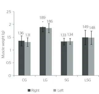
Muscle weight
No signii cant dif erence was found by comparing the RGM and the left muscle (LGM) (intragroup, p>0.05). In intergroup comparison, the RGM muscle weight in the LG was higher than in the SG (1.89±0.17
versus 1.33±0.13 g, p=0.001, Kruskall-Wallis). Results are demonstrated in Figure 2.
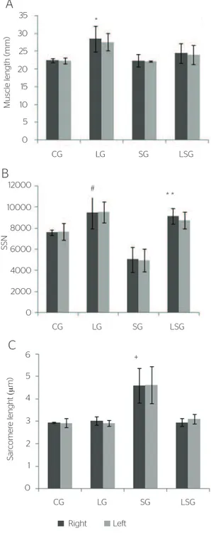
Muscle strength of the gastrocnemius
By analyzing the muscle strength of the RGM and the LGM, no intragroup signii cant dif erence was ob-served. In intergroup comparisons, the length of the RGM in the LG was higher in relation to the one in
the SG (28.53±3.63 versus 22.37±1.82 mm, p=0.01, ANOVA post hoc Tukey unequal HSD). Data are pre-sented in Figure 3A.
Estimation of the Serial
Sarcomere Number
In the comparison between the serial sarcomere number of the RGM and the LGM, no statistically signii cant dif erence was found (ANOVA post hoc Tukey).
In the intergroup comparison of the RGM, the LG was higher than the SG (9,455±1,540 versus
5,023±1,188; p=0.0001, ANOVA post hoc Tukey un-equal HSD), and the LSG was higher than the SG (9,123±720 versus 5,023±1,188; p=0.0002). Data are demonstrated in Figure 3B.
Length of sarcomeres
In relation to intragroup comparison, no statistically signii cant dif erence was found in any group.
In the length of sarcomeres of the RGM, a sig-nificant increase in the SG in was observed in re-lation to the LG (4.60±0.77 versus 3.03±0.18 µm, p=0.0008), in relation to the LSG (4.60±0.77 ver-sus 2.96±0.17 µm, p=0.001) and in relation to CG (4.60±0.77 versus 2.95±0.03 µm, p=0.001) (ANOVA
post hoc Tukey unequal HSD). Data are presented in Figure 3C.
Results are mean±standard deviation. CG: Control Group; LG: Lesion Group; SG: Stretching Group; LSG: Lesion and Stretching Group. *p=0.001 when compared to the SG. Right: right gastrocne-mius muscle; Left: left gastrocnegastrocne-mius muscle.
Figure 2. Ef ect of stretching on the gastrocnemius muscle weight
Muscle w
eight (g)
Right CG 1.36
1.89
1.33
1.49 1.48
1.34 1.86
1.31 2.5
1.5 2
1
0.5
0
LG SG LSG
Left *
Table. Ef ect of stretching on the body weight of rats
Groups
initial weight (g)
Mean±SD
Final weight (g)
Mean±SD
Relative diff erence
(%)
p-value
ANOVA
GC 196±19 286±19* 46,3 0,0006
LG 264±18 337±28*# 28,6 0,0001
SG 190±12 275±25* 45,1 0,0001
LSG 197±21 281±28* 43,7 0,0001
DISCUSSION
h e results in this study showed increased muscle weight, muscle length and estimation of SSN in the LG in relation to the SG. h e stretching protocol of the injured muscle provided an increment in the number of sarcomeres in relation to the non-injured group.
Muscle i ber is able to adapt to new stimuli, there-fore, it can change its volume, muscle length, number and length of the sarcomeres19.
In relation to i nal body weight, it was possible to observe increase in all of the groups, which is compat-ible with the regular body weight gain of the animal14. However, the LG presented higher body weight when compared to the SG and to the LSG. h is outcome sug-gests that daily stretching may have interfered in body weight gain. However, relative body weight gain was not signii cant between groups, and this fact demon-strates that stretching did not interfere in this variable.
Muscle weight of the LG was higher when com-pared to the SG, which may have occurred because of the acute phase after contusion, characterized by an inl ammatory period. In this case, the presence of edema is common, which may have led to increased muscle weight20.
Increased muscle weight after lesion is not in accor-dance with literature. While some authors found similar results in relation to this study, that is, increased muscle weight in the injured muscle20-22, others did not i nd any increase in the weight of the gastrocnemius muscle 48 hours after the lesion, and such lack of change was justi-i ed by the protejusti-in depletjusti-ion caused by the justi-injury23.
On the other hand, it is important to consider that body weight gain occurred in all of the groups, and both muscle weight and length increased with the increment of body weight. Besides, muscle length is not the best measure to establish the longitudinal muscle length. In that case, it is recommended to count the serial sarco-mere number.
In this study, a higher number of serial sarcomeres was observed in the RGM in the LG when compared to the SG. h erefore, it is assumed that the higher i nal absolute body weight in the LG may have caused in-creased muscle weight and length, thus resulting in more serial sarcomeres24. However, relative weight gain between groups did not present signii cant dif erences. Anyway, it is not possible to suggest that the higher number of serial sarcomeres of the LG was a result of muscle contusion.
In the analysis of the length of sarcomeres in the LG, they were shorter when compared to the SG. h is i nding corroborates the study by Williams and Goldspink25, who reported that changes in the serial sarcomere number imply adjustments in the length of sarcomeres. h erefore, the shorter length of sarcomeres found in the LG may have been a result of the higher Results are mean±standard deviation. CG: Control Group; LG: Lesion Group; SG: Stretching Group;
LSG: Lesion and Stretching Group. SSN: Estimation of the serial sarcomere number. Right: right gastrocnemius muscle; Left: left gastrocnemius muscle; (A) *p<0.05 compared to the SG (p=0.03); (B) #p<0.05 compared to the SG and **when compared to the SG (p=0.001); (C) +p<0.05 compared to the LG (p=0.001), LSG (p=0.001) and CG (p=0.04)
Figure 3. Muscle strength, estimation of number and length of sarcome-res in the gastrocnemius of rats
B
CG LG SG LSG
(μ
SSN
12000
10000
8000
6000
4000
2000
0
#
* *
C
CG LG SG LSG
S
ar
comer
e lenght
(μ
m)
0 1 2 3 4 5
6 +
(μ
Right Left CG
A
B
LG SG LSG
Muscle length (mm)
(μ
35
30
25
20
15
10
0 5
serial sarcomere number, so there could be a great su-perposition of contractile ilaments25,26.
On the other hand, the LSG presented higher se-rial sarcomere number when compared to the SG, how-ever, these groups did not present statistical diferences in relation to the CG. his outcome suggests that the injured muscle may respond diferently to stretching when compared to the healthy muscle. Other research-ers assessed the efect of stretching, twice a week, in the healthy soleus muscle of rats, and, after 3 weeks, they observed increased serial sarcomere number27.
In this study, it is possible to assume there were not time or stimuli enough to increment the serial sarco-mere number in non-injured muscles. Previous studies showed that the immobilization stimulus in stretched muscles, for 3 to 4 weeks, led to increased muscle length by the addition of serial sarcomere number28,29. However, in this study, muscles were not immobilized in the stretched position, and they also were not main-tained for 3 to 4 weeks, which is time enough for the important addition of serial sarcomere number, even when stretching is intermittent, that is, muscles are not maintained immobilized in the stretching position14.
It was surprising to ind more sarcomeres in the LSG when compared to the SG. Until now, no article was found that assessed the number and length of sarcomeres after muscle lesion. Besides, there are not many articles about the efects of stretching after muscle injury.
Hwang et al.9 observed the efects of passive stretch-ing (5 seconds of maintenance and 5 seconds of interval for 15 minutes) in muscles of rats in the inlammatory, regenerative and proliferative phases. hese authors concluded that all of the groups submitted to stretch-ing (initiated on the 2nd, 7th, and 14th days) presented decreased ibrosis, signiicant improvement of regenera-tion and increased muscle strength, which were more expressive on the 14th day.
Some studies assessed the efects of immobiliza-tion in the process of muscle regeneraimmobiliza-tion22,30. Järvinen et al.22 reported that mobilization leads to the faster and more intense re-epithelization of lesion borders, espe-cially at early stages. Faria et al.30 investigated diferent mobilization protocols after muscle lesion in rats. he proposed protocols were addressed at mobilizing during 5 to 8 days, with 15 to 45-minute sessions, beginning 1 and 3 hours after the lesion. In this study, the conclu-sion is that the process of muscle regeneration is related to the beginning and the time of mobilization, being higher in groups that began earlier and, therefore, were mobilized for longer.
herefore, the sarcomerogenesis observed in the LSG compared to the SG may have responded to the early beginning of mobilization, since in this study stretching began 72 hours after contusion. As to the SG, since the muscle was at normal state, that is, not shortened, not immobilized, not injured, it is assumed that the necessary stimulus to induce the addiction of serial sarcomeres should be stronger and for a longer period, as performed in other studies14,27.
In this study, longer length of sarcomeres was found in the SG in comparison to the LSG, which is in ac-cordance with the hypothesis that the adjustment of sarcomere length agrees with the number of sarcomeres for the muscle to develop maximum tension25.
Some limitations in this study can be indicated, such as the absence of an analysis of the cross-sec-tion area, the conjunctive tissue, the gene expression and proteins, in order to investigate the mechanisms of plasticity of injured muscles in response to the stretching stimulus.
In this study, it was observed that contusion and stretching did not interfere in the inal body weight gain of the animals. Besides, the stretching protocol was suicient to induce sarcomerogenesis in injured muscles, without interfering in the healthy muscle. However, muscles that have been only stretched, that is, not-injured, presented longer sarcomere length when compared to other groups, demonstrating that the absence of sarcomerogenesis leads to increased sarcomere length.
CONCLUSION
he outcomes of this study indicate the importance of stretching, even at the acute phase after muscle injury, in order to gain serial sarcomeres. With the proper extrapolation restrictions, the employed stretching protocol allows to suggest the clinical indication to prevent the loss of movement range, usually observed after muscle injury.
REFERENCES
2. Järvinen TAH, Järvinen TLN, Kääriäinen M, Kalimo H, Järvinen M. Muscle injuries: biology and treatment. Am J Sports Med. 2005;33(5):745-64.
3. Oliveira NML, Gava AD, Salvini TF. O efeito da crioterapia e compressão intermitente no músculo lesado de ratos: uma análise morfométrica. Rev Bras Fisioter. 2007;11(5):403-9.
4. Lapointe BM, Frémont P, Côté CH. Influence of nonsteroidal anti-inflammatory drug treatment duration and time of onset on recovery from exercise-induced muscle damage in rats. Arch Phys Med Rehabil. 2003;84(5):651-5.
5. Mendias CL, Tatsumi R, Allen RE. Role of cyclooxygenase-1 and -2 in satellite cell proliferation, diferentiation and fusion. Muscle Nerve. 2004;30(4):497-500.
6. Mackey AL, Mikkelsen UR, Magnusson SP, Kjaer M. Rehabilitation of muscle after injury- the role of anti-inflammatory drugs. Scand J Med Sci Sports. 2012;22(4):8-14.
7. Reurink G, Goudswaard GJ, Tol JL, Verhaar JA, Weir A, Moen MH. Therapeutic interventions for acute hamstring injuries: a systematic review. Br J Sports Med. 2012;46(2):103-9.
8. Mason DL, Dickens V, Vail A. Rehabilitation for hamstring injuries. Scand J Med Sci Sports. 2007;17(2):45-75.
9. Hwang JH, Ra Y, Lee KM, Lee JY, Ghil SH. Therapeutic efect of passive mobilization exercise on improvement of muscle regeneration and prevention of fibrosis after laceration injury of rat. Arch Phys Med Rehabil. 2006;87(1):20-6.
10. Kannus P. Immobilization or early mobilization after an acute soft-tissue injury? Phys Sport Med. 2000;28(3):1-8.
11. Järvinen MJ, Lehto MU. The efects of early mobilisation and immobilisation on the healing process following muscle injuries. Sports Med. 1993;15(2):78-89.
12. Kannus P, Jozsa L, Kvist M, Järvinen T, Järvinen M. Efects of immobilization and subsequent low-and high-intensity exercise on morphology of rat calf muscles. Scand J Med Sci Sports. 1998;8(3):160-71.
13. Gomes ARS, Coutinho EL, França CN, Polonio J, Salvini TF. Efect of one stretch a week applied to the immobilized soleus muscle on rat muscle fiber morphology. Braz J Med Biol Res. 2004;37(10):1473-80.
14. Coutinho EL, Gomes ARS, França CN, Oishi J, Salvini TF. Efect of a passive stretching on the immobilized soleus muscle fiber morphology. Braz J Med Biol Res. 2004;37(12):1853-61.
15. Minamoto V, Bunho SR, Salvini TF. Regenerated rat skeletal muscle after periodic contusions. Braz J Med Biol Res. 2001;34(11):1447-52.
16. Polizello JC, Carvalho LC, Freitas FC, Padula N, Shimano AC, Matiello- Sverzut AC. Propriedades mecânicas do músculo gastrocnêmio
de ratas, imobilizado e posteriormente submetido a diferentes protocolos de alongamento. Rev Bras Med Esp. 2009;15(3):195-9.
17. Taylor DC, Dalton JC, Seaber AV, Garret WE. Viscoelastic properties of muscle-tendon units. The biomechanical efects of stretching. Am J Sports Med. 1990;18(3):300-8.
18. Williams PE, Goldspink G. Longitudinal growth of striated muscle fibers. J Cell Sci. 1971;9(3):751-67.
19. Burkholder TJ, Lieber RL. Sarcomere number adaptation after retinaculum transection in adult mice. J Exp Biol. 1998;201(Pt 3):309-16.
20. Crisco JJ, Jokl P, Heinen GT, Connell MD, Panjabi MM. A muscle contusion injury model. Biomechanics, physiology and histology. Am J Sports Med. 1994;22(5):702-10.
21. Salvini TF, Coutinho EL, Russo TL, Deluca C. One-minute bouts of passive stretching after immobilization increase sarcomerogenesis in rat soleus muscle. Braz J Morphol Sci. 2006;23(2):271-7.
22. Järvinen M. Healing of a crush in rat striated muscle. A histological study of the efect of early mobilization and on the repair processes. Acta Pathol Microbiol Scand. 1975;83(3):269-82.
23. Fisher BD, Baracos VE, Shnitka TK, Mendryk SW, Reid DC. Ultrastructural events following acute muscle trauma. Med Sci Sports Exerc. 1990;22(2):185-93.
24. Menon T, Casarolli LM, Cunha BC, Souza L, Andrade PHM, Albuquerque CE, et al. Influência do alongamento passivo em três repetições de 30 segundos a cada 48 horas em músculo sóleo imobilizado de ratos. Rev Bras Med Esporte [on-line]. 2007;13(6):407-10.
25. Williams PE, Goldspink G. Changes in sarcomere length and physiological properties in immobilized muscle. J Anat. 1978;127(3):459-68.
26. Gordon AM.; Huxley AF, JULIAN FJ. The variation in isometric tension with sarcomere lenght in vertebrate muscle fibers. J Physiol. 1966;184(1):170-19.
27. Secchi KV, Morais CP, Cimatti PF, Tokars E, Gomes ARS. Efects of stretching and resistive exercise in rat skeletal muscle. Rev Bras Fisioter. 2008;12(3): 228-34.
28. Williams PE, Goldspink G. Connective tissue changes in immobilised muscle. J Anat. 1984;138(2):343-50.
29. Williams PE, Catanese T, Lucey EG, Goldspink G. The importance of stretch and contractile activity in the prevention of connective tissue accumulation in muscle. J Anat. 1988;158:109-14.