Supramolecular hydrogen-bonding network
in 1-(diaminomethylene)thiouron-1-ium 4-hydroxybenzenesulfonate
crystal
Genivaldo J. Perpétuo
a, Rafael S. Gonçalves
a, Jan Janczak
b,⇑aDepartamento de Física, Instituto de Ciências Exatas e Biológicas, Universidade Federal de Ouro Preto, 35400-000 Ouro Preto, MG, Brazil
bInstitute of Low Temperature and Structure Research, Polish Academy of Sciences, PO Box 1410, 50950 Wrocław, Poland
h i g h l i g h t s
Crystal of 1-(diaminomethylene)
thiouron-1-ium 4-hydroxybenzene-sulfonate was obtained.
Hydrogen bonding motif R22(8) of 1:1
forms supramolecular complex.
The characteristic vibrational bands of
the H and D analogue are discussed.
g r a p h i c a l
a b s t r a c t
The solid-state organisation of 1-(diaminomethylene) thiouron-1-ium 4-hydroxybenzenesulfonate has been analysed with respect to cation–anion and hydrogen bonding interactions. The oppositely charged units interactviahydrogen bonds with the graph of R22(8).
a r t i c l e
i n f o
Article history:
Received 16 January 2015
Received in revised form 18 March 2015 Accepted 27 April 2015
Available online 6 May 2015
Keywords:
1-(diaminomethylene)thiouron-1-ium 4-hydroxybenzenesulfonate Crystal structure
Hydrogen bonds DFT calculations Vibrational spectroscopy
a b s t r a c t
The single crystals of 1-(diaminomethylene)thiouron-1-ium 4-hydroxybenzenesulfonate were grown using a solution growth technique. The compound crystallises in the centrosymmetricP21/cspace group
of the monoclinic system. The conformation of the 1-(diaminomethylene)thiouron-1-ium cation is not strictly planar, but twisted. Both arms of the cation are oppositely rotated by 8.5(1)°around the CAN
bonds involving the central N atom. The arrangement of oppositely charged components, i.e. 1-(diamino methylene)thiouron-1-ium cations and 4-hydroxybenzenesulfonate anions in the crystal is mainly deter-mined by ionic and hydrogen-bonding interactions forming supramolecular network. The possible hydrogen-bonding interactions between cation and anion units were analysed on the basis of molecular orbital calculations. The obtained deuterated analogue crystallises similar as H-compound in the mono-clinic system (P21/c) with quite similar lattice parameters. The compound was also characterised by the
FT-IR and Raman spectroscopies. The characteristic bands of the functional and skeletal groups of the pro-tiated and deuterated analogue of 1-(diaminomethylene)thiouron-1-ium 4-hydroxybenzenesulfonate are discussed.
Ó2015 Elsevier B.V. All rights reserved.
http://dx.doi.org/10.1016/j.molstruc.2015.04.037 0022-2860/Ó2015 Elsevier B.V. All rights reserved.
⇑Corresponding author.
E-mail address:j.janczak@int.pan.wroc.pl(J. Janczak).
Contents lists available atScienceDirect
Journal of Molecular Structure
Introduction
Supramolecular chemistry and crystal engineering are closely related fields[1]. Both have been rapidly extending research area over the past two decades [2]. The non-covalent intermolecular interactions are of fundamental importance for understanding of
molecular recognition and self-organisation supramolecular syn-thesis of new solids with designated architecture and desired phys-ical and chemphys-ical properties [3]. A productive strategy in the crystal engineering is to build supramolecular structures from molecules containing complementary arrays of the hydrogen bonding sites [4]. The supramolecular synthon of recurring
N
N
S
N
N
H
H
H
H
H
H
N
N
S
N
N
H
H
H
H
H
H
O
S
O
O
O
H
H
(a)
(b)
(c)
Scheme 1.2-imino-4-thiobiuret (a) and it tautomeric form of 1-(diaminomethylene)-thiourea (b), and 4-hydroxybenzenesulfonic acid (c).
N
N
S
N
N
H
H
H
H
H
H
H
O
S
O
OH
O
N
N
S
N
N
H
H
H
H
H
H
H
O
S
O
OH
O
N
N
S
N
N
H
H
H
H
H
H
H
O
S
O
OH
O
N
N
S
N
N
H
H
H
H
H
H
H
O
S
O
OH
O
N
N
S
N
N
H
H
H
H
H
H
H
N
N
S
N
N
H
H
H
H
H
H
H
O
S
O
HO
O
O
S
O
OH
O
N
N
S
N
N
H
H
H
H
H
H
H
O
S
O
OH
O
N
N
S
N
N
H
H
H
H
H
H
H
O
S
O
OH
O
R (8)
22(a)
(b)
(e)
R (8)
22(c)
R (8)
22
(d)
R (6)
R (6)
2 2
1 1
(f)
(g)
(h)
R (6)
22R (6)
2 2R (6)
22R (8)
22R (8)
22Scheme 2.Possible hydrogen-bonding interactions between 1-(diaminomethylene)-thiouron-1-ium cation and 4-hydroxybenzenesulfonate anion with graphs R22(8), R21(6)
and R22(6).
hydrogen-bonding pattern is an effective approach for structural design of solids[5].
The commercially available 2-imino-4-thiobiuret (Aldrich, CAS No. 2114-20-05) is, as has been shown by the X-ray single crystal analysis[6], the tautomeric form of 1-(diaminomethylene)thiourea (Scheme 1). Both tautomers are already considered to be useful building blocks in crystal engineering, since they contain hydrogen-bonding sites. Additionally, both tautomers can act as
N,N- or N,S-coordinating ligands forming several types of com-plexes with metal ions[7].
The 1-(diaminomethylene)-thiourea contains the basic N atom with the lone pair of electrons, therefore it can forms salts with organic and inorganic acids forming extended hydrogen-bonding networks in solids[8]. In particular, the supramolecular aggrega-tion pattern of 1-(diaminomethylene)-thiourea with tartaric acid and its deuterated analogue are examples of supramolecular hydrogen-bonding networks in solids that can be utilised as mate-rials for non-linear optics[9].
In the present work, we investigate the supramolecular architec-ture formed by self-assembly of 1-(diaminomethylene)-thiourea with 4-hydroxybenzenesulfonic acid (Scheme 1c). This study is aimed into the interactions between oppositely charged units, i.e. 1-(diaminomethylene)-thiouron-1-ium cations and 4-hydroxyben-zenesulfonate anions, and the formation of the supramolecular structure. The possible hydrogen-bonding interaction between 1-(d iaminomethylene)-thiouron-1-ium cation and 4-hydroxybenzene-sulfonate anion with different graphs illustrates Scheme 2. The
compound was also characterised by vibrational spectroscopy, and the results are discussed and compared with the data obtained for a neutral 1-(diaminomethylene)-thiourea molecule.
Experimental
All materials were commercially available and used as received. Elemental analysis was carried out with a Perkin–Elmer 240 ele-mental analyser.
Preparation of 1-(diaminomethylene)thiouron-1-ium 4-hydroxybenzenesulfonate and its deuterated D8analogue
Commercially available 2-imino-4-thiobiuret (Aldrich, CAS No. 2114-02-05), which is in fact the tautomeric form 1-(diaminome-thylene)thiourea and the 4-hydroxybenzenesulfonic acid were added to hot water in a molar proportion of 1:1. When the solution became homogeneous it was cooled slowly and kept at room temperature. After several days, transparent colourless crystals of C6H4(OH)SO3C2H7N4S were formed. Analysis: calculated for
C8H12N4S2O4: C, 32.86; N, 19.17; O, 21.89; S, 21.94 and H, 4.14%.
Found: C, 32.66; N, 19.10; O, 22.04; S, 22.09 and H, 4.09%. Deuterated d8 analogue of 1-(diaminomethylene)thiouron-1-ium
4-hydroxybenzenesulfonate was prepared by the usual reaction with heavy water. The crystals of 1-(diaminomethylene)thiouro n-1-ium 4-hydroxybenzenesulfonate were dissolved in heavy water, and left in the atmosphere saturated with heavy water for one weak, in order to avoid the contamination of the crystals. The procedure was repeated twice.
X-ray data collection
X-ray intensity data for the crystals were collected using gra-phite monochromatic Mo K
a
radiation on a four-circlej
geometry KUMA KM-4 diffractometer with a two-dimensional area CCD detector. Thex
-scan technique with Dx
= 1.0° for each imagewas used for data collection. The 930 images for six different runs covering over 99% of the Ewald sphere were performed. One image was used as a standard after every 50 images for monitoring of the crystal stability and the data collection. No correction on the rela-tive intensity variations was necessary. Data collections were made using the CrysAlis CCD program[10]. Integration, scaling of the reflections, correction for Lorentz and polarisation effects and absorption corrections were performed using the CrysAlis Red pro-gram[10]. The structures were solved by the direct methods using SHELXS-97 and refined using SHELXL-97 program[11]. The hydro-gen atoms involving in the hydrohydro-gen bonds were located in differ-ence Fourier maps and were refined. The hydrogen atoms joined to aromatic carbon atoms were introduced in their geometrical Table 1
Crystallographic data for 1-(diaminomethylene)thiouron-1-ium 4-hydroxy-benzenesulfonate.
Empirical formula (C2H7N4S)(C6H4(OH)SO3)
Formula weight (g mol 1) 292.34
Crystal system, space group Monoclinic,P21/c(No. 14)
a(Å) 5.646(1)
b(Å) 7.601(1)
c(Å) 29.405(5)
b(°) 93.66(2)
V(Å3) 1259.3(4)
Z 4
Dcalc/Dobs(g cm 3) 1.542/1.54
l(mm1) 0.436
Crystal size (mm) 0.380.320.20
Radiation type, wavelength,k(Å) MoKa, 0.71073
Temperature (K) 295(2)
hrange (°) 3.01–29.40
Absorption correction Numerical, CrysAlis Red[10]
Tmin/Tmax 0.8526/0.9191
Reflections collected/unique/observed 13522/3015/2004
Rint 0.0248
Refinement on F2
R[F2> 2r(F2)] 0.0336
wR(F2all reflections) 0.0699
Goodness-of-fit,S 1.002
Dqmax,Dqmin(e Å3) +0.161, 0.223
wR= {R[w(Fo2–Fc2)2]/RwFo4}1/2;w1= 1/[r2(Fo2) + (0.0325P)2] whereP= (Fo2+ 2Fc2)/3.
Table 2
Selected bond lengths (Å) and angles (°) for 1-(diaminomethylene)thiouron-1-ium 4-hydroxy-benzenesulfonate.
S1AC1 1.6716(15) C1AN2 1.3107(19)
C1AN1 1.3831(19) N1AC2 1.3621(18)
C2AN3 1.3032(19) C2AN4 1.2950(20)
S2AC3 1.7579(14) S2AO1 1.4438(11)
S2AO2 1.4643(10) S2AO3 1.4554(11)
N2AC1AN1 112.81(13) N4AC2AN3 121.64(16)
N2AC1AS1 122.00(13) N4AC2AN1 121.14(16)
N1AC1AS1 125.15(11) N3AC2AN1 117.21(14)
C2AN1AC1 130.85(13)
Table 3
Hydrogen-bond geometry (Å,°) for 1-(diaminomethylene)thiouron-1-ium 4-hydroxy-benzenesulfonate.
DAH A DAH H A D A DAH A
N1AH1 O3a 0.80 (2) 2.27 (2) 3.016 (2) 155 (2)
N2AH2 S1b 0.90 (2) 2.53 (2) 3.414 (2) 168 (2)
N2AH3 O3a 0.84 (2) 2.23 (2) 2.994 (2) 153 (2)
N3AH4 O1 0.85 (2) 2.08 (2) 2.921 (2) 170 (2)
N3AH5 O2a 0.88 (2) 2.01 (2) 2.880 (2) 172 (2)
N4AH6 O3 0.86 (2) 2.17 (2) 3.018 (2) 169 (2)
N4AH7 S1 0.93 (2) 2.20 (2) 2.988 (2) 141 (2)
O4AH44 O2c 0.82 (2) 1.94 (2) 2.732 (2) 162 (2) Symmetry codes.
ax,y+ 1,z.
b x 1, y+ 2, z+ 1. c x+ 1,y+ 1/2, z+ 1/2.
positions. The final difference Fourier maps showed no peaks of chemical significance. Details of the data collection parameters, crystallographic data and final agreement parameters are collected
in Table 1. Visualisations of the structures were made with the Diamond 3.0 program [12]. Selected geometrical parameters are listed inTable 2and the geometry of hydrogen bonding interac-tions is collected in Table 3. The obtained deuterated analogue crystallises similar as H-compound in monoclinic system (P21/c)
with quite similar lattice parameters.
Vibrational spectra measurements
The vibrational measurements of H-compound and its deuter-ated (D8) analogue were carried out at room temperature. The
Fourier transform infrared spectrum was recorded from nujol mulls between 4000 and 400 cm 1on a Bruker IFS 113 V FTIR
spec-trometer. Resolution was set up to 2 cm 1. The Fourier Transform
Raman spectrum was recorded on a FRA-106 attached to the Bruker 113 V FTIR spectrometer equipped with Ge detector cooled to liquid nitrogen temperature. Resolution was set up to 2 cm 1,
signal/noise ratio was established by 32 scans. Nd3+-YAG
air-cooled diode pumped laser of power ca. 200 mW was used as an exciting source. The incident laser excitation was 1064 nm. The scattered light was collected at the angle of 180°in the region of 3600–80 cm 1, resolution 2 cm 1, 256 scans.
Theoretical calculation
Theoretical calculations with geometry optimisation of possible hydrogen-bonding interaction between 1-(diaminomethyle ne)-thiouron-1-ium cation and 4-hydroxybenzenesulfonate anion with different graphs illustrated inScheme 2were performed with Gaussian09 program package [13]. All calculations were carried out with the DFT method using the Becke3–Lee–Yang–Parr corre-lation functional (B3LYP) [14–16]with the 3-21+G⁄basis set. As convergence criterions the threshold limits of 0.00045 and Table 4
Difference between the energies of most stable hydrogen-bonded unit c and the other units of 1-(diaminomethylene)-thiouron-1-ium cation and 4-hydroxybenzenesul-fonate anion.
Hydrogen bonded units as inScheme 2 DE(kJ mol 1)
a 0
b 2.03
c 6.88
d 7.03
e 6.72
f 70.10
g 69.22
h 70.08
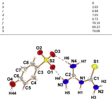
Fig. 1.View of 1-(diaminomethylene)thiouron-1-ium 4-hydroxybenzenesulfonate
showing displacement ellipsoids at the 50% probability level and H atoms as a sphere of arbitrary radii. Dashed lines represent the hydrogen bonds.
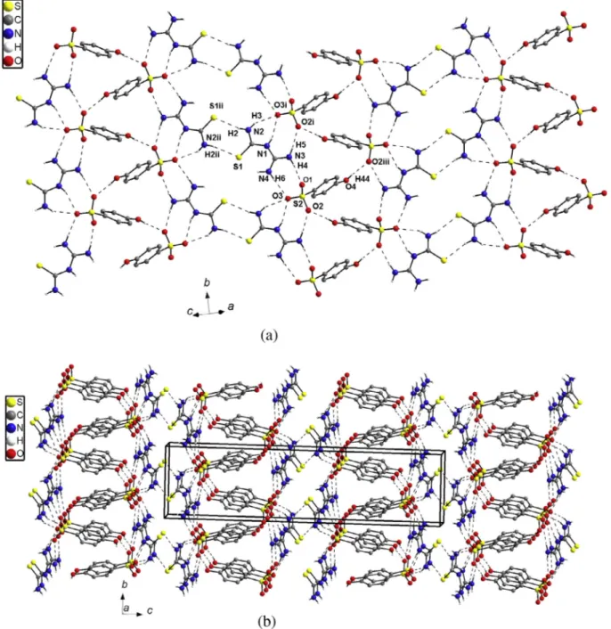
Fig. 2.View of the heat-to-tail hydrogen bonded chain of 4-hydroxybenzenesulfonate (a) and one-dimensional ribbon of 4-hydroxybenzenesulfonate(-) anions with
assistance of 1-(diaminomethylene)thiouron-1-ium cations (b).
0.0018 a.u. were applied for the maximum force and the displace-ment, respectively. Energies were calculated for optimised geome-tries of hydrogen-bonded units of 1-(diaminomethylene)-thiouron-1-ium cation and 4-hydroxybenzenesulfonate anion. The difference between the energy of most stable hydrogen-bonded unit a (see Scheme 2) and the energy of the other hydrogen-bonded units of 1-(diaminomethylene)-thiouro n-1-ium cation and 4-hydroxybenzenesulfonate anion are listed inTable 4.
Results and discussion
Good quality single crystals of 1-(diaminomethylene) thiouron-1-ium 4-hydroxybenzenesulfonate suitable for the X-ray single crystal analysis were obtained from water solutions at ambi-ent temperature. The 1-(diaminomethylene) thiouron-1-ium
4-hydroxybenzenesulfonate crystallises in the centrosymmetric space groupP21/cof monoclinic system. The deuterated D8analogue
was obtained by usual reaction of protiated crystals with heavy water. The deuterated analogue also crystallises in the monoclinic system (P21/c) with quite similar lattice parameters, so both
proti-ated and deuterated crystals of 1-(diaminomethylene) thiouron-1-ium 4-hydroxybenzenesulfonate are isostructural.
The X-ray single crystal analysis of 1-(diaminomethylene) thiouron-1-ium 4-hydroxybenzenesulfonate shows that the sul-fonate group is deprotonated, and the proton is transferred to the central N1 atom of 1-(diaminomethylene) thiourea molecule form-ing 1-(diaminomethylene) thiouron-1-ium cation. The oppositely charged units interact via two almost linear hydrogen bonds with a graph of R22(8) forming molecular complex as illustrateFig. 1. The
R22(8) motif is formed by donation to the sulfonate group of
4-hydroxybenzenesulfonate anion from the amino groups bonded to the C2 atom as showsScheme 2e.
Fig. 3.View of two-dimensional supramolecular layer (a) and the crystal packing of 1-(diaminomethylene)thiouron-1-ium 4-hydroxybenzenesulfonate (b).
The conformation of the 1-(diaminomethylene)thiouron-1-ium cation in the crystals is not strictly planar, but twisted. Both arms of the cation are oppositely rotated around the CAN bonds involv-ing the central N1 atom. The dihedral angle between the N1/C1/S1/N2 and N1/C2/N3/N4 planes is equal to 8.5(1)°. The dihe-dral angle in the present structure is significantly smaller than that in the crystal of neutral 1-(diaminomethylene)thiourea (22.2(1)°) [6]. The gas-phase conformation of the 1-(diaminomethylene) thiouron-1-ium cation as show theab initioMO calculations is also twisted with similar dihedral angle of 6.2° [8a]. A search in the Cambridge Structural Data Base (version 5.35, November 2013) [17] for structures containing 1-(diaminomethylene)thiouro n-1-ium cation yield 23 structures, in which the cation exhibits also twisted conformation. The 1-(diaminomethylene)thiouro n-1-ium cation twisting may differ (1.4°) for 1-(diaminomethyle ne)thiouron-1-ium perchlorate [8b] to 22.9(1) for 1-(diaminome-thylene)thiouron-1-ium chloride [8a] and depends on the anions and is undoubtedly dependent on the hydrogen bonding system formed with the oppositely charged units. Protonation of the neu-tral 1-(diaminomethylene)thiourea molecule causes a decrease of the steric effect of the lone pair of electron at the central N1 atom and reduce the twisting angle between the arms of the 1-(diamino methylene)thiouron-1-ium cation, when compared with that of
neutral molecule. The C1AS1 bond (Table 2) is slightly longer than the typical C@S double bond as observed in the thioformaldehyde CH2C@S (1.6019(8) Å) [18], which represents 100% double-bond
character. The three CAN bonds linking the amine groups are shorter than the CAN bonds involving the central N1 atom (Table 2). The planarity of the amine groups points to the sp2
hybridisation of the orbitals on the amine nitrogen atoms and the lone pair of electron localised on theporbital. Therefore the partial delocalisation of the lone pair onporbital of the amine groups and of the
p
bond of the double C1@S1 and C2@N1 bonds is possible and leads to shortening of other CAN bonds linking the amine groups and to the elongation of the C1@S1 and C2@N1 bonds (Table 2).The geometrical parameters of the anionic part of the crystal, i.e. 4-hydroxybenzenesulfonate(-) do not deviate significantly from the reported values in the other structures containing this anion [17]. The hydroxyl group of 4-hydroxybenzenesulfonate(-) is coplanar with the aromatic ring, whereas the SO3group is oriented
with the O2 atom almost perpendicular to the ring plane (O2AS2AC3AC4 torsion angle is equal to 92.0(1)°). The CAS bond linking the SO3group with a distance of 1.7579(14) Å is typical for
the C(sp2)
AS bond[19].
The 4-hydroxybenzenesulfonate(-) anions related by a screw axis (21parallel tob) are linked head to tail forming OAH O hydrogen
(a)
(b)
4000 3500 3000 2500 2000 1500 1000 500
0.0 0.2 0.4 0.6 0.8 1.0
1112
1376
1438
710
1126
1270
2323
492
642
587
569
1682
695
834
949
974
1007
1030
1155
1169
1210
1277
1316
1500
1524
1540
1604
1643
2295
2374
2441
2497
2543
N
3386
3184
3286
Transmittance
4000 3500 3000 2500 2000 1500 1000 500
0.0 0.2 0.4 0.6 0.8 1.0
1112
710
3102
644
3334
986
1007
1156
1277
1346
1501
1517
725
568
691
834
1030
1126
1206
1376
1438
1657
1606
1705
N
3134
3184
3286
3386
Transmittance
Wavenumber [cm-1]
Wavenumber [cm-1]
Fig. 4.IR-spectrum of 1-(diaminomethylene)thiouron-1-ium 4-hydroxybenzene-sulfonate (a) and its deuterated analogue (b).
bonded zigzag chains along [0 1 0] direction (Fig. 2a). The one-dimensional arrangement of 4-hydroxybenzenesulfonate(-) anions form ribbon with assistance 1-(diaminomethylene)thiouro n-1-ium cations and the NAH O hydrogen bonds (Fig. 2b). Within the ribbon the hydrogen bonded 1-(diaminomethylene) thiouron-1-ium 4-hydroxybenzenesulfonate(-) molecules are related by the screw axis 21and translation. Neighbouring, inversion
related ribbons are combined via NAH S hydrogen bonds with a graph of R22(8) (Table 3) to form two-dimensional supramolecular
structure parallel to the (1 04) plane (Fig. 3a). The importance of such interactions has been questioned[20]. However, the DAH S inter-actions (D = donor) are important in the biological systems due to presence of high content of S atom in biological molecules. In addition, the NAH S interactions have been utilised for design supramolecular arrangement of thiourea derivatives [21]. Therefore, such NAH S interactions seem to be important in the present structures (Table 3), where the formation of the
intramolecular NAH S interactions is favoured by the six-membered hydrogen-bonded ring with a graph of S(6) and the intermolecular eight-membered hydrogen-bonded ring with a graph of R22(8); the presence of C@S bond makes it
some resonance-assisted stabilisation. The possibility of resonance-induced hydrogen bond ring formation with the S atom of a C@S group with N substituents has been mentioned by Allen et al.[22]. However, in this structure the hydrogen bonds involving the S atom seem to be driven by the stronger NAH O and OAH O hydrogen bonds (Table 3). Neighbouring, NAH S hydrogen bonding two-dimensional layers related via translation along the
a-axis interact mainly via van der Waals forces, since there are no directional interactions between the successive layers. Transitionally related along a-axis aromatic rings of 4-hydroxybenzenesulfonate(-) anions are overlapped (Fig. 3b). The distance between the ring centroids Cg(x,y,z) Cg(1 +x,yz) is
5.643(1) Å. This value is too large for the effective
p
–p
interactionsFig. 5.Raman spectrum of 1-(diaminomethylene)thiouron-1-ium 4-hydroxybenzene-sulfonate (a) and its deuterated analogue (b).
between the
p
-cloud of the aromatic rings of the 4-hydroxybenzenesulfonate(-) anions.Molecular orbital calculations with geometry optimisation and the energies of possible hydrogen-bonding interactions between 1-(diaminomethylene)-thiouron-1-ium cation and 4-hydroxyben-zenesulfonate anion are fall into three groups. The first with the lower energy is observed for hydrogen bonded units a and b (Scheme 2) with graphs of R21(6) and R22(8). However, the unit a
with the lowest energy represents the most possible hydrogen-bonding interactions between 1-(diaminomethyle ne)-thiouron-1-ium cation and 4-hydroxybenzenesulfonate anion and therefore it is observed in the structure of the crystal (see Figs.2b and3a). The other hydrogen bonded unit b with R21(6)
and R22(8) graphs with comparable energy to the unit a (with
DE= 2.03 kJ mol 1) is not present in the structure of the crystal.
The second group with intermediate energy was found for the hydrogen bonded units with a graph of R22(8) (c, d and e in
Scheme 2). Within these three units with comparable energy only one of them (hydrogen bonded unit e) is observed in the structure of the crystal (see Figs. 1 and 2b and 3a). The third group of hydrogen bonding interactions with significantly greater energy is found for the units with a graph of R22(6) formed
by donation to the sulfonate group from only one amine group (units f, g and h inScheme 2) and they are no found in the crystal. The difference (DE) between the energy of the most stable unit a and the energy of other hydrogen bonded units are listed in Table 4.
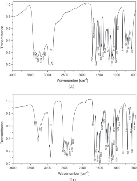
The FT-IR spectra of 1-(diaminomethylene)thiouron-1-ium 4-hydroxybenzenesulfonate and its deuterated analogue are shown inFig. 4a and b, respectively, whereas the Raman spectra are shown inFig. 5a and b. The title compound has several func-tional and skeletal groups such as three NH2 C@S, CANAC,
NACAN and NACAS groups in the cation and the SO3, OH and
the six-membered aromatic ring in the anion. The bands corre-sponding to the vibration of these groups were identified with the aid of infrared correlation charts[23]. The IR spectra of neutral 1-(diaminomethylene)thiourea [8g] and of p-phenolsulfonic acid [24]will be helpful for assignment of the bands observed in the spectrum of the title compound. A careful inspection of the IR trum (Fig. 4a) shows medium-strong intensity bands in the spec-tral range of 3400–3100 cm 1. These bands can be attributed to
the OH group of 4-hydroxybenzenesulfonate(-) anion and to the asymmetric and symmetric stretching of three NH2groups of the
1-(diaminomethylene)thiouron-1-ium cation. These bands, as expected, are shifted in the IR spectrum of deuterated analogue (Fig. 4b) to the spectral region of 2550–2250 cm 1. The isotopic
ratio between 1.331 and 1.352 points on the vibration anhar-monicity. The strong band at 2543 cm 1should be attributed to
the OD and the other to the asymmetric and symmetric stretching of three ND2 groups. In the spectrum of deuterated analogue
(Fig. 4b), the bands of the of the protiated compound with signifi-cantly lower intensities are observed. Theses bands resulting from Table 5
FT-IR spectral data for 1-(diaminomethylene)thiouron-1-ium 4-hydroxy-benzenesul-fonate and its deuterated analogue.
Protiated,m cm1 Deuterated,m cm 1 Assignment Isotopic ratio
3386s 2543s ma(NH2) orma(ND2) asym stretch, m(OH) or v(OD)
1.331
3334s 2497s ma(NH2) orma(ND2) asym stretch 1.335
3286s 2441s ma(NH2) orma(ND2) asym stretch 1.346 3184s 2374s ms(NH2) orms(ND2) sym stretch 1.341 3134s 2323m ms(NH2) orms(ND2) sym stretch 1.349 3102s 2295m ms(NH2) orms(ND2) sym stretch 1.352 Broad
Band 2800
2200 NAH...O or NAD...O hydrogen bonds
1705s 1210s Imine bond stretch 1.409
1657w 1682w,
1643s
1606vs 1604s NACAN bend + ring def.
1517m 1540s, 1524s
1501m 1500s
1438s 1438w m(CAN)
1376m 1376w c(CAC) overlapped with nujol
1346m 1316w
1277m 1277m ma(SO3) asym stretch 1206s
1156s 1155s ma(SO3) asym stretch
1126s 1126w ma(SO3) asym stretch
1118m 1112w ms(SO3) sym stretch
1030s 1030s ms(SO3) sym stretch
1007m 1007m ms(SO3) sym stretch., overlapped withc(CAN)
986w 974w, 949w m(CAC)
834m 834s CACAC def. of phenol ring 725m
710m 710m m(C@S)
691m 695s CACAO def. of phenol ring
644m 642w
587m
568m 569s Skeletal CANAC, NACAN, SO3 asym. def.
492w
Weak, w; medium, m; strong, s; very, v.
Table 6
Raman spectral data for 1-(diaminomethylene)thiouron-1-ium 4-hydroxy-benzene-sulfonate and its deuterated analogue.
Protiatedm cm1
Deuterated mcm 1
Assignment Isotopic
ratio
3290w 2546m ma(NH2) orma(ND2) asym stretch 1.292
3254w 2437m ma(NH2) orma(ND2) asym stretch 1.335
3187w 2375s; and
2355sh
ms(NH2) orms(ND2) sym stretch and OAH(D) O stretch
1.342
3076m 3075s CAH stretch
3057w 3056m CAH stretch
1706vw 1210w Imine bond stretch 1.399
1658w 1641w
1601m NACAN bend + ring def.
1590w 1596m NACAN bend + ring def. 1550w
1510w 1469w
1403w
1377w c(CAC)
1336w 1313w
1280w 1277w ma(SO3) asym stretch
1206w
1173w 1173w ma(SO3) asym stretch 1118vs 1120vs ms(SO3) sym stretch 1031vs 1032s ms(SO3) sym stretch
1008w 1009m ms(SO3) sym stretch
988w 949w m(CAC)
841m 839m CACAC def. out of plane of phenol ring
827m 826m Ring-sextant out-of-plane bend
747s 732w
712w 713m m(C@S)
653w 650w CACAO def. of phenol ring
636m 636m
576w 589m Skeletal CANAC, NACAN, SO3asym. def.
519w
508w 508w
431m 432w SO3sym. def.
401w 399w
360w
336w 337w
299s 299s
242m 242m
Weak, w; medium, m; strong, s; very, v; shoulder, sh.
the equilibrium between protiated and deuterated analogue and point on the not fully exchange of H to D. The degree of deuteration is estimated to 75%. In the Raman spectrum (Fig. 5a)
m
(NH2)stretching vibrations appear in the 3300–3000 cm 1region as a
very weak ones, while in the Raman spectrum (Fig. 5b) of deuter-ated analogue these bands, as expected, are shifted to the region of 2550–2350 cm 1 (with the isotopic ratio of 1.292–1.352). The
m
(NH2) stretching vibrations bands are overlapped with thevibra-tion stretching of the OH group of 4-hydroxybenzenesulfonate(-) anion that form OAH(D) O hydrogen bond forming head-to-tail hydrogen bonded chain (Fig. 2a). In addition in this spectral region the two narrow medium-strong bands at 3075 and 3057 cm 1are
observed in the Raman spectrum of both, protiated and deuterated, samples and are assigned to the CAH stretching of the 4-hydroxybenzenesulfonate anion. The strong narrow band at 1705 cm 1in the spectrum of protiated compound is assigned to
the stretching of imine group of the 1-(diaminomethylene)thiouro n-1-ium cation, since it is not observed in the IR spectrum of neu-tral 1-(diaminomethylene)thiourea [8g]. A similar band is observed in some imines and their salts[25]. In addition, the band of imine group of the deuterated sample is shifted, as expected, to the 1210 cm 1. Its Raman counterpart is observed at 1707 cm 1in
spectrum of protiated sample as a weak band (Fig. 5a) and at 1210 cm 1in the deuterated analogue (Fig. 5b). The isotopic ratio
for the band of imine group is equal to1.4 and points on the almost harmonic vibration. The X-ray data reveal that the all amine groups of the 1-(diaminomethylene)thiouron-1-ium cation are involved in two types of hydrogen bonds: NAH O with N O dis-tance between 2.880(2) Å and 3.018(2) Å, and NAH S with N S of 2.908(2) Å and 3.414(2) Å in intra- and intermolecular interac-tion, respectively. This reveals as a shoulder around2800 cm 1 that is shifted to 2200 cm 1 in the spectrum of deuterated analogue. In addition the broad band in the region of 1400–1100 cm 1that overlapped with the other bands points the
presence of the NAH O hydrogen bonds. In the spectrum of
p-phenolsulfonic acid, three very strong bands at 1288, 1172 and 1125 cm 1 are present [24], which originate from asymmetric
stretching vibrations of the SO3 group. For isolated SO3
group with C3v symmetry one expected four normal modes:
v3(E) = 1291 cm 1 (asym. stretch), v1(A1) = 1053 cm 1 (sym.
stretch), v4(E) = 551 cm 1 (asym. def.) and v2(A2) = 535 cm 1
(sym. def.). In the present structure, due to lowering of the symme-try of the SO3 group from the idealC3vsymmetry, the length of
SAO bonds ranging from 1.4438(11) to 1.4643(11) Å, and the crystal field effect, the splitting can be observed for the double degenerated v3 and v4 modes. Thus the bands of asymmetric
stretching of SO3 group are observed at the 1277, 1156 and
1126 cm 1, whereas the strong narrow bands at 1030 and
1007 cm 1 originate from symmetric stretching of SO 3
group in the spectrum of protiated and deuterated analogue (Fig. 4a and b). The respective asymmetric and symmetric stretch-ing of SO3group are also observed in the Raman spectrum of
pro-tiated and deuterated analogue (Fig. 5a and b) at, as expected, almost the same wavenumbers. The strong IR band at 834 cm 1
originates from the CACAC deformation of the aromatic ring, and the band at 695 cm 1can be attributed to the C
ACAO of the phe-nol moiety. Similar bands are also observed in the Raman spectrum of protiated and deuterated samples. These bands with similar wavenumbers are also observed in the spectrum of melaminium
p-phenolsulfonate[24]. The band of the C@S group of 1-(diamino methylene)thiouron-1-ium cation is observed at 716 cm 1 in both H and D samples, since a similar band is observed in the spec-trum of other salts containing 1-(diaminomethylene)thiouro n-1-ium cation [8g–j,9] as well as in the spectrum of thiourea and several thiourea metal complexes, in which thev(C@S) band is observed in the range of 720–700 cm 1[22,26]. Thev(C
@S) band
is also observed in the Raman spectrum of protiated and deuter-ated samples at almost the same wavenumber. The relatively strong band at568 cm 1observed in the infrared spectrum of protiated and deuterated analogue having a weak Raman counter-part at576 cm 1 was attributed to SO3 deformation, which is
overlapped with the CANAC, NACAN skeletal vibrations. The observed IR and Raman frequencies of the most prominent bands for protiated and deuterated sample are listed inTables 5 and 6.
Conclusion
This study confirms the usefulness of 1-(diaminomethylene) 0thiourea as a building block in the crystal engineering and demonstrates its interaction withp-phenolsulfonic acid forming of extended supramolecular structure. Head to tail OAH O hydrogen bonded interactions betweenp-phenolsufonate anions lead to formation of zigzag chains that with assistance of 1-(diami nomethylene)thiouron-1-ium cations forming ribbons. The ribbons interact each other via NAH S hydrogen bonds forming two-dimensional supramolecular architecture. Supramolecular architecture is formed by the two relatively stable hydrogen bond-ing interactions between the 1-(diaminomethylene)-thiouro n-1-ium cation and 4-hydroxybenzenesulfonate anion; one with graphs of R21(6) and R22(8) and the other with a graph of R22(8).
Comparison of the IR spectra of protiated 1-(diaminomethylene) thiouron-1-ium 4-hydroxybenzenesulfonate with its deuterated analogue clearly evidenced marked differences in the regions of vibrations of the amine, imine and hydroxyl groups as well as in the region of the hydrogen bonds (seeTable 6).
Appendix A. Supplementary material
Additional material comprising full details of the X-ray data col-lection and final refinement parameters including anisotropic ther-mal parameters and full list of the bond lengths and angles have been deposited with the Cambridge Crystallographic Data Center in the CIF format as supplementary publications no. CCDC 1043925. Copies of the data can be obtained free of charge on the application to CCDC, 12 Union Road, Cambridge, CB21EZ, UK, (fax: (+44) 1223-336-033; email: deposit@ccdc.cam.ac.uk).
Supplementary data associated with this article can be found, in the online version, athttp://dx.doi.org/10.1016/j.molstruc.2015.04. 037.
References
[1](a) G.R. Desiraju, Crystal Engineering. The Design of Organic Solids, Elsevier, Amsterdam, the Nederlands, 1989;
(b) J.M. Lehn, Supramolecular Chemistry: Concepts and Perspectives, VCH, Weinheim, 1995;
(c) G.R. Desiraju, Perspectives in Supramolecular Chemistry, Vol. 2, Wiley, Chichester, U.K, 1996;
(d) G.R. Desiraju, J. Am. Chem. Soc. 135 (2013) 9952–9967. [2](a) C.B. Aakeröy, Acta Cryst. B53 (1997) 569–586;
(b) D. Braga, Chem. Commun. (2003) 2751–2754;
(c) C.B. Aakeröy, J. Desper, H. Faluso, L. Hussain, L. Brock, N. Schultheiss, CrystEngComm 10 (2008) 1816–1821;
(d) C.B. Aakeröy, P. Chopade, C. Gasner, J. Desper, Chem. Commun. (2011) 4688–4690.
[3](a) G.R. Desiraju, Chem. Commun. (1997) 1475–1482;
(b) J. Starbuck, N.C. Norman, A.G. Orpen, New J. Chem. 23 (1997) 969–972; (c) D. Braga, F. Grepioni, Coord. Chem. Rev. 183 (1999) 19–41;
(d) A.M. Beatly, CrystEngComm. 3 (2001) 243–255; (e) C. Janiak, Dalton Trans. (2003) 2781–2784; (f) L. Brammer, Chem Soc. Rev. 33 (2004) 476–489;
(g) C.B. Aakeröy, D.J. Salomon, CrystEngComm. 7 (2005) 439–448; (h) G.R. Desiraju, Angew. Chem. Int. Ed. 46 (2007) 8342–8356;
(i) C.B. Aakeröy, V.S. Panikkattu, B. DeHaven, J. Desoer, Cryst. Groth Des. 12 (2012) 2579–2587.
[4](a) J.C. MacDonald, G.M. Withesides, Chem. Rev. 94 (1994) 2382–2420; (b) G.M. Whitesides, E.E. Simanek, J.P. Mathias, C.T. Seto, D. Chin, M. Mammen,
D.M. Gordon, Acc. Chem. Res. 28 (1995) 37–44;
(c) G.R. Desiraju, Angew. Chem. Int. Ed. 34 (1995) 2311–2327; (d) T. Steiner, Angew. Chem. Int. Ed. 48 (2002) 49–76;
(e) C.C. Seaton, K. Chadwick, G. Sadig, K. Guo, R.J. Davey, Cryst. Growth Des. 10 (2010) 726–733.
[5](a) M.C. Etter, J. MacDonald, J. Bernstein, Acta Cryst. B46 (1990) 256–262; (b) M.C. Etter, Acc. Chem. Res. 23 (1990) 120–126;
(c) M.C. Etter, J. Phys. Chem. 95 (1991) 4601–4610. [6]J. Janczak, G.J. Perpetuo, Acta Cryst. C64 (2008) o114–o116. [7](a) K. Chakrabaty, S.P.S. Gupta, Indian J. Phys. A57 (1983) 205–209;
(b) K. Chakrabaty, T. Kar, S.P.S. Gupta, Acta Cryst. C46 (1990) 2065–2068; (c) E. Doxiadi, R. Vilar, A.J.P. White, D.J. Williams, Polyhedron 22 (2003) 2991– 2998;
(d) M. Hołyn´ska, M. Korabik, M. Kubiak, Polyhedron 29 (2010) 530–538. [8](a) G.J. Perpétuo, J. Janczak, Acta Cryst. C64 (2008) o264–o268;
(b) J. Janczak, G.J. Perpétuo, Acta Cryst. C64 (2008) o330–o334; (c) J. Janczak, G.J. Perpétuo, Acta Cryst. C65 (2009) o118–o120; (d) M. Hołyn´ska, M. Kubiak, Acta Cryst. C64 (2008) o609–o612; (e) M. Hołyn´ska, M. Kubiak, Acta Cryst. C65 (2009) o191–o194; (f) M. Hołyn´ska, M. Kubiak, Acta Cryst. C65 (2009) o410–o413; (g) J. Janczak, G.J. Perpétuo, J. Mol. Struct. 975 (2010) 166–172; (h) J. Janczak, G.J. Perpétuo, J. Mol. Struct. 988 (2011) 73–81; (i) G.J. Perpétuo, J. Janczak, J. Mol. Struct. 1007 (2012) 74–80; (j) G.J. Perpétuo, J. Janczak, J. Mol. Struct. 10541 (2013) 127–138. [9]G.J. Perpétuo, J. Janczak, J. Mol. Struct. 1031 (2013) 14–21.
[10] CrysAlis CCD and CrysAlis Red, Version 171.32.6, Oxford Diffraction Poland, Wrocław, Poland (2006).
[11](a) G.M. Sheldrick, SHELXS97, SHELXL97, Programs for Crystal Stuctures Solution and Refinement, University of Göttingen, Göttingen, Germany, 1997; (b) G.M. Sheldrick, Acta Cryst. A 64 (2008) 112–122.
[12] K. Brandenburg, H. Putz, DIAMOND Version 3.0, Crystal Impact GbR, Bonn, Germany, 2006.
[13] M.J. Frisch, G.W. Trucks, H.B. Schlegel, G.E. Scuseria, M.A. Robb, J.R. Cheeseman, G. Scalmani, V. Barone, B. Mennucci, G.A. Petersson, H. Nakatsuji, M. Caricato, X. Li, H.P. Hratchian, A.F. Izmaylov, J. Bloino, G. Zheng, J.L. Sonnenberg, M. Hada, M. Ehara, K. Toyota, R. Fukuda, J. Hasegawa, M. Ishida, T. Nakajima, Y. Honda, O. Kitao, H. Nakai, T. Vreven, J.A. Montgomery, Jr., J.E. Peralta, F. Ogliaro, M. Bearpark, J.J. Heyd, E. Brothers, K. N. Kudin, V.N. Staroverov, T. Keith, R.
Kobayashi, J. Normand, K. Raghavachari, A. Rendell, J.C. Burant, S.S. Iyengar, J. Tomasi, M. Cossi, N. Rega, J.M. Millam, M. Klene, J.E. Knox, J.B. Cross, V. Bakken, C. Adamo, J. Jaramillo, R. Gomperts, R.E. Stratmann, O. Yazyev, A.J. Austin, R. Cammi, C. Pomelli, J.W. Ochterski, R.L. Martin, K. Morokuma, V.G. Zakrzewski, G.A. Voth, P. Salvador, J.J. Dannenberg, S. Dapprich, A.D. Daniels, O. Farkas, J.B. Foresman, J.V. Ortiz, J. Cioslowski, D.J. Fox, Gaussian Inc, Wallingford CT, 2013. Gaussian09, Revision D.01, Programme, Gaussian Inc, Wallingford, CT, 2013. [14]A.D. Becke, J. Chem. Phys. 104 (1996) 1040–1046.
[15]C. Lee, W. Yang, R.G. Parr, Phys. Rev. B 37 (1988) 785–789.
[16]B.G. Parr, W. Yang, Density-functional Theory of Atoms and Molecules, Oxford University Press, New York, 1989.
[17]F.H. Allen, Acta Cryst. B58 (2002) 380–388.
[18]D.R. Johnson, F.X. Powell, W.H. Kirchoff, J. Mol. Spectrosc. 39 (1971) 136–145. [19]F.H. Allen, O. Kennard, D.G. Watson, L. Brammer, A.G. Orpen, R. Taylor, J. Chem.
Soc., Perkin Trans. 2 (1989) S1–S19.
[20] G.Y. Bai, H.S. Ning, J. Simpson, X.Y. Qin, N. Li, Acta Cryst. E62 (2006) o4567– o4568.
[21]F.H. Allen, C.H. Bird, R.S. Rowland, P.R. Raithby, Acta Cryst. B53 (1997) 696– 701.
[22]F.H. Allen, C.H. Bird, R.S. Rowland, P.R. Raithby, Acta Cryst. B53 (1997) 680– 695.
[23](a) G. Socrates, Infrared Characteristic Group Frequencies, Wiley-Interscience, Chichester, U.K., 1980;
(b) G. Socrates, Infrared and Raman Characteristic Group Frequencies, 3rd ed., Wiley, New York, USA, 2001.
[24]M.K. Marchewka, Acta Chim. Slov. 50 (2003) 239–250.
[25](a) J. Favrot, J.M. Leclercq, R. Roberge, S. Sandorfy, D. Vocelle, Photochem. Photobiol. 29 (1979) 99–108;
(b) J. Favrot, D. Vocelle, C. Sandrofy, Photochem. Photobiol. 30 (1979) 417–421. [26](a) P.A. Ajibade, N.H. Zulu, Int. J. Mol. Sci. 12 (2011) 7186–7198;
(b) S.M.S. Jambi, S.S. Kandil, J. Mater. Environ. Sci. 3 (2012) 591–604; (c) P.M. Ushasree, R. Jayavel, C. Subramanian, P. Ramasamy, J. Cryst. Growth 197 (1999) 216–220;
(d) R. Rajasekaran, P.M. Ushasree, R. Jayavel, P. Ramasamy, J. Cryst. Growth 229 (2001) 563–567;
(e) R. Rajasekaran, R.M. Kumar, R. Jayavel, P. Ramasamy, J. Cryst. Growth 252 (2001) 317–327.