Hydrogen-bonding network in the crystal of
1-(diaminomethylene)thiouron-1-ium picrate
Jan Janczak
a,*, Genivaldo Julio Perpétuo
b aInstitute of Low Temperature and Structure Research, Polish Academy of Sciences, P.O. Box 1410, 50950 Wrocław, Poland
b
Departamento de Física, Instituto de Ciências Exatas e Biológicas, Universidade Federal de Ouro Preto, 35400-000 Ouro Preto, MG, Brazil
a r t i c l e
i n f o
Article history:
Received 25 February 2010
Received in revised form 10 April 2010 Accepted 12 April 2010
Available online 24 April 2010
Keywords:
1-(Diaminomethylene)thiouron-1-ium picrate
Crystal structure Hydrogen bonds Vibrational spectroscopy
a b s t r a c t
The single crystals of 1-(diaminomethylene)thiouron-1-ium picrate were grown using a solution growth technique. The compound crystallises in the centrosymmetric C2/c space group of the monoclinic system. The conformation of the 1-(diaminomethylene)thiouron-1-ium cation is almost planar, while the confor-mation of the picrate( ) anion is non-planar. Both NO2groups linked in ortho positions in relation to the phenolate oxygen are oppositely turned in relation to the ring plane. Arrangement of the oppositely charged components, i.e. 1-(diaminomethylene)thiouron-1-ium cations and picrate( ) anions in the crystal is mainly determined by ionic and hydrogen bonding interactions forming pseudo-one dimen-sional chains. The compound was also characterised by the FT-IR and Raman spectroscopy. The charac-teristic bands of the NH2, NO2, C@S and CarAO groups as well as of skeletal groups are discussed.
Ó2010 Elsevier B.V. All rights reserved.
1. Introduction
Strong OAH Y and NAH Y (Y = O, N) hydrogen bonds play an
important role in molecular recognition and self-organisation in crystal engineering and biology [1–3]. On the other hand, much weaker interactions like CAH Y (Y = O, N) hydrogen bonds as well
as the interactions between the
p
-clouds of aromatic rings have been recently recognized as equally important factors determining the organisation of molecules in crystals and have found a broad application in the crystal engineering for the design of new func-tional materials[4–8].The commercially available crystalline product described as 2-imino-4-thiobiuret (amidinothiourea) by Aldrich (CAS No. 2114-02-5) is in fact the tautomeric form of 1-(diaminomethylene)thio-urea (Scheme 1), as has been identified by X-ray single crystal anal-ysis[9]. Both tautomers are potentially interesting compounds that can be used in the crystal engineering to built up extended frame-works, since they contain complementary arrays of hydrogen bonding sites.
Additionally, both tautomers possess several potential coordi-nation modes, since they can act asN,N- or N,S-coordinating li-gands and can form several different types of complexes with metal ions. The coordination of the metals by these tautomers is feasible with both neutral and deprotonated (anionic) forms
[10–12]. Besides the known Pt and Pd complexes with these tau-tomers, the 2-imino-4-thiobiuret or 1-(diaminomethylene)thio-urea can form salts, since they contain the N atom with lone pair of electrons that can accept the H atom, forming positively charged cations[13–17].
Picric acid (2,4,6-trinitrophenol) is an organic acid, which is used, in the dyeing industry and as an explosive. The presence of three electron withdrawing nitro groups makes it as a good
p
-acceptor for neutral carrier donor molecule[18–20]. In addition, picric acid is used at human theraphy (treatment of burns, anisep-tic and astringent agent)[21]. Several complexes or salts of the pic-ric acid with organic molecules exhibit non-linear optical applications[22].
In the present work, we investigate the crystal structure of 1-(diaminomethylene)thiouron-1-ium picrate by the X-ray diffrac-tion. The compound was also characterized by vibrational (FT-IR and Raman) spectroscopy, and the results are discussed in compar-ison with the data obtained for the neutral molecule of 1-(diaminomethylene)thiourea.
2. Experimental
All materials were commercially available and used as received. The Fourier transform infrared spectra were recorded from Nujol mulls between 400 and 4000 cm 1on a Bruker IFS 113V FTIR spec-trometer at room temperature. Elemental analyses were carried out with a Perkin-Elmer 240 elemental analyzer.
0022-2860/$ - see front matterÓ2010 Elsevier B.V. All rights reserved. doi:10.1016/j.molstruc.2010.04.015
*Corresponding author.
E-mail address:[email protected](J. Janczak).
Contents lists available atScienceDirect
Journal of Molecular Structure
2.1. Preparation of 1-(diaminomethylene)thiouron-1-ium picrate
Commercially available 2-imino-4-thiobiuret (amidinothiourea, Aldrich, CAS No. 2114-02-5), which is in fact the tautomeric form 1-(diaminomethylene)thiourea and picric acid (Aldrich, purity of 99%) were added to hot water in a molar proportion of 1:1. When the solution became homogenous it was cooled slowly and kept at room temperature. After several days, transparent yellowish crys-tals were formed. Anal. Calculated for C8H9N7O7S: C, 27.67; H, 2.60; N, 28.24, O, 32.27 and S, 9.22%. Found: C. 27.54; H, 2.57; N, 28.32; O, 32.33 and S, 9.24%.
2.2. X-ray data collection
X-ray intensity data for the crystal were collected using graph-ite monochromatic Mo K
a
radiation on a four-circlej
geometryKUMA KM-4 diffractometer with a two-dimensional area CCD detector. The
x
-scan technique with Dx
= 1.0° for each imagewas used for data collection. The 760 images for six different runs covering over 95% of the Ewald sphere were performed. The unit cell parameters were refined by the least-squares methods on the basis of 1826 reflections. One image was used as a standard after every 40 images for monitoring the crystal stability and data col-lection, and no correction on the relative intensity variations was necessary. 15,262 reflections (3364 independent, Rint= 0.0231) were measured up to 57.90°in 2h. Data collections were made
using the CrysAlis CCD program[23]. Integration, scaling of the reflections, correction for Lorenz and polarisation effects and absorption corrections were performed using the CrysAlis Red program [23]. The structure was solved by the direct methods using SHELXS-97 and refined using SHELXL-97 programs [24]. The hydrogen atoms joined to aromatic ring were located in their geometrical positions and refined as riding model with the U i-so= 1.2Ueqof the C atoms joining H, the other H atoms were lo-cated from difference Fourier maps and were refined. The final difference Fourier map showed no peaks of chemical significance. The largest peaks on the final D
q
map were +0.176 and0.269 eÅ 3. Details of the data collection parameters, crystallo-graphic data and final agreement parameters are collected in Table 1. Visualisation of the structure was made with the Dia-mond 3.0 program[25]. The geometrical parameters are collected inTable 2.
2.3. Quantum calculations
Ab initio molecular orbital calculations full geometry optimiza-tion were performed with the Gaussian98 program package[26]. All calculations were performed by the density functional three-parameter hybrid (B3LYP) methods[27,28]with the 6 – 31 + G* ba-sis set starting from the X-ray geometry. As convergence criterions the threshold limits of 0.00025 and 0.0012 a.u. were applied for the maximum force and the displacement, respectively. The geometri-cal parameters of the MO-claculated structure are contained in Table 2.
3. Results and discussion
Good quality single crystals of 1-(diaminomethylene)thiouron-1-ium picrate suitable for the X-ray single crystal analysis were ob-tained from water solution at room temperature.Fig. 1shows the molecular structure of the title compound. The X-ray single crystal analysis revealed that the proton of the hydroxyl group of picric acid is transferred to the N1 atom of 1-(diaminomethylene)thio-urea forming the 1-(diaminomethylene)thiouron-1-ium cation. These oppositely charged units interact via NAH O hydrogen
bonds forming the cation–anion complex (Fig. 1).
The 1-(diaminomethylene)thiouron-1-ium cation is almost pla-nar (Table 2). This is in contrast to the neutral molecule of 1-(diaminomethylene)thiourea in the crystal[9], where it is non-pla-nar; both arms are turned in opposite directions around the CAN bonds involving the central N1 atom. Thus, in the crystal the con-formation of the neutral 1-(diaminomethylene)thiourea is twisted (22.2(1)°), in contrast to the conformation of the
1-(diaminometh-ylene)thiouron-1-ium cation in the present structure, which is al-most planar. A similar non-planar conformation of the cation as for neutral 1-(diaminomethylene)thiourea molecule is observed in all known crystal structure of the 1-(diaminomethylene)thiou-ron-1-ium salts: chloride, bromide and iodide [13], perchlorate, hydrogen sulfate, dihydrogen phosphate and dihydrogen arsenate [14], nitrate and phosphite[15]. The C1AS1 bond in the cation is longer by 0.05 Å than the typical double bond as observed in
the thioformaldehyde CH2C@S (1.6108(9) Å) [29], which repre-sents 100% double-bond character, and is shorter than the value of1.74 Å as observed in the thiolate anions that represents 50%
double-bond character[30]. Thus the bond order of the C1AS1 is intermediate between 2 and 1.5. Three CANH2 bond distances are in the range 1.309(2)1.321(2) Å, are thus shorter than the
typical single bond C(sp2)
ANH2of 1.3411.363 Å[31]. The CAN
bond lengths involving the central N1 atom are significantly longer than the three CAN bonds linking the amine groups (Table 2). The C1AN1AC2 angle of 129.73(12)°is higher than an expected value of sp2hybridization due to the steric hindrance of S and NH2group. The planarity of the amine groups indicates the sp2hybridization of the orbitals on the amine nitrogen atoms with the lone pair of
N
N
S
N
N
H
H
H
H
H
H
N
N
S
N
N
H
H
H
H
H
H
(I) (II)
Scheme 1.
Table 1
Crystallographic data for 1-(diaminomethylene)thiouron-1-ium picrate.
Empirical formula C8H9N7O7S Formula weight (g mol 1) 347.28
Crystal system, space group Monoclinic, C2/c (No. 15)
a(Å) 12.698(2)
b(Å) 10.184(2)
c(Å) 21.354(4)
b(°) 101.03(1)
V(Å3) 2710.4(9)
Z 8
Dcalc/Dobs(g cm3) 1.702/1.70
l(mm1) 0.293
F(000) 1424
Crystal size (mm) 0.380.260.20 Radiation type, wavelength,k(Å) Mo Ka, 0.71073 Temperature (K) 295(2) hrange (°) 3.0428.95
Absorption correction Numerical, CrysAlis Red[23]
Tmin/Tmax 0.8997/0.9476 Reflections collected/unique/observed 15,262/3364/2350
Rint 0.0231
Refinement on F2
R[F2> 2r(F2)] 0.0361
wR(F2all reflections) 0.0988 Goodness-of-fit,S 1.004
Dqmax,Dqmin(e Å3) +0.176, 0.269
wR= {R[w(F2o F2c)
2]/R
wF4o}
½;
w 1=r2(
F2o) + (0.0577P) 2where
electrons on theporbital that is perpendicular to the plane of the NH2 groups. Additionally, theporbitals of the C, S and N atoms forming the
p
bond of the C1AS1 and C2AN1 double bonds, are also perpendicular to the plane, therefore the partial delocalisation of thep
-bonds over the CANH2bonds linking the amine groups,due to symmetry of theporbitals, is possible. Therefore, the double C1@S1 and C2@N1 bonds are longer and the other single CAN bonds linking the amine groups shorter than the typical values of the double C@S and C@N bonds or single CAN bond[32]. A similar correlation between the bond lengths and angles is observed in the gas phase, as predicted for an isolated 1-(diaminomethylene)thi-ouron-1-ium cation by the density functional theory[13], however the molecular orbital calculation, in contrast to the crystal, results
the twisted conformation of the cation (Table 2). Thus the interac-tions with the picrate counter-ion with the formation of NAH O
hydrogen bonds make the 1-(diaminomethylene)thiouron-1-ium cation almost planar.
The asymmetric unit 1-(diaminomethylene)thiouron-1-ium picrate contains the 1-(diaminoethylene)thiouron-1-ium cation and the picrate anion where the hydroxyl group is deprotonated. Deprotonation of the hydroxyl group in picrate anion results in the shortening of the C3AO1 bond (1.2446(18) Å) close to the value of the double C@O bond (1.214–1.224 Å)[31], as well as in the opposite rotation of both NO2 groups attached in orto positions in relation to the carbon atom linking the deprotonated hydroxyl group, due to the repulsive interaction between the negative polar-ized NO2groups and the negative charged oxygen (O1). The torsion O51AN5AC4AC3 and O72AN7AC8AC3 angles describing the ori-entation of the NO2groups in relation to the plane of the ring are 36.5(2)°and 29.9(2)°, respectively. Thus the conformation of the
picrate anion is non-planar and plays a pivotal role in hydrogen bonding with the 1-(diaminomethylene)thiouron-1-ium cation Table 2
Bond lengths (Å) and angles (°) for 1-(diaminomethylene)thiouron-1-ium picrate. 1-(diaminomethylene)thiouron-1-ium cation X-ray ab initio
S1AC1 1.6640(15) 1.662
C1AN2 1.321(2) 1.348
C1AN1 1.3837(18) 1.421
N1AC2 1.3720(18) 1.368
C2AN4 1.3087(19) 1.343
C2AN3 1.3096(18) 1.319
N2AC1AN1 112.87(13) 111.4
N2AC1AS1 121.21(12) 122.9
N1AC1AS1 125.91(11) 125.7
C2AN1AC1 129.73(12) 129.9
N4AC2AN3 122.36(14) 121.3
N4AC2AN1 115.80(13) 117.0
N3AC2AN1 121.84(14) 121.8
S1AC1AN1AC2 0. 9(2) 9.9
N3AC2AN1AC1 0.6(2) 6.1
picrate( ) anion
C3AC4 1.4523(19) 1.475
C4AC5 1.3678(18) 1.378
C5AC6 1.3810(19) 1.406
C6AC7 1.3855(18) 1.406
C7AC8 1.3734(18) 1.378
C3AC8 1.4461(18) 1.475
O1AC3 1.2446(16) 1.257
C4AN5 1.4514(17) 1.449
C6AN6 1.4427(16) 1.426
C8AN7 1.4476(18) 1.449
N5AO51 1.2251(17) 1.265
N5AO52 1.2257(17) 1.282
N6AO62 1.2231(16) 1.282
N6AO61 1.2256(15) 1.282
N7AO71 1.2259(16) 1.282
N7AO72 1.2267(16) 1.265
O1AC3AC8 124.84(13) 124.0
O1AC3AC4 123.78(12) 124.0
C8AC3AC4 111.38(12) 112.0
C5AC4AN5 116.07(12) 116.3
C5AC4AC3 124.91(12) 123.9
N5AC4AC3 119.01(12) 119.8
C4AC5AC6 118.71(12) 119.9
C5AC6AC7 121.32(12) 120.3
C5AC6AN6 119.63(12) 119.9
C7AC6AN6 119.05(12) 119.8
C8AC7AC6 119.15(12) 119.9
C7AC8AC3 124.33(12) 123.9
C7AC8AN7 115.85(12) 116.3
C3AC8AN7 119.82(12) 119.8
O51AN5AO52 122.61(13) 122.5
O51AN5AC4 118.94(13) 119.5
O52AN5AC4 118.43(13) 118.0
O62AN6AO61 122.64(12) 121.9
O62AN6AC6 118.68(12) 119.0
O61AN6AC6 118.68(12) 119.0
O71AN7AO72 122.82(13) 122.5
O71AN7AC8 118.65(12) 118.0
O72AN7AC8 118.51(12) 119.5
C3AC4AN5AO51 36.5(2) 35.0
C3AC8AN7AO72 29.9(2) 34.9
O52
H7
N5
H5
N4
H6
H51
N3
O51
C2
C4
H4
C5
O62
O1
N1
C3
H1
C6
N6
C1
S1
O72
C8
C7
H2
N2
O61
H3
N7
H71
O71
Fig. 1.A view of 1-(diaminomethylene)thiouron-1-ium picrate showing displace-ment ellipsoids at the 50% probability level and H atoms as a sphere of arbitrary radii. Dashed lines represent the hydrogen bonds.
Table 3
Hydrogen-bond geometry (Å,°).
DAH A DAH H A D A DAH A
N1AH1 O1 0.91(2) 1.93(2) 2.793(2) 157.3(14) N2AH2 O72 0.84(2) 2.08(2) 2.909(2) 172.3(19) N2AH3 O71i 0.89(2) 2.43(2) 3.149(2) 138.0(16) N2AH3 O62ii 0.89(2) 2.59(2) 3.366(2) 145.9(16) N3AH4 S1 0.91(2) 2.26(2) 2.978(2) 135.6(15) N3AH5 O61iii 0.86(2) 2.42(2) 3.084(2) 134.9(15) N3AH5 O52iv 0.86(2) 2.47(2) 3.198(2) 142.6(16) N4AH6 O1 0.92(2) 1.94(2) 2.771(2) 148.4(15) N4AH6 O51 0.92(2) 2.35(2) 3.024(2) 129.6(14) N4AH7 O51iv 0.78(2) 2.25(2) 3.032(2) 176.6(18)
Symmetry code:i= x+ ½, y ½, z+ 1;ii=x, y,z+ ½; iii=x ½, y+ ½, z+ ½;iv= x, y+ 1, z+ 1.
and leads to the formation of intermediate NAH O hydrogen
bonds (Table 3). The conformation of the picrate anion is different from the conformation of the picric acid, in which one of two NO2 groups in orto position is coplanar with the ring due to the forma-tion of intramolecular hydrogen bond with the hydroxyl group [33]. The third NO2 group joined in para position in relation to the hydroxyl or deprotonated hydroxyl group is coplanar with the ring in both neutral picric acid molecule and deprotonated pic-rate( ) anion. The orientation of this NO2group is stabilised by the intramolecular interaction with the H atoms (O61 H71 and
O62 H51). The CAC bonds involving the C atom linking the
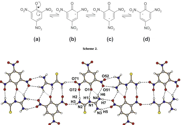
deprotonated hydroxyl group are significantly longer than the remaining four CAC bonds within the ring (Table 2). The repulsive interactions of the deprotonated oxygen atom O1 with the electron withdraw NO2groups attached in the ortho positions in relation to the O1 are responsible for the lengthening of the C3AC4 and C3AC8 bonds as well as for the significant differences between the internal CACAC angles within the ring. Both CACAC angles joining the NO2 groups (C3AC4AC5 and C3AC8AC7) are signifi-cantly greater and the C4AC3AC8 angle joining the deprotonated hydroxyl group is significantly smaller than the expected angle for C sp2hybridization (Table 2). The partially double in character of C'O bond linked the phenolic oxygen as well as the relatively long C3AC4 and C3AC8 bonds can be explained by the resonance structures presented inScheme 2.
Ab-initio full-optimised molecular orbital calculation per-formed for an isolated picrate anion shows similar correlation be-tween the bond lengths and distances as found in the crystal (Table 2). However, the small differences between the X-ray and MO cal-culated values are noticeable but understable if we take into ac-count the interactions with oppositely charged partner in the crystal. The ab initio MO calculations confirm the non-planar con-formation of picrate anion observed in the crystal. In the gas phase structure the NO2groups attached to the ring in ortho position in relations to deprotonated O1 atom are turned around the C4AN7 and C8AN7 bonds by35.0°. The deprotonation of the hydroxyl
group (O1) increases the steric effect of the lone electron pairs at O1 and causes the rotation of the ortho NO2group. The steric effect of the lone pair of electrons at the O1 atom makes the internal C4AC3AC8 angle significantly smaller than 120° and both
O1AC3AC4 and O1AC3AC8 angles greater than 120° and is in agreement with the valence shell electron pair repulsion model (VSEPR), according to which the lone-pair of electrons occupies a wider region as the bonding pair[34]. In addition, the steric effect of the lone pair of electrons at O1 together with the interaction the negatively polarized NO2groups are responsible for the angle dis-tortions within the ring from 120° as expected for C sp2
hybridization.
The oppositely charged units, i.e. 1-(diaminomethylene)thiou-ron-1-ium cation and picrate anion interact via NAH O hydrogen
bonds forming hydrogen bonded cation–anion complex. These cation–anion complexes related by an inversion interact via addi-tional NAH O hydrogen bonds forming pseudo-one dimensional
hydrogen bonded polymer (Fig. 2). In the crystal, the hydrogen-bonded polymers are located almost parallel to the (212) and (2-12) planes forming layers parallel to the (0 0 1) crystallographic plane (Fig. 3). Within one layer the almost planar 1D-polymers interact via
p
–p
clouds between the aromatic six-membered ringof picrate anions and the planar 1-(diaminomethylene)thiouron-1-ium cations in which the
p
electrons of the double C1AS1 and C2AN1 bonds are delocalized over the whole cation. The distance between the ring of picrate anion and the mean plane of the 1-(diaminomethylene)thiouron-1-ium cation are 3.56 Å indicating on the weakp
–p
interactions that stabilise the three dimensionalstructure.
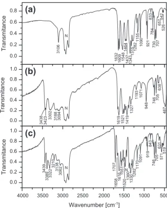
The FT-IR and Raman spectra of the 1-(diaminomethylene)thiou-ron-1-ium picrate are show inFigs. 4 and 5, respectively. Bands cor-responding to vibration of the functional groups were identified with the aid of infrared correlation charts[35,36]. In addition the spectra were compared with the spectra of 1-(diaminomethyl-ene)thiourea and picric acid that are also illustrated inFigs. 4 and 5. The title compound has several functional and skeletal groups
O71
H3 N2 H2
N3 H5 N1 H1
O51
N4 H7 O1
O72
H6 O52
Fig. 2.Pseudo-one dimensional hydrogen bonded chain of 1-(diaminomethylene)thiouron-1-ium picrate.
O
NO2 O2N
NO2
O
NO2 NO2 O2N
O
NO2 NO2 O2N
O
NO2 NO2 O2N
_
(a)
(b)
(c)
(d)
Scheme 2.
such as three NH2, C@S, CANAC, NACAN and NAC@S in the cation and three NO2, CarANO2, CarAO and six-membered skeletal aro-matic ring in the anion. A careful inspection of the IR spectrum shows three medium-strong intensity bands in the range between 3439 and 3322 cm 1that can be attributed to the asymmetric stretching of the three NH2groups of 1-(diaminomethylene)thiouron-1-ium cation and three weaker bands in the spectral region of 3210– 3062 cm 1attributed to the symmetric stretching of the NH2groups. The strong intensity band at 1616 cm 1in IR with weak intensity Ra-man counterparts at 1611 cm 1is attributed to NH2asymmetric deformation mode. The X-ray data reveal that all NH2groups of the 1-(diaminomethylene)thiouron-1-ium cation are involved in nine NAH O hydrogen bonds with bond lengths ranging from 2.77 to3.37 Å (the distance between donor and acceptor). This
is reflected in the infrared spectrum as a broad band in the range 3300–2750 cm 1. Additionally, the broad band in the range of 1400–1100 cm 1 overlapped with the
msym
stretching NO2points to the presence of the NAH O hydrogen bonds. The vibrations ofthe NAC(S)AN group in the thiourea are observed in the range be-tween 1480 and 1130 cm 1. The band at 1419 cm 1observed in the IR spectrum of 1-(diaminomethylene)thiourea (Fig. 4b) corre-sponds to the 1414 cm 1of thiourea, and is a contributions of the NH2rocking and NACAN vibrations[37]. The
m(C
@S) in thiourea is observed at 730 cm 1, while in several thiourea metal complexes them(C
@S) band is observed in the range 715–700 cm 1[32]. Vibra-tion of the C@S bond is coupled with the bending vibration of the pic-rate( ) ring and the band is observed at the 705 cm 1. Similar band is also observed in other picrate slats.The picrate being the 2,4,6-trinitrophenolate( ) anion has char-acteristic bands of the deprotonated phenol, the bands of three NO2 groups and the bands of the six-membered aromatic ring. As can be seen fromFig. 4a, the band at 3106 cm 1observed in the spectrum of pure picric acid disappeared in the spectrum of picrate (Fig. 5c). The relatively low wavenumber (3106 cm 1) points to the presence of the intramolecular OAH O interaction that was
a
b
c
Fig. 3.Molecular packing of 1-(diaminomethylene)thiouron-1-ium picrate in the unit cell.
4000 3500 3000 2500 2000 1500 1000 500 0.0 0.2 0.4 0.6 0.8 1.0 1520 519 571 631 705 746 843 919 1091 1170 1282 1330 1360 1551N 1616 1658 1707 N 3062 3138 3210 3322 3389 3439 Transmitance
Wavenumber [cm-1]
(c)
0.0 0.2 0.4 0.6 0.8 1.0 487 705 638 746 948 1071 1193 1327 1419 1521 1619 N 3188 3094 3302 3423 3438 Transmitance(b)
0.0 0.2 0.4 0.6 0.8 526 542 657 784 707 730 833 921 1090 1155 1282 1315 1343 1437 1464 1548 1609 1632 N 3106 Transmitance(a)
Fig. 4.Room temperature FTIR spectra of picric acid (a), 1-(diaminomethyl-ene)thiourea (b) and 1-(diaminomethylene)thiouron-1-ium picrate (c).
3500 3000 2500 2000 1500 1000 500 0.0 0.6 1.2 1.8 2.4 336 423 512 604 745 704 824 949 1084 1170 1299 1344 1366 1485 1553 1611 1712 Intensity
(c)
0.00 0.15 0.30 0.45 0.60 367 443 493 629 656 698 746 956 1075 1200 1325 1403 1483 1503 1614 1631 3091 3200 3303 3412 Intensity(b)
0 2 4 6 329 349 402 544 741 831 942 1089 1178 1279 1345 1530 1582 1611 1633 3109 Intensity(a)
Wavenumber [cm-1]
Fig. 5.Room temperature Raman spectra of picric acid (a), 1-(diaminomethyl-ene)thiourea (b) and 1-(diaminomethylene)thiouron-1-ium picrate (c).
confirmed by the X-ray single crystal analysis of picric acid[33]. Thus the absence of the OAH stretching confirms a proton transfer from picric acid to 1-(diaminomethylene)thiourea molecule with the formation of the picrate( ) anion and 1-(diaminomethyl-ene)thiouron-1-ium cation. Apart from this, the other functional groups existing in the picrate( ) anion are the nitro NO2groups and the phenolic CarAO group. The CAC ring stretching modes are observed as expected at 1616, 1488 and 1467 cm 1(Table 4) in the spectrum of 1-(diaminomethylene)thiouron-1-ium picrate as well as in the spectrum of picric acid. The CAH bending mode is assigned as a medium intensity band at 824 cm 1 in Raman spectrum and at 843 cm 1in the IR spectrum of the salt. Picric acid absorbs strongly at 1345 cm 1as a result of symmetric stretching vibration of nitro groups. The acid also displays two closely spaced bands at 1562 and 1530 cm 1due to the NO2asymmetric stretch-ing vibration mode. Besides the mentioned above bands, in the present crystal, the Raman spectrum exhibits, a band at 1368 cm 1 and a very strong band at 1299 cm 1; both are not observed in the spectrum of pure picric acid. Comparing the con-formation of the picrate( ) anion in the 1-(diaminomethylene)thi-ouron-1-ium picrate with the conformation of molecule of pure picric acid[33] it should be stated that they are different. Both NO2groups of the picrate( ) anion are linked to the ring in orto positions in relation to the phenolic oxygen (CO ) and are oppo-sitely rotated in relation to the plane of the ring, whereas in picric
acid only one of two orto NO2group is rotated, the other is copla-nar with the aromatic ring due to the formation of the intramolec-ular OAH O hydrogen bond between the hydroxyl group and the
oxygen atom of the NO2group. This conformation together with the intermolecular NAH O hydrogen bonds leads to the splitting
of the
msym
(NO2) band. The band at 1282 cm 1in the IR spectrum is assigned to phenolic O vibration,m(Car
AO) mode as was assigned in several picrate salts[38–43]. The appearance of a medium inten-sity band at 1285–1255 cm 1 in the metal picrate complexes is attributed to them(Car
AO) mode due to the coordination of pheno-lic oxygen after deprotonation [21]. For the lanthanide picrates, this band is observed at1275 cm 1[44–46]. The origin andnat-ure of the relatively strong and narrow band at 1707 cm 1in the IR spectrum of the salt (Fig. 4c), which is not observed in pure picric acid (Fig. 4a) and pure 1-(diaminomethylene)thiourea (Fig. 4b) are not clear. In the 1:1 and 2:1 complexes of N-methylmorpholine betaine with picric acid the band in this spectral region (1730 cm 1in the complex of 1:1 and 1727 cm 1in the complex
of 2:1) has been attributed to the
m(Car
@O) of picrate( ) anion[47]. However, as shown by the X-ray analysis in both complexes the CAO bond lengths of 1.260(5) and 1.221(6) Å[47]are very close to the value of 1.2446(16) Å observed in the present structure as well as in other picrate salts[48]. Since in the spectra of several picrate salts the band above 1700 cm 1is not observed[38–46], therefore the band at 1707 cm 1can not be assigned to the pheno-lic oxygen vibration. Nevertheless, this band could be ascribed to the stretching of an imine bond of the 1-(diaminomethylene)thiou-ron-1-ium cation, since a similar band is observed in some imines and their picrate slats [49]. The stretching vibration of an imine bond frequency goes from 1667 cm 1 in some free imines to 1704 cm 1in their picrates[49]. The observed frequencies andtheir assignments are listed inTable 4.
4. Conclusion
The single crystals of 1-(diaminomethylene)thiouron-1-ium picrate were grown using a solution growth technique. The 1-(diaminomethylene)thiouron-1-ium cation is planar. This is in con-trast to the neutral molecule of 1-(diaminomethylene)thiourea in the crystal as well as in other known crystals of 1-(diaminometh-ylene)thiouron-1-ium salts, in which the conformation is non-pla-nar. In the picrate( ) anion both orto NO2groups in relation to the deprotonated hydroxyl group are oppositely turned in relation to the six-membered aromatic ring, due to the repulsive interaction between the negatively charged phenolic oxygen and the oxygen atoms of NO2group. Deprotonation of the picric acid leads to dis-appearance of the OAH stretching vibration band at 3106 cm 1 (observed in picric acid,Fig. 4a) and to appearance of the phenolic O stretching vibration band at 1170 cm 1 (observed in the salt, Fig. 4c). In the crystal, nine NAH O hydrogen bonds exist, which
are manifested in the IR spectrum as a broad band between the 3300 and 2750 cm 1and in the range of 1400–1100 cm 1. These bands overlapped with the
msym
stretching NO2 as well as with the NAC(S)AN skeletal group of the cation and with the skeletal aromatic ring of picrate anion. An arrangement of the oppositely charged components, i.e. 1-(diaminomethylene)thiouron-1-ium cations and picrate( ) anions in the crystal is mainly determined by the ionic and hydrogen bonding interactions forming pseudo-one dimensional chains.5. Supplementary material
Additional material comprising full details of the X-ray data col-lection and final refinement parameters including anisotropic ther-mal parameters and full list of the bond lengths and angles have Table 4
FT IR and Raman spectral data for 1-(diaminomethylene)thiouron-1-ium picrate.
Infrared,m (cm 1)
Raman,m (cm 1)
Assignment
3439m NH2asym stretch. 3389s NH2asym stretch. 3322s NH2sym stretch. 3210w NH2sym stretch.
3138w NAH
3082vw NAH
3062m NAH
2618vw Overtones and combination bands 2262vw Overtones and combination bands 1712vw Imine bond stretch.
1707s Imine bond stretch. 1658m NH2asym def. 1616vs NH2asym def.
1611w
1551s 1553m m(CAN) and NO2asym stretch. 1520m 1522w m(CAN) and NO2asym stretch. 1498m m(CAC) ring stretch.
1485w m(CAC) ring stretch.
1467s m(CAC) ring stretch. overlapped with
Nujol 1366m m(CAO)
1360s NO2sym stretch. overlapped with Nujol 1344vs NO2sym stretch.
1330vs NO2sym stretch. 1299vs NO2sym stretch.
1282s Phenolic O
1170m 1170m
1091 1084m CAN stretch.
948m CAC stretch.
919m CAC stretch.
843m NO2def.
824s NO2def., CAH def.
746m 745w x(NO2)
705m 705w C@S stretch. overlapped withd(ring)
631w
604
571m CAC@O def.
519w
512w CACACAC ph ring def.
336m CACACAC ph ring def.
vs, very strong; s, strong; m, medium; w, weak.
been deposited with the Cambridge Crystallographic Data Center in the CIF format as supplementary publications No. CCDC 767502. Copies of the data can be obtained free of charge on the application to CCDC, 12 Union Road, Cambridge, CB21EZ, UK, (fax: +44 1223 336 033; email: [email protected]).
References
[1] J.C. MacDonald, G.R. Whitesides, Chem. Rev. 94 (1994) 2382.
[2] G.R. Desiraju, T. Steiner, The Weak Hydrogen Bond in Structural Chemistry and Biology, Oxford University Press, Oxford, 1999.
[3] G.R. Desiraju, Angew. Chem. Int. Ed. 34 (1995) 2311. [4] G.R. Desiraju, Chem. Commun. (1997) 1475. [5] T. Steiner, Angew. Chem. Int. Ed. 41 (2002) 48.
[6] C.A. Hunter, K.R. Lawson, J. Perkins, C.J. Urch, J. Chem. Soc. Perkin Trans. 2 (2001) 651.
[7] J.A. Zerkowski, J.C. MacDonald, G.M. Whitesides, Chem. Mater. 6 (1994) 1250. [8] G.R. Desiraju, Acc. Chem. Res. 29 (1996) 441.
[9] J. Janczak, G.J. Perpétuo, Acta Crystallogr. C64 (2008) o114.
[10] K. Chakrabarty, T. Kar, S.P.S. Gupta, Acta Crystallogr. C46 (1990) 2065. [11] M.K. Kabir, K. Yamada, K. Adachi, M. Kondo, S. Kawata, Acta Crystallogr. E58
(2002) m580.
[12] E. Doxiadi, R. Vilar, A.J.P. White, D.J. Williams, Polyhedron 22 (2003) 2991. [13] G.J. Perpétuo, J. Janczak, Acta Crystallogr. C64 (2008) o264.
[14] J. Janczak, G.J. Perpétuo, Acta Crystallogr. C64 (2008) o330. [15] J. Janczak, G.J. Perpétuo, Acta Crystallogr. C65 (2009) o118. [16] M. Hołyn´ska, M. Kubiak, Acta Crystallogr. C64 (2008) o609. [17] M. Hołyn´ska, M. Kubiak, Acta Crystallogr. C65 (2009) o191. [18] P.G. Farrell, F. Terrier, R. Schaal, Tetrahedron Lett. 26 (1985) 2435.
[19] G.C. Franchini, A. Marchetti, L. Tassi, G. Tosi, J. Chem. Soc. Faraday Trans. 1 84 (1988) 4427.
[20] M.A.F. Elmosallamy, Anal. Sci. 20 (2004) 285.
[21] R.C. Maurya, P. Sharma, S. Roy, Synth. React. Inorg. Metal. Org. Chem. 33 (2003) 683.
[22] R.P. Sharma, R. Sharma, R. Bala, P. Venugopalan, J. Coord. Chem. 58 (2005) 899. [23] CrysAlis CCD and CrysAlis Red, Version 171.32.8, Oxford Diffraction
Poland,Wrocław, Poland, 2006.
[24] G.M. Sheldrick,SHELXS97,SHELXL97, Programs for Crystal Sturctures Solution and Refinement, University of Göttingen, Göttingen, Germany, 1997. [25] K. Brandenburg, H. Putz, DIAMOND Version 3.0, Crystal Impact GbR, Bonn,
Germany, 2006.
[26] J.M. Frisch, G.W. Trucks, H.B. Schlegel, P.M.W. Gill, B.G. Johnson, M.A. Robb, J. Cheeseman, T. Keith, G.A. Petersson, J.A. Montgomery, K. Raghavachari, M.A. Al-Laham, V.G. Zakrzewski, J.V. Ortiz, J.B. Foresman, J. Cislowski, B.B. Stefanov, A. Nanayakkara, M. Challacombe, C.Y. Peng, P.Y. Ayala, W. Chen, W.M. Wong, J.L. Andres, E.S. Replogle, R. Gomperts, R.L. Martin, D.J. Fox, J.S. Binkley, D.J. Defrees, J. Baker, B.B. Stewart, M. Head-Gordon, C. Gonzales, J.A. Pople, Gaussian98, Revision A.3, Gaussian, Inc., Pittsburgh, PA, 1998.
[27] A.D. Becke, J. Chem. Phys. 104 (1996) 1040. [28] C. Lee, W. Yang, R.G. Parr, Phys. Rev. B37 (1988) 785.
[29] D.R. Johnson, F.X. Powell, W.H. Kirchhoff, J. Mol. Spectrosc. 39 (1971) 136. [30] R. Fausto, L.A.E. Batista de Carvalho, J.J.C. Teixeira-Dias, M.N. Ramos, J. Chem.
Soc. Faraday Trans. 2 (1989) 1945.
[31] F.H. Allen, O. Kennard, D.G. Watson, L. Brammer, A.G. Orpen, R. Taylor, J. Chem. Soc. Perkin Trans. 2 (1987) S1–S19.
[32] F.H. Allen, C.M. Bird, R.S. Rowland, P.R. Raithby, Acta Crystallogr. B53 (1997) 680.
[33] M. Soriano-Garcia, T. Srikrishnan, R. Parthasarathy, Acta Crystallogr. A34 (1978) S114.
[34] R.J. Gillespie, Chem. Soc. Rev. 21 (1992) 59.
[35] G. Socrates, Infrared Characteristic Group Frequencies;, Wiley-Interscience, Chichester, UK, 1980.
[36] G. Socrates, Infrared and Raman Characteristic Group Frequencies, third ed., Wiley, New York, USA, 2001.
[37] G.M.S. El-Bahy, B.A. El-Sayed, A.A. Shabana, Vib. Spectrosc. 31 (2003) 101. [38] M.B. Mary, V. Sasirekha, V. Ramakrishnan, Spectrochim. Acta A65 (2006) 955. [39] Y.L. Guo, Y.W. Wang, W. Dou, J.R. Zheng, W.S. Liu, C.Y. Su, Inorg. Chim. Acta 360
(2007) 3361.
[40] E. Pinto Marinho, D.M. Araújo Melo, L.B. Zinner, E.E. Castellano, J. Zukerman-Schpector, P.C. Isolani, G. Vicentini, Polyhedron 16 (1997) 3519.
[41] V. Tomašic´, Lj. Tušek, A. Višnjevac, B. Kojic´-Prodovic´, N. Filipovic´-Vincekovic´, J. Colloid Interface Sci. 227 (2000) 427.
[42] Y.L. Guo, Y.W. Wang, W.S. Liu, W. Dou, X. Zhong, Spectrochim. Acta A67 (2007) 624.
[43] A. Chandramohan, R. Bharathikannan, J. Chandrasekaran, P. Maadeswaran, R. Renganathan, V. Kandavelu, J. Cryst. Growth 310 (2008) 5409.
[44] T. Yongchi, L. Yingqiu, N. Jiazan, J. Mol. Sci. (Int. Ed.) 5 (1987) 83. [45] B. Ji, Q. Du, K. Ding, Y. Li, Z. Zhou, Polyhedron 15 (1996) 403.
[46] Y.W. Wang, W.S. Liu, N. Tang, M.Y. Tan, K.B. Yu, J. Mol. Struct. 660 (2003) 41. [47] Z. Dega-Szafran, A. Katrusiak, M. Szafran, E. Sokołowska, J. Mol. Struct. 615
(2002) 73.
[48] F.H. Allen, Acta Crystallogr. B58 (2002) 380.