Structural and spectroscopic characterization of
1-(diaminomethylene) thiouron-1-ium benzoate and
bis(1-(diaminomethylene)thiouron-1-ium) phthalate trihydrate
Genivaldo Julio Perp
etuo
a, Jan Janczak
b,*aDepartamento de Física, Instituto de Ci^encias Exatas e Biologicas, Universidade Federal de Ouro Preto, 35400-000, Ouro Preto, MG, Brazil bInstitute of Low Temperature and Structure Research, Polish Academy of Sciences, PO Box 1410, 50950, Wrocław, Poland
a r t i c l e
i n f o
Article history:
Received 9 July 2015 Received in revised form 21 October 2015 Accepted 26 October 2015 Available online 30 October 2015
Keywords:
1-(diaminomethylene)thiouron-1-ium benzoate
Bis(1-(diaminomethylene)-thiouron-1-ium) phthalate trihydrate
Crystal structure Hydrogen bonds Vibrational spectroscopy
a b s t r a c t
Two single crystals of 1-(diaminomethylene) thiouron-1-ium benzoate (1) and bis(1-(diamino-methylene)thiouron-1-ium) phthalate trihydrate (2) were grown using a solution growth technique. The compound1crystallises in the centrosymmetricP21/c space group of the monoclinic system, whereas the compound2 in the centrosymmetricPbcn space group of orthorhombic system. The solid-state organisation of1 and 2has been analysed with respect to cation-anion and hydrogen bonding in-teractions. The oppositely charged units interact via almost linear hydrogen bonds with the graphs of R22(8) and R21(6) forming molecular complexes. In the crystal1the R22(8) motif is formed by donation to the carboxylate group from amine group joined to C1 and from imine group and R21(6) motif is formed by donation to the O2 from amine group joined to C2 and from imine group, whereas in crystal2the graphs are formed oppositely. Interactions between the hydrogen-bonded molecular complexes in1 lead to formation of layered 2D structure, whereas in2, due to presence of hydrated water molecules lead to formation of 3D hydrogen-bonded supramolecular network. The obtained deuterated analogues of1and 2crystallise similar as H-compound in the monoclinic and orthorhombic system with quite similar lattice parameters. The compounds were also characterised by the FT-IR and Raman spectroscopies. The characteristic bands of the functional and skeletal groups are discussed.
©2015 Elsevier B.V. All rights reserved.
1. Introduction
Crystal engineering involving a combination of synthesis and structural chemistry is a branch of material science that rapidly expanding over the past two decades[1,2]. The accurate prediction of a structure and the properties of a product from the structures of basic substrates, which can be considered a designing process, re-mains the ultimate goal[3]. The exploration of new supramolecular structures and interactions responsible for such an arrangement brings much useful information concerning factors important in structure formation that are helpful in the design process[4]. The hydrogen bonds and other non-covalent intermolecular in-teractions are of fundamental key for molecular recognition in supramolecular synthesis of new solids[5].
A productive strategy in crystal engineering and control of
crystal architecture is to build supramolecular structures from molecules containing complementary arrays of hydrogen bonding sites[6]. The supramolecular synthon of hydrogen-bonding pattern is an effective approach for structural design of solids [7]. Commercially available 2-imino-4-thiobiuret (Aldrich, CAS No. 2114-20-05) is, as has been shown by the X-ray analysis, it tauto-meric form of 1-(diaminomethylene)thiourea (Scheme 1)[8].
Both tautomers are useful in crystal engineering as building blocks, since they contain hydrogen-bonding sites. Additionally, both tautomers can act asN,N- orN,S-coordinating ligands forming several types of complexes with metal ions[9]. The 1-(diamino-methylene)-thiourea contains the basic N atom, therefore it can forms slats with organic and inorganic acids forming extended hydrogen-bonding networks in solids [10]. The supramolecular aggregation pattern of 1-(diaminomethylene)-thiourea with tar-taric acid and its deuterated analogue are examples of supramo-lecular hydrogen-bonding networks in solids that can be utilised as materials for non-linear optics[11].
To explore the usefulness of 1-(diaminomethylene)-thiourea as *Corresponding author.
E-mail address:[email protected](J. Janczak).
Contents lists available atScienceDirect
Journal of Molecular Structure
j o u r n a l h o m e p a g e : h t t p : / / w w w . e l s e v i e r . c o m / l o c a t e / m o l s t r u c
http://dx.doi.org/10.1016/j.molstruc.2015.10.080 0022-2860/©2015 Elsevier B.V. All rights reserved.
a building block in supramolecular synthesis, in the present work we investigate the supramolecular architecture formed by self-assembly of 1-(diaminomethylene)-thiourea with benzoic and phthalic acids (Scheme 2). This study is aimed into the interaction between the building blocks formed the supramolecular architec-ture in solids. Both compounds were also characterised by
vibrational spectroscopy. Assignment of the bands has been sup-ported by the comparison of the IR-spectra of protiated compounds with the IR-spectra of deuterated analogues.
2. Experimental
2-imino-4-thiobiuret, benzoic acid and phthalic acid were commercially available and used as received. Elemental analysis was carried out with a PerkineElmer 240 elemental analyser.
2.1. Preparation of 1-(diaminomethylene)thiouron-1-ium benzoate (1) and its deuterated analogue
Commercially available 2-imino-4-thiobiuret (Aldrich, CAS No. 2114-02-05), which is in fact the tautomeric form 1-(diamino-methylene)thiourea (0.118 g) and the benzoic acid (0.122 g) were added to hot water in a molar proportion of 1:1. When the solution became homogeneous it was cooled slowly and kept at room temperature. After several days, transparent colourless crystals of C6H5COO$C2H7N4S (1) were formed. Analysis: calculated for C8H12N4SO2: C, 44.98; N, 23.32; O, 13.32; S, 13.34 and H, 5.03%. Found: C,45.11; N, 23.22; O, 13.45; S, 13.22, and H, 5.00%. Deuterated analogue of 1-(diaminomethylene)thiouron-1-ium benzoate was prepared by the usual reaction with heavy water. The crystals of 1-(diaminomethylene)thiouron-1-ium benzoate were dissolved in heavy water, and left in the atmosphere saturated with heavy water for one weak, in order to avoid the contamination of the crystals. Next the procedure was repeated twice.
2.2. Preparation of bis(1-(diaminomethylene)thiouron-1-ium) phthalate trihydrate (2) and its deuterated analogue
2-imino-4-thiobiuret (0.118 g) and phthalic acid (0.166 g) were dissolved in hot water in a molar proportion of 1:1. The solution was cooled and kept at the room temperature. After several days, colourless single crystals of bis(1-(diaminomethylene)thiouron-1-ium) phthalate trihydrate (2) were obtained. Analysis: calculated Scheme 1.Tautomeric equilibrium between 2-imino-4-thiobiuret (a) and
1-(diamino-methylene)-thiourea (b).
Scheme 2.Benzoic acid (a) and phthalic acid (b).
Table 1
Crystallographic data for 1-(diaminomethylene)thiouron-1-ium benzoate (1) and bis(1-(diaminomethylene)thiouron-1-ium) phthalate trihy-drate (2).
1 2
Empirical formula C9H12N4O2S C12H24N8O7S2
Formula weight (g mol 1) 240.29 456.51
Crystal system Monoclinic Orthorhombic
Space group P21/c(No. 14) P b c n(No. 60)
a(Å) 17.400 (4) 7.9170 (16)
b(Å) 4.9390 (10) 14.130 (3)
c(Å) 13.424 (3) 18.293 (4)
b() 99.13 (3)
V(Å3) 1139.0 (4) 2046.4 (7)
Z 4 4
Dcalc/Dobs(g cm 3) 1.401/1.40 1.482/1.48
m(mm 1) 0.276 0.313
Crystal size (mm) 0.280.140.12 0.320.240.10
Radiation type, wavelength,l(Å) MoKa, 0.71073 MoKa, 0.71073
Temperature (K) 295 (2) 295 (2)
qrange() 3.07÷29.54 3.09÷29.59
Absorption correction Numerical, CrysAlis Red Numerical, CrysAlis Red
Tmin/Tmax 0.9292/0.9651 0.9112/0.9708
Refls collected/unique/observed 12998/2958/1546 21484/2711/1685
Rint 0.0572 0.0534
Refinement on F2 F2
R[F2>2s(F2)] 0.0437 0.0374
wR(F2all reflections) 0.0627 0.0677
Goodness-of-fit,S 0.989 1.001
Drmax,Drmin(e Å 3) þ0.162, 0.201 þ0.218, 0.189
wR¼{S[w(Fo2eFc2)2]/SwFo4}½;w1¼1/[s2(Fo2)þ(aP)2] wherea¼0.01 anda¼0.024 for 1 and 2, respectively, andP¼(Fo2þ2Fc2)/3.
for C12H24N8O7S2: C, 31.57; N, 24.55; O, 24.53; S, 14.05; and H, 5.30%. Found: C, 31.48; N, 24.60; O, 24.65; S 14.00, and H, 5.27%. Deuterated analogue of bis(1-(diaminomethylene)thiouron-1-ium) phthalate trihydrate was prepared by the usual reaction with heavy water, and left in the atmosphere saturated with heavy water for one weak, in order to avoid the contamination of the crystals. Next the procedure was repeated twice.
2.2.1. X-ray data collection
X-ray intensity data for the1and2single crystals were collected using graphite monochromatic Mo K
a
radiation on a four-circlek
geometry KUMA KM-4 diffractometer with a two-dimensional area CCD detector. Theu
-scan technique withD
u
¼1.0for each image was used for data collection. The 900 images for six different runs covering over 99% of the Ewald sphere were performed. One image was used as a standard after every 50 images for monitoring of the crystals stability and the data collection. No correction on therelative intensity variations was necessary. Data collections were made using the CrysAlis CCD program[12]. Integration, scaling of the reflections, correction for Lorentz and polarisation effects and absorption corrections were performed using the CrysAlis Red program[12]. The structures were solved by the direct methods using SHELXS-97 and refined using SHELXL-97 program[13]. The hydrogen atoms involving in the hydrogen bonds were located in difference Fourier maps and were refined. The hydrogen atoms joined to aromatic carbon atoms were introduced in their geometrical positions. Thefinal difference Fourier maps showed no peaks of chemical significance. Details of the data collection pa-rameters, crystallographic data andfinal agreement parameters are collected inTable 1. Visualisations of the structures were made with the Diamond 3.0 program[14]. Selected geometrical parameters are listed inTable 2 and the geometry of hydrogen bonding in-teractions is collected in Table 3. The obtained deuterated ana-logues of1and2crystallise similar as H-compounds in the same crystal system with quite similar lattice parameters.
2.2.2. X-ray powder diffraction
The protiated and deuterated samples of1and2were measured on a PANanalytical X'Pert diffractometer equipped with a Cu-K
a
radiation source (l
¼1.54182 Å) at room temperature.2.2.3. Vibrational spectra measurements
The vibrational measurements of1and2and theirs deuterated Table 2
Selected bond lengths (Å) and angles () 1-(diaminomethylene)thiouron-1-ium benzoate (1) and bis(1-(diaminomethylene)thiouron-1-ium) phthalate trihydrate (2).
1 2
C1eS1 1.679 (2) 1.6640 (15)
C1eN1 1.381 (3) 1.3837 (18)
C1eN2 1.312 (3) 1.3216 (19)
C2eN1 1.366 (3) 1.3609 (18)
C2eN3 1.309 (3) 1.3214 (19)
C2eN4 1.311 (3) 1.3008 (19)
C3eO1 1.267 (2) 1.2706 (16)
C3eO2 1.244 (2) 1.2492 (16)
C3eC4 1.511 (3) 1.5025 (19)
N2eC1eN1 112.7 (2) 112.72 (13)
N2eC1eS1 122.8 (2) 121.42 (12)
N1eC1eS1 124.5 (2) 125.86 (12)
C2eN1eC1 131.4 (2) 130.07 (13)
N3eC2eN4 121.5 (2) 120.43 (15)
N3eC2eN1 115.5 (2) 116.51 (14)
N4eC2eN1 123.1 (2) 123.06 (14)
O2eC3eO1 125.2 (2) 124.25 (14)
Table 3
(a) Hydrogen-bond geometry (Å,
) for 1-(diaminomethylene)thiouron-1-ium ben-zoate (1) and (b) for bis(1-(diaminomethylene)thiouron-1-ium) phthalate trihydrate (2).
DeH/A DeH H/A D/A DeH/A
(a)1
N1eH1/O2 0.89 (2) 1.89 (2) 2.769 (2) 165 (2)
N2eH21/O1 0.97 (2) 1.85 (2) 2.806 (2) 169 (2)
N2eH22/S1i 0.88 (2) 2.50 (2) 3.368 (2) 168 (2) N3eH31/O1ii 0.87 (2) 1.98 (2) 2.776 (3) 152 (2)
N3eH32/O2 0.84 (2) 2.17 (2) 2.896 (3) 145 (2)
N4eH41/O1ii 0.84 (2) 2.16 (2) 2.900 (3) 148 (2)
N4eH42/S1 0.87 (2) 2.32 (2) 3.025 (2) 138 (2)
(b)2
N1eH1/O1 0.83 (2) 1.94 (2) 2.762 (2) 170 (2)
N2eH12/O1 0.86 (2) 2.46 (2) 3.190 (2) 143 (2)
N2eH22/O2i 0.90 (2) 2.09 (2) 2.966 (2) 165 (2)
N3eH13/O2 0.89 (2) 2.06 (2) 2.945 (2) 177 (2)
N3eH23/O3ii 0.85 (2) 2.36 (3) 3.078 (2) 143 (2) N3eH23/S1iii 0.85 (2) 2.90 (3) 3.443 (2) 124 (2) N4eH14/O3ii 0.86 (2) 2.06 (2) 2.880 (2) 159 (2)
N4eH24/S1 0.86 (2) 2.29 (2) 3.008 (2) 140 (2)
O3eH2/O4 0.87 (1) 1.94 (1) 2.801 (2) 170 (2)
O3eH3/O2iv 0.86 (1) 2.16 (1) 3.013 (2) 171 (2)
O4eH4/O1 0.87 (1) 1.86 (1) 2.721 (2) 171 (2)
Symmetry codes for crystal1: (i) xþ1, yþ2, zþ1; (ii)x, yþ3/2,z 1/2. Symmetry codes for crystal2: (i)x 1,y,z; (ii) xþ1/2, yþ1/2,zþ1/2; (iii)xþ1,
y,z; (iv)x 1/2,yþ1/2, zþ1/2.
Fig. 1.A view of 1-(diaminomethylene)thiouron-1-ium benzoate (a), bis(1-(diamino-methylene)thiouron-1-ium phthalate trihydrate (b) showing displacement ellipsoids at the 50% probability level and H atoms as a sphere of arbitrary radii. Dashed lines represent the hydrogen bonds. Symmetry code: (i) 2 x, y, 1.5 z.
analogues were carried out at room temperature. The Fourier transform infrared spectra were recorded from nujol mulls be-tween 4000 and 400 cm 1on a Bruker IFS 113 V FTIR spectrometer. Resolution was set up to 2 cm 1. The Fourier Transform Raman spectrum was recorded on a FRA-106 attached to the Bruker 113 V FTIR spectrometer equipped with Ge detector cooled to liquid ni-trogen temperature. Resolution was set up to 2 cm 1, signal/noise ratio was established by 32 scans. Nd3þ-YAG air-cooled diode pumped laser of power ca. 200 mW was used as an exciting source. The incident laser excitation was 1064 nm. The scattered light was collected at the angle of 180in the region of 3600e80 cm 1, res-olution 2 cm 1, 256 scans.
3. Results and discussion
Good quality single crystals of 1-(diaminomethylene) thiouron-1-ium benzoate (1) and bis(1-(diaminomethylene)thiouron-1-ium) phthalate trihydrate (2) suitable for the X-ray single crystal analysis were obtained from water solutions at ambient temperature. The 1-(diaminomethylene) thiouron-1-ium benzoate crystallises in the centrosymmetric space groupP21/cof monoclinic system whereas bis(1-(diaminomethylene)thiouron-1-ium) phthalate trihydrate crystallises in the centrosymmetric space group Pbcn of the orthorhombic system. The deuterated analogues were obtained by usual reaction of protiated crystals with heavy water. The deuter-ated analogues also crystallise as the protideuter-ated compounds in the
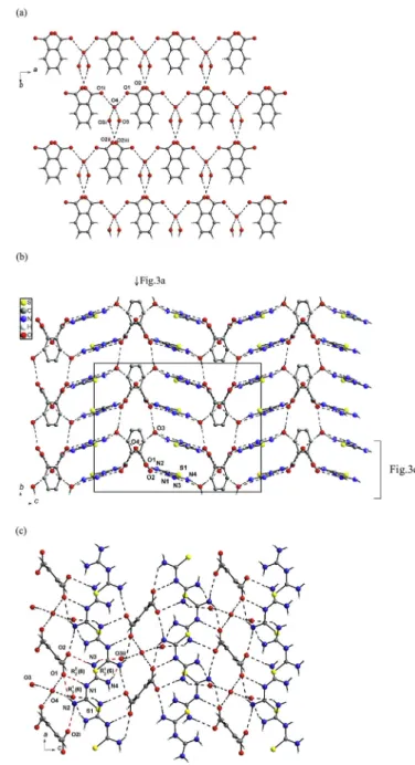
Fig. 2.A view of hydrogen bonded chain of 1-(diaminomethylene)thiouron-1-ium benzoate (a) and the two dimensional hydrogen-bonding layers of 1-(diamino-ethylene)hiouron-1-ium benzoate viewed alongb-axis (b) andc-axis (c).
Fig. 3.The anionic 2D-layer of hydrogen bonded by water molecules of phthalate(2-) anions viewed alongc-axis, symmetry codes (i)ex, y, 0.5-z; (ii) 0.5þx, 0.5þy, 0.5þz; (iii) 0.5 x, 0.5þy, z (a), 3D hydrogen bonded supramolecular network of bis(1-(diaminomethylene)thiouron-1-ium phthalate trihydrate viewed alonga-axis (b) and the undulating layer viewed along b-axis, symmetry codes: (i) x 1, y, z; (ii) xþ0.5, yþ0.5,zþ0.5 (c).
same space groups with quite similar lattice parameters, so the protiated and deuterated crystals of1and2are isostructural and was confirmed by the powder X-ray diffraction experiments (Figs. S1 and S2in supplementary material). The microscopic im-ages of1and2crystals are shown inFigs. S3 and S4in supple-mentary material.
The X-ray single crystal analysis of 1 and 2 shows that the carboxylate groups are deprotonated, and the proton is transferred to the central N1 atom of 1-(diaminomethylene) thiourea molecule forming 1-(diaminomethylene) thiouron-1-ium cation. The oppo-sitely charged units interact via almost linear hydrogen bonds with the graphs of R22(8) and R21(6) forming molecular complexes as illustrate in Fig. 1. In the crystal 1the R22(8) motif is formed by donation to the carboxylate group from amine group joined to C1 and from imine group and R12(6) motif is formed by donation to the O2 from amine group joined to C2 and from imine group (Fig. 1a), whereas in crystal2the graphs are formed oppositely. In addition, in the hydrated crystals2, hydrogen bonded water dimer interacts with O1 atom of COO group via OeH/O hydrogen bond (Fig. 1b). The conformation of the 1-(diaminomethylene)thiouron-1-ium cation in the crystals1and2is not strictly, but twisted. Both arms
of the cation are oppositely rotated around the CeN bonds involving the central N1 atom. The dihedral angle between the N1/ C1/S1/N2 and N1/C2/N3/N4 planes is equal to 3.1 (1)in crystal1 and 1.8 (1) in crystal2. The dihedral angle in the present structures is significantly smaller than that in the crystal of neutral 1-(dia-minomethylene)thiourea (22.2(1)) [8]. The currently available data on 1-(diaminomethylene)thiouron-1-ium salts[15]show that the cation twisting may differ when different anions are used (1.4 (1)for 1-(diaminomethylene)thiouron-1-ium perchlorate[10b]to 22.9 (1)for 1-(diaminomethylene)thiouron-1-ium chloride[10a]) and is undoubtedly dependent on the hydrogen bonding system formed by the oppositely charged units. The gas-phase conforma-tion of the 1-(diaminomethylene)thiouron-1-ium caconforma-tion as show theab-initioMO calculations is also twisted with similar dihedral angle of 6.2[10a]. The C1
eS1 bond (Table 2) is slightly longer than the typical C]S double bond as observed in the thioformaldehyde CH2C]S (1.6019 (8) Å)[16], which represents 100% double-bond character. The three CeN bonds linking the amine groups of the 1-(diaminomethylene)thiouron-1-ium cation in both crystals (1 and2) are shorter than the CeN bonds involving the central N1 atom (Table 2). The planarity of the amine groups points to thesp2
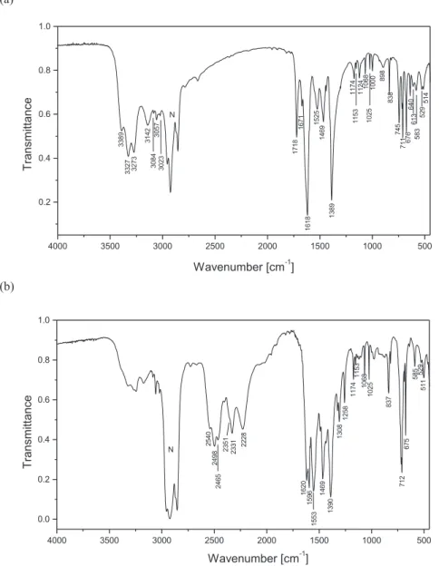
Fig. 4.IR-spectrum of protiated (a) and deuterated analogue (b) of 1-(diaminomethylene)thiouron-1-ium benzoate.
hybridisation of the orbitals on the amine nitrogen atoms and the lone pair of electron localised on theporbital. Therefore the partial delocalisation of the lone pair onporbital of the amine groups and of the
p
bond of the double C1¼S1 and C2¼N1 bonds is possible and leads to shortening of other CeN bonds linking the amine groups and to the elongation of the C1¼S1 and C2¼N1 bonds (Table 2).The geometrical parameters of the anionic parts of the crystals, i.e. benzoate( ) and phthalate (2-), do not deviate significantly from the reported values in the other structures containing these anions[15]. The CeO bond lengths of the COO groups (Table 2) point on delocalisation of the
p
-bond and the charge over both CeO bonds. The COO group of benzoate( ) anion is not coplanar with the benzene ring. It is slightly rotated around the C3eC4 bond. The dihedral angle between the planes of COO and benzene ring is equal to 10.2 (1). This is in contrast to the planar conformation of benzoic acid in crystal due to formation of the hydrogen-bonded dimer[17]. The non-planar phthalate (2-) anion has twofold sym-metry axis running in the centre of the C4eC4iand C6eC6ibonds (symmetry code as in Fig. 1) with the COO groups oppositely rotated around the C3eC4 or C3ieC4iby±48.8 (1). The rotationangles are greater than that observed in the crystal of pure phthalic acid ~30.5[18]due to the repulsive forces between deportonated COO groups.
In the crystal1, the hydrogen bonding interactions (Table 3a) between the c-glide plane related cation-anion molecular com-plexes lead to formation of chains along thec-axis (Fig. 2a). Inver-sion related chains are arranged parallel to (100) plane forming two dimensional supramolecular layers (Fig. 2b and c). Within the layers the inversion-related chains interact each other via NeH/S hydrogen bonds with a graph of R22(8). The importance of such interactions has been questioned[19]. However, the DeH/S in-teractions (D¼donor) are important in the biological systems due to presence of high content of S atom in biological molecules. In addition, the NeH/S interactions have been utilised for design supramolecular arrangement of thiourea derivatives[20]. There-fore, such NeH/S interactions seem to be important in the present structure (Table 3), where the formation of the intramolecular NeH/S interactions is favoured by the six-membered hydrogen-bonded ring with a graph of S(6) and the intermolecular eight-membered hydrogen-bonded ring with a graph of R22(8); the presence of C]S bonds makes it some resonance-assisted
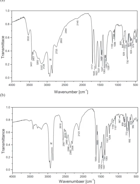
Fig. 5.IR-spectrum of protiated (a) and deuterated analogue (b) of bis(1-(diaminomethylene)thiouron-1-ium phthalate trihydrate.
stabilisation. The possibility of resonance-induced hydrogen bond ring formation with the S atom of a C]S group with N substituents has been mentioned by Allen et al.[21]. However, in this structure the hydrogen bonds involving the S atom seem to be driven by the stronger NeH/O and OeH/O hydrogen bonds (Table 3). Neigh-bouring, NeH/S hydrogen bonded two-dimensional tapes related via translation along thea-axis interact mainly via van der Waals forces, since there are no directional interactions between the successive layers (Fig. 2c). There are no
p
ep
interactions between the aromatic rings of the benzoate anions, since the centre gravity of the rings are separated by 4.939 (4) Å.In the crystal2, due to presence of the water molecules, the phthalate(2-) anions are interconnected by water molecules via OeH/O into 2D-layers. Transitionally related alonga-axis phtha-late(2-) anions interact as acceptors with water molecules (O4) forming hydrogen bonded chains aligned along the [100] direction (O1/HeO4eH/O1i, seeFig. 3a). Neighbouring chains related by
n-glide plane are combined together by the water molecules (O3). The water molecules O3 are donors in the OeH/O hydrogen bonds, whereas as acceptors play the oxygen atoms (O1) of carboxylate groups of one chain and the water molecules (O4) of
the other chain. The interconnected chains in such manner form anionic OeH/O hydrogen bonding layers parallel to (001) plane (Fig. 3a). Neighbouring, inversion related water-phthalate(2-) anionic layers are located parallel to (001) plane at z¼¼and¾ (Fig. 3b). The successive OeH/O hydrogen bonding anionic layers are interconnected by 1-(diaminomethylene)thiouron-1-ium cat-ions via NeH/O hydrogen bonds (Table 3b) into three-dimensional supramolecular network (Fig. 3b). The 1-(diamino-methylene)thiouron-1-ium cations are alternatively linked via
NeH/O hydrogen bonds with R22(8) and R12(6) graphs to the one anionic layer and with R21(6) graph to the other anionic layer as illustrated in Fig. 3c forming undulating layer. Repetitive occur-rence of O3eH3/O2ivhydrogen bonds link the undulating layers forming three-dimensional supramolecular network (Fig. 3b). Be-tween the aromatic rings of phthalate anions there are no
p
ep
interactions, since the center gravity of the rings are separated by 7.917 (3) Å.The FT-IR spectra of protiated and deuterated analogue of 1-(diaminomethylene)thiouron-1-ium benzoate and bis(1-(dia-minomethylene)thiouron-1-ium) phthalate trihydrate are shown inFigs. 4 and 5, respectively, whereas the Raman spectra of the
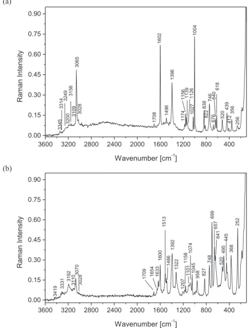
Fig. 6.Raman spectrum of 1-(diaminomethylene)thiouron-1-ium benzoate (a) and bis(1-(diaminomethylene)thiouron-1-ium phthalate trihydrate (b).
protiated compounds are shown inFig. 6. The title compounds have several functional and skeletal groups such as three NH2, C]S, CeNeC, NeCeN and NeCeS groups in the cation and one or two COO groups and the six-membered aromatic ring in the anions. The bands corresponding to the vibration of these groups were identified with the aid of infrared correlation charts[22]. The IR spectra of neutral 1-(diaminomethylene)thiourea [10 g] and of benzoic acid[23], phthalic acid[24]as well as benzoate( ) and phthalate(2-) [25] will be helpful for assignment of the bands observed in the spectra of the title compounds (Tables 4 and 5). The IR spectrum of protiated 1-(diaminomethylene)thiouron-1-ium benzoate (Fig. 4a) shows medium-strong intensity bands in the spectral range of 3400 and 3000 cm 1. These bands can be attrib-uted to the asymmetric and symmetric stretching of the three NH2 groups of the 1-(diaminomethylene)thiouron-1-ium cation. These bands, as expected, are shifted in the IR-spectrum of deuterated sample to the spectral region of 2600e2200 cm 1 (Fig. 4b). The isotopic ratio between 1.310 and 1.372 points on the vibration anharmonicity. The IR-spectra of protiated and deuterated samples of bis(1-(diaminomethylene)thiouron-1-ium) phthalate trihydrate show similar correlation between the observed
n
asym(NH2) andn
sym(NH2) andn
asym(ND2) andn
sym(ND2) (Fig. 5a and b). The IR spectra of deuterated analogues (Figs. 4b and 5b), the bands of the protiated compounds with significantly lower intensities are observed. These bands resulting from the equilibrium between the protiated and deuterated analogues and point on the not fully ex-change of H to D. The degree of deuterization is estimated to ~80% in1and ~90% in2. In the Raman spectrum of protiated compounds (Fig. 6) then
(NH2) stretching vibration bands appear in the 3350e3000 cm 1as a very weak once. The Raman spectra exhibit two narrow bands at 3065 and 3028 cm 1in1and at 3070 and 3028 cm 1 in2and they are assigned to CeH stretching of thebenzoate and phthalate anions (Table 6). The medium strong in-tensity band at 1718 cm 1and 1717 cm 1in the IR spectrum of benzoate and phthalate salts, respectively, is assigned to the stretching of imine bond of 1-(diaminomethylene)thiouron-1-ium cation, since it is not observed in the IR spectrum of neutral 1-(diaminomethylene)thiourea [10 g]. Its counterpart is observed at 1708 and 1709 cm 1 in the Raman spectrum of benzoate and phthalate salts (Fig. 6). A similar band is observed in the IR-spectrum of some imines and their salts[26]. This assignment of the imine stretching vibration band is confirmed by IRespectra of deuterated samples, in which the deuterated imine stretching is observed at 1258 cm 1in benzoate and at 1254 cm 1in phthalate (Figs. 4b and 5b,Tables 4 and 5). The isotopic ratio for the band of imine group is equal to 1.366 (for1) and 1.369 (for2) and points on the almost the same anharmonic vibration. The
n
(C]S) band of the 1-(diaminomethylene)thiouron-1-ium cation is observed in the spectral region of 720e710 cm 1in both salts. Then
(C]S) band in the spectrum of several thiourea metal complexes is observed in the range of 715-700 cm 1[21]. The 1-(diaminomethylene)thio-uron-1-ium cation contains CeN and the CeNeC, NeCeN skeletal groups and the respective vibrational bands are also observed (Tables 4 and 5).The assignment of the IR bands of benzoate( ) and phthalate(2-) anions of the samples were made based on the literature[27]. The notation used in the Tables 4 and 5for benzoate( ) and phtha-late(2-) anions for the vibrational modes associated with the ben-zene ring is commonly used for substituted benben-zene derivatives and is made by analogy to the notation established for the modes of benzene by Wilson[28]. For benzoate( ) and phthalate(2-) anions the characteristic
n
(CeC)ar,n
asym(COO ) andn
sym(COO ) bands are observed in the spectral region of 1620e1380 cm 1, and theb
(CeH) andg
(CeH) vibrational bands are observed in the range ofTable 4
FT - IR spectral data for protiated and deuterated analogue of 1-(diaminomethylene)thiouron-1-ium benzoate.
Protiated,ncm1 Deuterated,ncm1 Assignment Isotopic ratio
3389m 2540s nasym(NH2)/nasym(ND2) asym stretch. 1.334
3327s 2498s nasym(NH2)/nasym(ND2) asym stretch. 1.332
3273s 2465s nasym(NH2)/nasym(ND2) asym stretch. 1.328
3142m 2351m nsym(NH2)/nsym(ND2) sym stretch 1.336
3084w 2331m nsym(NH2)/nsym(ND2) sym stretch 1.323
3057w 2228m nsym(NH2)/nsym(ND2) sym stretch 1.372
3023w broad
band ~2800 ~2200 NeH/O/NeD/O hydrogen bonds
1718m 1258m Imine bond stretch. 1.366
1671w
1618vs 1620s,1596s 8a, 8b (Benzenen(CC))a
1525w 1553s nasym(COO )
1469m 1469s 19a (Benzenen(CC)) overlapped withn(CN)
1389vs 1390s nsym(COO )
1308m 14 (Benzenen(CC))
1174w 1174w 9a (Benzenen(CC))
1153w 1153w n(CN)
1124w 1123w n(CN)
1068w 1068w 18b (Benzened(CH))
1025w 1025w 18a (Benzenen(CC))
1000w 998w
898w
838m 837m bsym(COO )
745m
711m 712s n(C]S)
676m 675m Skeletal CeNeC, NeCeN
640w t(NH2),u(NH2)
613w Skeletal CeNeC, NeCeN
583w 585w Skeletal CeNeC, NeCeN
529w 529w basym(COO )
514w 511w
vs, very strong; s, strong; m, medium; w, weak.
aNotation for the modes of the benzene ring according to Willson[28].
1360e1000 cm 1and 970e740 cm 1, respectively. The assignment of the characteristic
n
(CeC)ar,n
asym(COO ) andn
sym(COO ) bands for phthalate(2-) anion has been supported by theoretical calcula-tion performed by Loring et al.[29]. Since the phthalate salt con-tains hydrated water molecules, in the IR-spectrum the bands ofn
asym(OH) andn
sym(OH) are observed as medium bands at 3522 and 3431 cm 1(Fig. 5a), which are shifted to 2618 and 2567 cm 1in IR-spectrum of the deuterated analogue (Fig. 5b). In the Raman spectrum of hydrated compound2the stretching vibration of OeH of water are attributed to the weak band at 3419 cm 1(Fig. 6b). The X-ray data reveal that the NH2groups of the 1-(diaminomethylene) thiouron-1-ium cation are involved in NeH/O and NeH/S hydrogen bonds. In addition, in the crystal2the hydrated water molecules are involved as donors and as acceptors in the O(water)-eH/O(water)and NeH/O(water)hydrogen bonds. These hydrogen bonds are relatively weak, with the lengths between the 2.721 (1) and 3.078 (2) Å (Table 3). This reveals as a broad band in the range of 3300e2500 cm 1in both protiated samples, which is shifted to ~2200 cm 1in the deuterated analogues.
4. Conclusion
This study confirms the usefulness of 1-(diaminomethylene) thiourea as a building block in the crystal engineering and dem-onstrates its interaction with benzoic and phthalic acids forming of extended supramolecular hydrogen bonding structures. The R22(8)
and R21(6) motifs describe the interaction between the oppositely charged units of the crystals. The hydrogen bonding interactions lead to formation of layered 2D supramolecular structure in 1-(diaminomethylene)thiouron-1-ium benzoate (1), whereas in bis(1-(diaminomethylene)-thiouron-1-ium) phthalate trihydrate (2) the hydrogen bonding interactions lead to formation of 3D su-pramolecular network. In crystal2, the hydrated water molecules are contributed in the formation with phthalate(2-) anions of OeH/O hydrogen bonding anionic 2D layered substructure. In both crystals are no
p
ep
interactions between the aromatic rings of anions. Comparison of the IR spectra of 1-(diaminomethylene)thi-ouron-1-ium benzoate and bis(1-(diaminomethylene)-thiouron-1-ium) phthalate trihydrate with the spectra of theirs deuterated analogues shows marked differences in the region of vibrations of the amine groups as well as in the region of NeH/O and OeH/O hydrogen bonds.Supplementary material
Additional material comprising X-ray powder diffraction pat-terns of protiated and deuterated samples of 1 and 2 as well as the microscopic images of these two crystals. Full details of the X-ray data collection andfinal refinement parameters including aniso-tropic thermal parameters and full list of the bond lengths and angles have been deposited with the Cambridge Crystallographic Data Center in the CIF format as supplementary publications no. Table 5
FT-IR spectral data for protiated and deuterated analogue of bis(1-(diaminomethylene)thiouron-1-ium) phthalate trihydrate.
Protiated,ncm 1 Deuterated,ncm 1 Assignment Isotopic ratio
3532m 2618w nasym(OH)/vasym(OD) (water) 1.349
3431s 2567m nasym(NH2)/nasym(ND2) asym stretch.,
overlapped withnsym(OH) orvsym(OD) (water)
1.337
3401s 2515w nasym(NH2)/nasym(ND2) asym stretch. 1.352
3263s 2460m nasym(NH2)/nasym(ND2) asym stretch. 1.326
3157s 2386s nsym(NH2)/nsym(ND2) sym stretch 1.323
3126s 2360m nsym(NH2)/nsym(ND2) sym stretch 1.310
3102s 2306m nsym(NH2)/nsym(ND2) sym stretch 1.345
broad
band ~2100 NeH/O/NeD/O hydrogen bonds
~2800 2123m NeH/O/NeD/O hydrogen bonds
2725m OeH/O/OeD/O hydrogen bonds
2603w OeH/O/OeD/O hydrogen bonds
2464w 2145w
1717s 1254m Imine bond stretch. 1.369
1645s 1640s
1621vs 1600m 8a, 8b (Benzenen(CC))a
1537vs 1550vs nasym(COO )
1466m 1454s 19a (Benzenen(CC)) overlapped withn(CN)
1447s 1447s n(CN)
1397s,
1383vs 1383s nsym(COO )
1355m 967w d(NH2)/d(ND2)
1328m 1321w n(CN)
1155m 1155w 9a (Benzenen(CC))
1108w 1107w n(CN)
1088m
859m 858w
845m
829m 829w
772m 772m basym(COO )
730m 722w n(C]S)
691 692m 4 (Benzenen(CC))
640w t(NH2),u(NH2)
624w 624w Skeletal CeNeC, NeCeN
579w 582w Skeletal CeNeC, NeCeN
521w 520w basym(COO )
482w 483w
vs, very strong; s, strong; m, medium; w, weak.
aNotation for the modes of the benzene ring according to Willson[28].
CCDC 1409699 and 1409700. Copies of the data can be obtained free of charge on the application to CCDC, 12 Union Road, Cam-bridge, CB21EZ, UK, (fax: (þ44) 1223-336-033; email: deposit@ ccdc.cam.ac.uk).
Appendix A. Supplementary data
Supplementary data related to this article can be found athttp:// dx.doi.org/10.1016/j.molstruc.2015.10.080.
References
[1] (a) G.R. Desiraju, Crystal Engineering. The Design of Organic Solids, Elsevier, Amsterdam, 1989;
(b) J.C. MacDonald, G.R. Whitesides, Chem. Rev. 94 (1994) 2382e2420;
(c) G.R. Desiraju, Angew. Chem. Int. Ed. 46 (2007) 8342e8352.
[2] (a) J.M. Lehn, Supramolecular Chemistry: Concepts and Perspectives, VCH, Weinheim, 1995;
(b) C.B. Aaker€oy, Acta Cryst. B53 (1997) 569e586;
(c) D. Braga, Chem. Commun. (2003) 2751e2754;
(d) G.R. Desiraju, Acc. Chem. Res. 35 (2002) 565e573.
[3] (a) B. Moulton, M.J. Zaworotko, Chem. Rev. 101 (2001) 1629e1658;
(b) G.R. Desiraju, J. Am. Chem. Soc. 135 (2013) 9952e9967.
[4] (a) G.R. Desiraju, Chem. Commun. (1997) 1475e1482;
(b) C.B. Aaker€oy, N.R. Champness, C. Janiak, CrystEngComm 12 (2010) 22e43.
[5] (a) G.R. Desiraju, Perespectives in Supramolecular Chemistry, vol. 2, Wiley, Chichester, U.K, 1996;
(b) C.B. Aaker€oy, P.D. Chopade, C. Gasner, J. Desper, Chem. Commun. 47 (2011) 4688e4690;
(c) O. Altintas, D. Schulze-Suenninghausen, B. Luy, C. Barner-Kowollik, ACS
Macro. Lett. 2 (2013) 211e216;
(d) C.T. Seto, G.M. Whitesides, J. Am. Chem. Soc. 115 (1993) 905e916;
(e) J.I. Arenas-Garcia, D. Herrera-Ruiz, K. Mondragon-Vasquez, H. Morales-Rojas, H. H€opfl, Cryst. Growth Des. 12 (2012) 811e824;
(f) G.R.Desiraju, CrustEngComm 5 (2003) 466e467.
[6] (a) G.M. Whitesides, E.E. Simanek, J.P. Mathias, C.T. Seto, D. Chin, M. Mammen, D.M. Gordon, Acc. Chem. Res. 28 (1995) 37e44;
(b) G.R. Desiraju, Angew. Chem. Int. Ed. 34 (1995) 2311e2327;
(c) T. Steiner, Angew. Chem. Int. Ed. 48 (2002) 49e76;
(d) G.J. Perpetuo, J. Janczak, Acta Cryst. C63 (2007) o301eo302;
(e) C.C. Seaton, K. Chadwick, G. Sadig, K. Guo, R.J. Davey, Cryst. Growth Des. 10 (2010) 726e733;
(f) B.A. Blight, C.A. Hunter, D.A. Leigh, H. McNab, P.I.T. Thomson, Nat. Chem. 3 (2011) 244e248;
(g) A.J. Wilson, Nat. Chem. 3 (2011) 193e194;
(h) C.C. Cheng, Y.C. Yen, F.C. Chang, RSC Adv. 1 (2011) 1190e1194;
(i) R. Patra, H.M. Titi, I. Goldberg, Cryst. Growth Des. 13 (2013) 1342e1349.
[7] (a) M.C. Etter, J. MacDonald, J. Bernstein, Acta Cryst. B46 (1990) 256e262;
(b) M.C. Etter, Acc. Chem. Res. 23 (1990) 120e126;
(c) M.C. Etter, J. Phys. Chem. 95 (1991) 4601e4610.
[8] J. Janczak, G.J. Perpetuo, Acta Cryst. C64 (2008) o114eo116.
[9] (a) K. Chakrabaty, S.P.S. Gupta, Indian J. Phys. A57 (1983) 205e209;
(b) K. Chakrabaty, T. Kar, S.P.S. Gupta, Acta Cryst. C46 (1990) 2065e2068;
(c) E. Doxiadi, R. Vilar, A.J.P. White, D.J. Williams, Polyhedron 22 (2003) 2991e2998;
(d) M. Hołynska, M. Korabik, M. Kubiak, Polyhedron 29 (2010) 530 e538.
[10] (a) G.J. Perpetuo, J. Janczak, Acta Cryst. C64 (2008) o264eo268;
(b) J. Janczak, G.J. Perpetuo, Acta Cryst. C64 (2008) o330eo334;
(c) M. Hołynska, M. Kubiak, Acta Cryst. C64 (2008) o609eo612;
(d) J. Janczak, G.J. Perpetuo, Acta Cryst. C65 (2009) o118eo120;
(e) M. Hołynska, M. Kubiak, Acta Cryst. C65 (2009) o191 eo194;
(f) M. Hołynska, M. Kubiak, Acta Cryst. C65 (2009) o410 eo413;
(g) J. Janczak, G.J. Perpetuo, J. Mol. Struct. 975 (2010) 166e172;
(h) J. Janczak, G.J. Perpetuo, J. Mol. Struct. 988 (2011) 73e81;
(i) G.J. Perpetuo, J. Janczak, J. Mol. Struct. 1007 (2012) 74e80;
(j) G.J. Perpetuo, J. Janczak, J. Mol. Struct. 10541 (2013) 127e138;
(k) G.J. Perpetuo, R.S. Gonçalves, J. Janczak, J. Mol. Struct. 1096 (2015) 74 e83;
(l) J. Janczak, Cryst. Growth Des. 15 (2015) 5097e5111.
[11] G.J. Perpetuo, J. Janczak, J. Mol. Struct. 1031 (2013) 14e21.
[12] C.C.D. CrysAlis, CrysAlis Red, Version 171.32.6, Oxford Diffraction Poland, Wrocław, Poland, 2006.
[13] G.M. Sheldrick, SHELXS97, SHELXL97, Programs for Crystal Structures Solution and Refinement, University of Gottingen, G€ €ottingen, Germany, 1997. [14] K. Brandenburg, H. Putz, DIAMOND Version 3.0, Crystal Impact GbR, Bonn,
Germany, 2006.
[15] F.H. Allen, Acta Cryst. B58 (2002) 380e388.
[16] D.R. Johnson, F.X. Powell, W.H. Kirchoff, J. Mol. Spectrosc. 39 (1971) 136e145.
[17] (a) G. Bruno, L. Randaccio, Acta Cryst. B36 (1980) 1711e1712;
(b) D. Feld, M.S. Lahmann, Z. Kristallogr. 157 (1981) 215e231.(c) C.C. Wilson, N. Shankland, A.J. Florence, J. Chem. Soc. Faraday Trans. 92 (1996) 5051e5067;
(d) W. Cai, A. Katrusiak, CrystEngComm 14 (2012) 4420e4424.
[18] O. Ermer, Helv. Chim. Acta 64 (1981) 1902e1909.
[19] G.Y. Bai, H.S. Ning, J. Simpson, X.Y. Qin, N. Li, Acta Cryst. E62 (2006) o4567eo4568.
[20] F.H. Allen, C.H. Bird, R.S. Rowland, P.R. Raithby, Acta Cryst. B53 (1997) 696e701.
[21] F.H. Allen, C.H. Bird, R.S. Rowland, P.R. Raithby, Acta Cryst. B53 (1997) 680e695.
[22] (a) G. Socrates, Infrared Characteristic Group Frequencies, Wiley-Interscience, Chichester, U.K, 1980;
(b) G. Socrates, Infrared, Raman Characteristic Group Frequencies, third ed., Wiley, New York, USA, 2001.
[23] (a) S. Hayashi, N. Kimura, Bulletin of the Institute for Chemical Research, Kyoto University (1966), 44(4), 335e340.(b) I.D. Reva, S.G. Stepanian, J. Mol. Struct. 349 (1995) 337e340;
(c) S.G. Stepanian, I.D. Reva, E.D. Radchenko, G.G. Sheina, Vibr. Spectrosc. 11 (1996) 123e133.
[24] (a) J.F. Arenas, J.I. Marcos, Spectrochim. Acta A36 (1980) 1075e1081;
(b) E.S. Blanca, J.L. Nú~nez, P. Martinez, J. Mol. Struct. 142 (1986) 45e48.
[25] (a) J.F. Arenas, J.I. Marcos, Spectrochim. Acta A35 (1979) 355e363;
(b) G.B. Deacon, R.K. Phillips, Coord. Chem. Rev. 33 (1980) 227e250;
(c) W. Lewandowski, B. Dasiewicz, P. Koczon, J. Skierski, K. Dobrosz-Teperek,
R.Swisłocka, L. Fuks, W. Priebe, A.P. Mazurek, J. Mol. Struct. 604 (2002) 189e193.
[26] (a) J. Favrot, J.M. Leclercq, R. Roberge, S. Sandorfy, D. Vocelle, Photochem. Photobiol. 29 (1979) 99e108;
(b) J. Favrot, D. Vocelle, C. Sandrofy, Photochem. Photobiol. 30 (1979) 417e421.
[27] G. Varsanyi, Assignments for Vibrational Spectra of 700 Benzene Derivatives, Akademiai Kiado, Budapest, 1972.
[28] E.B. Wilson, Phys. Rev. 45 (1934) 706e714.
[29] J.S. Loring, M. Karlsson, W.R. Fawcett, W.H. Casey, Spectrochim. Acta A57 (2001) 1635e1642.
Table 6
Raman spectral data for 1-(diaminomethylene) thiouron-1-ium benzoate (1) and bis(1-(diaminomethylene)thiouron-1-ium) phthalate trihydrate (2).
1,ncm1 2,ncm 1 Assignment
3419w na(H2O)
3345w 3331w na(NH2) asym stretch.
3314w na(NH2) asym stretch.
3249w 3240sh na(NH2) asym stretch.
3200w 3192w ns(NH2) sym stretch. and OeH/O stretch.
3156w ns(NH2) sym stretch.
3109w 3110w ns(NH2) sym stretch.
3065m 3070m CeH stretch.
3028w 3028w CeH stretch.
1708w 1709w Imine bond stretch.
1654w 1633w
1602m 1600m nasym(COO ), NeCeN bendþring def. 1513m
1496w 1486w NeCeN bendþring def.
1396m 1392m nsym(COO )
1322m d(NH2)
1174w 1207w
1156w 1158w n(CeN),n(CeC)
1139w
1126w 1101w G(CeC)
1074w
1027w 1045w n(CeC)
1004s
958w 838m
822w 827m basym(COO )
746m 748m n(CeC),n(C]S)
699m CeCeC def. out of plane of phenol ring
676w 657m n(CeC)
640w 641m t(NH2),u(NH2)
618m 610sh Skeletal CeNeC, NeCeN
520w 520w basym(COO)
495m
439m 445m
412w 414w
356w 368m Skeletal CeNeC, NeCeN,
256w 252m
weak, w; medium, m; strong, s; very, v; shoulder, sh.