267
CLINICS 2008;63(2):267-70
LETTER TO THE EDITOR
Department of Dermatology, Faculdade de Medicina da Universidade de São Paulo - São Paulo/SP, Brazil.
NECROLYTIC MIGRATORY ERYTHEMA ASSOCIATED
WITH GLUCAGONOMA: A REPORT OF 2 CASES
Renata Câmara Teixeira, Marcello Menta Simonsen Nico, Anelise Casillo Ghideti
INTRODUCTION
Glucagonoma syndrome is a rare disease that is usually associated with an underlying neuroendocrine tumor. Necro-lytic Migratory Erythema (NME) has been associated with intestinal malabsorption disorders, hepatic cirrhosis, chronic pancreatitis, inflammatory bowel disease, and non-pancreatic malignancies, but may not always be associated with gluca-gonoma. In 1979, Mallinson and co-workers coined the term glucagonoma syndrome to describe alpha-cell pancreatic tumors associated with a characteristic erosive skin eruption, termed Necrolytic Migratory Erythema by Wilkinson. NME is characterized by an irregular annular eruption with serpigi-nous advancing borders, erosion, and crusting, resulting in a scalded appearance. The eruption has a cyclical nature with concurrent lesions at different levels of healing.
Here, we report two patients with NME associated with hyperglucagonemia and neuroendocrine tumor.
CASE 1
This case involved a 66-year-old Brazilian male with an eight year history of recurrent cutaneous lesions.
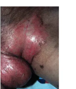
Abdominal surgery was performed due to colon cancer eight years before the dermatologic complaint. He also men-tioned a significant alcohol history. Physical examination revealed erythematous, erosive scaling and crusted patches on the genital and groin area (Figure 1). He also presented with similar lesions in the perioral and periocular areas as-sociated with angular cheilitis and a depapilated, bright red tongue (Figure 2). The patient denied intestinal symptoms and weight loss.
Laboratory testing showed normocytic normochromic anemia and slightly elevated levels of amylase and lipase. The patient had normal levels of serum zinc, folic acid, vitamin B12, albumin, globulins, alanine and aspartate tranferase. Hepatitis B and C and HIV serologies were
negative. An elevated level of blood glucose (125mg/ml) was observed. The patient’s plasma level of glucagon was greater than 1,280 pg/ml (normal range: <60 pg/ml).
An abdominal computed tomography (CT) scan revealed a hypervascularized tumor measuring 6.1 x 3.8 cm in the body and the caudal portion of the pancreas, and an absence of liver metastasis. The patient was then subjected to a pan-createctomy, and the cutaneous lesions vanished one week after surgery.
Figure 1 - Erosive scaling and crusted patches on the genital and groin area
268
CLINICS 2008;63(2):267-70 Necrolytic migratory erythema associated with glucagonoma
Teixeira RC et al.
CASE 2
A 62-year-old Brazilian man was referred to our Der-matology Department complaining of two years of eroded skin. The eruption had a cyclical pattern, with periods of resolution.
During a weight loss investigation six months prior, a CT scan was performed and a pancreatic tumor with hepatic metastasis was confirmed. Surgical removal of the tumor was not possible. The patient had an episode of deep vein throm-bosis associated with pulmonary embolism two months prior to being referred to our department earlier, and was taking marevan.
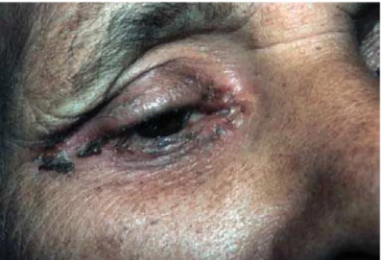
Physical examination revealed erythematous scaling and crusted plaques, with exulceration on the edges of the lesions, involving the groin and genital areas (Figure 3) and the periorbital area (Figure 4). He also presented with tense bullous lesions with purulent content and pellagroid eczematous plaques on the anterior surface of the legs and feet, associated with purpura and edema (Figure 5). Angular cheilitis and a depapilated red tongue were also present. In addition to the skin changes, the patient complained of weight loss and watery diarrhea.
Laboratory testing revealed normocytic normochromic anemia, hypoalbuminemia and elevated levels of amylase, lipase, alkaline phosphatase, gamma glutamyl transferase, alanine and aspartate transferase. Plasma levels of glucose and zinc were normal. The patient’s fasting plasma glucagon level was 1,280 pg/ml (normal range: <60 pg/ml), and amino acid levels were decreased.
An ultrasound-guided needle biopsy of a hepatic lesion was performed, and histopathological examination and im-munohistochemistry revealed a neuroendocrine tumor.
Histopathological examination of a purpuric skin lesion
showed extravasated erythrocytes on the superficial dermis and hyaline thrombus on the vascular lumen of on the deep dermis. These findings were attributed to the medication taken by the patient.
Palliative treatment with interferon alpha and octreotide LAR was proposed, but the patient presented an erythema-tous rash that was attributed to interferon alpha. Therefore, the offending drug was suspended. Octreotide LAR, 20 mg/ month, was continued with complete resolution of the skin lesions. The patient was discharged and prescribed ambula-tory chemotherapy.
DISCUSSION
Glucagonoma arises from alpha cells of pancreatic islets of Langerhans and may appear as either a benign, localized alpha cell adenoma, or a slow growing metastasizing malig-nant tumor. Glucagonoma is associated with striking system-ic clinsystem-ical manifestations, referred to as the “Glucagonoma
Figure 3 - Erythematous scaling and crusted plaques, with exulceration on the edges of the lesions, involving the groin and genital areas
Figure 4 - Erythematous scaling and crusted plaques, with exulceration on the edges of the lesions, involving the periorbital area
269
CLINICS 2008;63(2):267-70 Necrolytic migratory erythema associated with glucagonoma
Teixeira RC et al.
Syndrome.” Systemic manifestations of the syndrome are numerous, and include diabetes mellitus, anemia, venous thrombosis, skin rash (NME), weight loss, glossitis, cheilitis, diarrhea, steatorrhea, and psychiatric disorders.
The cause of skin changes in NME is unclear. Glucagon
per se has not been found to be the direct cause because there are patients who present with NME without neuroen-docrine tumors or hyperglucagonemia. However, normaliza-tion of glucagon concentranormaliza-tion by surgery or somatostatin analogs almost invariably results in a rapid resolution of the skin lesions. Furthermore, there is a recent report of a case of iatrogenic NME after intravenous administration of glucagon for the treatment of persistent hypoglycemia13 due
to an insulinoma. This case supports the hypothesis of glu-cagon leading to the skin lesions. Zinc, essential fatty acid, and amino acid deficiencies are all considered to be possible causes of NME. However, not all patients with skin lesions present with these metabolic changes, and not all patients with these metabolic changes present with resolution of the skin lesions after zinc, essential fatty acid, or amino acid supplementation.14
NME is a rare dermatosis that is usually associated with an underlying pancreatic islet cell tumor. The skin eruption may be the first manifestation of the disease, and its recogni-tion may lead to a diagnosis, as in case 1. The eruprecogni-tion has a cyclic nature, with periods of skin lesion resolution, as ob-served in both of our cases. The lesions consist of erythema-tous scaling and crusting patches most frequently observed in areas of trauma, such as the groin, intergluteal, and genital areas. Bullous lesions may occur. Cheilitis and glossitis are very common mucosal manifestations. It is commonly ob-served that these patients present with a history of antibiotic or antifungal treatments without improvements of their skin conditions before the correct diagnosis is made.
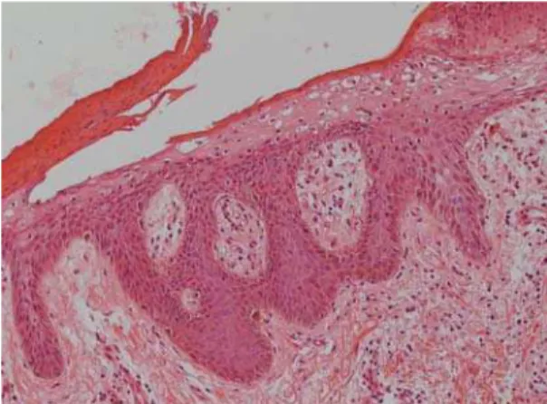
The histopathologic features of NME are nonspecific and may be seen in pellagra, necrolytic acral erythema, or zinc deficiency. Vacuolated, pale, swollen epidermal cells and necrosis of the superficial epidermis are characteristic (Figure 6). Biopsy specimens from the edges of active le-sions are most likely to show the characteristic upper epi-dermal necrosis; however, many biopsy specimens do not have features that are typical, or even suggestive, of NME. Therefore, multiple biopsies are recommended when this diagnosis is suspected.
Diagnostic criteria of glucagonoma syndrome have been proposed by Stacpole and include demonstration of a tumor producing increased levels of glucagon, as revealed by spe-cial staining and increased circulatory levels of glucagon. In addition, the patient must meet at least one of the following criteria: (1) skin eruption, (2) diabetes mellitus, and (3) hy-poaminoacidemia.
Glucagonomas show significant evidence of hypervas-cularity, so selective celiac and superior mesenteric arte-riographies are the most reliable ways to detect the primary neoplasm if the abdominal CT scan does not disclose the tu-mor. Histopatologic confirmation of the glucagonoma occurs via immunohistochemistry, electronic microscope analysis, andin situ hybridization of glucagon messenger RNA. The absence of immunoreactivity for glucagon or glucagon mes-senger RNA transcripts in a metastatic focus may be due to tumor heterogeneity since glucagon may not be expressed in all metastatic foci. This could explain the negative immuno-histochemistry for glucagon in case 2. Usually, the antigens used to assess neuroendocrine differentiation of tumors are synaptophysin and cromogranin. Hormones such as insulin, glucagon, vasointestinal polypeptide, somatostatin, and pancreatic polypeptide are used to determine the predomi-nant hormone produced by tumors. In some cases, tumors produce more than one hormone.
The prognosis of the disease varies greatly according to the stage in which the tumor is diagnosed. By the time of di-agnosis, 50-100% of patients already present with metastatic disease, and a cure is often impossible, as in case 2. The tumor is resistant to chemotherapy, and metastatic disease is often not amenable to surgical resection9. However, since this
islet cell tumor is slow-growing, prolonged survival (more than 20 years) is possible, and in metastatic disease, most causes of death appear to be unrelated to the tumor.17
Successful palliative treatment is possible with long-acting somatostatin analogs,22,26 and/or interferon alpha.
Supplementation with zinc, amino acids, and essential fatty acids appears to be beneficial in some cases.12,14
The patient in case 1 was a diagnostic challenge. Only skin lesions and mucosal changes were present at the time of diagnosis. In this case, the dermatologist was responsible for the pancreatic tumor diagnosis in the curable stage of disease.
270
CLINICS 2008;63(2):267-70 Necrolytic migratory erythema associated with glucagonoma
Teixeira RC et al.
In case 2, it was not possible to perform a surgical resec-tion of the tumor, so only an improvement in the skin lesions was feasible.
We describe two cases that illustrated good outcomes of
skin lesions in two different situations: case 1 was a patient with an early diagnosis and a curable disease and case 2 was a patient with a metastatic tumor. Both patients presented with resolution of skin lesions after beginning treatment.
REFERENCES
1. Kovacs RK, Korom I, Dobozy A, Farkas G, Ormos J, Keminy L. Necrolytic migratory erythema. J Cutan Pathol. 2006;33:242-5. 2. Echenique-Elizondo M, Lizarduy IM. Glucagonoma and necrolytic
migratory erythema. Rev Esp Enferm Dig. 2005;6:455-7.
3. Schanz S, Schaefer J, Fierlbeck G. Image of the month. Gastroenterology 2005;129:1816.
4. Technau K, Renkl A, Norgaeur J, Ziemer M. Necrolytic migratory erythema with myelodysplastic syndrome without glucagonoma. Eur J Dermatol. 2005;15:110-2.
5. Marco PB, Miljkovic J, Zemljic TG. Necrolytic migratory erythema associated with hyperglucagonemia and neuroendocrine hepatic tumor. Acta Dermatoven APA. 2005;14:161-6.
6. Kitamura Y, Sato M, Hatamochi A, Yamazaki S. Necrolitc migratory erythema without glucagonoma associated with hepatitis B. Eur J Dermatol. 2005;15:49-51.
7. Nofal AA, Nofal E, Attwa E,El-Assar O, Assaf M. Necrolytic acral erythema: a variant of necrolitc migratory erythema or a distinct entity? International Journal of dermatology 2005;44:916-21.
8. Pujol RM, Wang CYE, el-Azhary RA, Su WPD, Gibson LE, Schroeter L. Necrolytic migratory erythema: clinicopathologic study of 13 cases. Internacional Journal of Dermatology. 2004;43:12-8.
9. van Beek AP, Haas ERM, van Vloten WA, Lips CJM, Roijers JFM, Dijk MRCv. The glucagonoma syndrome and necrolytic migratory erythema: a clinical review. European journal of endocrinology. 2004;151:531-7.
10. Echenique-Elizondo M, Valls AT, Orue JLE, Lizarduy IM, Aguirre JI. Glucagonoma and pseudoglucagonoma syndrome. Journal of the pancreas, 2004;5:179-85.
11. Alexander EK, Robinson M, Staniec M, Dluhy RG. Peripheral amino acid and fatty acid infusion for the treatment of necrolytic migratory erythema in the glucagonoma syndrome. Clinical endocrinology. 2002;57:827-31.
12. Mullans EA, Cohen PR. Iatrogenic necrolytic migratory erthema: A case report and review of nonglucagonoma-associated necrolytic migratory erythema. Journal of the American Academy of Dermatology. 1998; 38:866-73.
13. Sinclaie SA, Reynolds NJ. Necrolytic migratory erythema and zync deficiency. British Journal of Dermatology.1997;136:783-85. 14. Delaporte E, Catteau B, Piette F. Necrolytic migratory erythem-like
eruption in zinc deficiency associated with alcoholic liver disease. British Journal of Dermatology. 1997;137:1027-28.
15. Wermers RA, Fatourechi V, Kvols LK. Clinical spectrum of Hyperglucagonemia Associared With Malignant Neuroendocrine Tumors. Mayo Clin Proc. 1996;71:1030-38.
16. Wern\mers RA, Fatourechi V, Wynne AG, Kvols LK, Lloyd RV. The Glucagonoma Syndrome. Clinical and Pathologic Features in 21 Patients. Medicine. 1996;75:53-63.
17. el Darouty M,Abu el Ela M. Necrolytic migratory erythema without Glucagonoma in patients with liver disease. J Am Acad Dermatol. 1996;1092-3.
18. Bencini PL, Vigo GP, Caputo R. Necrolytic Migratory Erythema without Glucagonoma in a Heroin- Depedent Patient. Dermatology. 1994;189:72-4.
19. Blackford S, Wright S, Roberts DL. Necrolytic migratory erythema without glucagonoma: the role of dietary essential fatty acids. British Journal of Dermatology.1992;125:460-62.
20. Wilkinson SM, Carthwright PH, Allen C, Reeves S, Alexander L, Byrne JPH. Necrolytic migratory erythema:association with neuroendocrine tumour with predominant insulin secretion. British Journal of Dermatology. 1990;123:801-05.
21. Rosenbaum A, Flourie B, Chagnon S, Blery M, Modigliani R. Octreotid(SMS201-995) in the treatment of metastatic glucagonoma: Report of one case and review of the literature. Digestion. 1989;42:116-20.
22. Govea JR, Holm A, Aldrete JS. Response of Glucagonomas to Surgycal Excision and Chemotherapy. Report of two cases and review of the literature. The American Surgeon. 1989;55:523-27.
23. van der Loss TLJM, Lambrecht ER, Lambers JCCA. Successful treatment of glucagonoma-related necrolytic migratory erythema with dacarbazine. Journal of the American Academy of Dermatology. 1987;16:468-72.
24. Vandersteen PR, Scheithauer BW. Glucagonoma syndrome. A clinicopathologic, immunocytochemical, and ultrastructural study. Journal of the American Academy of Dermatology. 1985;12:1032-39. 25. Elsborg L, Glenthoj A. Effect of Somatostatin in Necrolytic Migratory
Erythema of Glucagonoma. Acta Med Scand. 1985;218:245-9. 26. Doyle JA,Schroeter AL,Rogers RS 3rd. Hyperglucagonemia and
necrolotic migratory erythema in cirrhosis-possible pseudoglucagonoma syndrome.Br J Dermatol. 1979;100:581-7.