CLINICAL SCIENCE
I Department of Gastroenterology, Gastrointestinal Surgery Unit, Laparo-scopic Surgery Unit, Faculdade de Medicina da Universidade de São Paulo - São Paulo/SP, Brazil
II Faculdade de Medicina da Universidade de São Paulo - São Paulo/SP, Brazil
III Department of Pathology, Faculdade de Medicina da Universidade de São Paulo - São Paulo/SP, Brazil
Email: [email protected] Tel.: 55 11 5572 6435
Received for publication on August 07, 2009 Accepted for publication on November 03, 2009
ARE HISTOLOGICAL ALTERATIONS OBSERVED IN
THE GALLBLADDER PRECANCEROUS LESIONS?
Adriana Lúcia Agnelli Meirelles-Costa,I Claudio José Caldas Bresciani,I
Rodrigo Oliva Perez,I Barbara Helou Bresciani,II Sheila Aparecida C.
Siqueira,III Ivan CecconelloI
doi: 10.1590/S1807-59322010000200005
Meirelles-Costa ALA, Bresciani CJC, Perez RO, Bresciani BH, Siqueira SAC, Cecconello I. Are histological alterations observed in the gallbladder precancerous lesions? Clinics. 2010;65(2):143-50.
INTRODUCTION: Gallbladder cancer, which is characterized by rapid progression and a poor prognosis, is a complex disease to treat. Unfortunately, little is known currently about its etiology or pathogenesis. A better understanding of its carcinogenesis and determining risk factors that lead to its development could help improve the available treatment options.
METHOD: Based on this better understanding, the histological alterations (such as acute cholecystitis, adenomyomatosis, xan-thogranulomatous cholecystitis, polyps, pyloric metaplasia, intestinal metaplasia, dysplasia, cancer and others) in gallbladders from 1,689 patients who underwent laparoscopic cholecystectomy for cholecystolithiasis were analyzed. The association of these gallbladder histological alterations with clinical data was studied.
RESULTS: Gender analysis revealed a greater incidence of inlammatory changes in males, while dysplasia and cancer were only found in women. The incidence of cholesterolosis was greater in the patients 60 years of age and under, and the incidence of adeno-myomatosis and gangrene was greater in the elderly patients. A progressive increase in the average age was observed as alterations progressed through pyloric metaplasia, intestinal metaplasia, dysplasia and then cancer, suggesting that the metaplasia-dysplasia-carcinoma sequence may occur in gallbladder cancer. Gallbladder histological alterations were also observed in asymptomatic patients.
CONCLUSION: The results of this study suggest that there could be an association between some histological alterations of gallbladder and cancer, and they also suggest that the metaplasia-dysplasia-carcinoma sequence could in fact be true in the case of gallbladder cancer. Nevertheless, further studies directed towards a perfect understanding of gallbladder carcinogenesis are required.
KEYWORDS: Gallbladder neoplasms; Cholecystectomy; Laparoscopic; Metaplasia; Adenomyoma; Cholelithiasis.
INTRODUCTION
Gallbladder cancer is the most common cancer of the biliary tree and the ifth most common cancer of the gastrointestinal tract.1-,3 It is a disease known for its rapid evolution and high mortality rate. Its main prognostic factor
is the pathological stage of the disease.1,4,5
Because no speciic symptoms are produced during the early stages, gallbladder cancer is often only discovered during surgery or diagnosed at an advanced stage of the disease. The small group of patients with prolonged survival rates are those patients with tumors in early stages who were diagnosed by chance after a cholecystectomy for cholecystolithiasis.6,7 Nevertheless, incidental gallbladder cancer is discovered in 0.3 to 2% of all cholecystectomies performed for benign conditions.2,7-10
evidence relating these lesions to the cancer is determined indirectly.
Because there is unfortunately no efficient method to detect gallbladder cancer, a better understanding of the risk factors for gallbladder cancer development and pre-malignant lesions of the gallbladder could help select patients for prophylactic cholecystectomies, and, perhaps, reduce the mortality of this almost invariably fatal disease. There are two important carcinogenesis models known: the adenoma-carcinoma sequence and the metaplasia-dysplasia-carcinoma sequence. The observation that the gallbladder presents proper histological alterations raises the possibility of other carcinogenesis models in the gallbladder. Due to this observation, analysis of the behavior of the histological alterations observed in the gallbladder with cholecystolithiasis is an interesting topic of study.
METHODS
A retrospective analysis was conducted using the medical records of 1,689 patients who underwent laparoscopic cholecystectomies between November 1999 and December 2006 in two hospitals: Clinics’ Hospital of the University of São Paulo and Oswaldo Cruz German Hospital. The histopathological analysis - coordinated by a single pathologist - was performed in both hospitals.
An abdominal ultrasound examination was used to diagnose cholecystolithiasis in all patients. Gallbladder cancer was not conirmed in any of the patients during the preoperative stage.
Analyzed clinical data included gender, age and presence of symptoms before surgery. The patients were classiied according to their ages: 40 years of age or younger, 41 to 60 years of age and over 60 years of age. Regarding the symptoms before surgery, patients were divided in two groups: asymptomatic and symptomatic.
In all patients, the surgeon conducted a macroscopic examination after removing the gallbladder from the abdomen; if there was any abnormality, the specimen was sent for a frozen section test.
The histopathological examination consisted of an initial macroscopic examination of the specimen, where specific microscopic analysis of suspicious areas was performed. After that, the specimen was sent to a routine microscopic examination, which included a random sample of the fundus, body and neck of the gallbladder. These samples were then embedded in parafin, sectioned, stained with hematoxylin-eosin and examined under an optical microscope. All histological alterations described on the pathologists’ reports were included in the study. Analyzed pathological data included the presence of
acute cholecystitis, gangrene, abscess, fibrosis, sclero-atrophic gallbladder, adenomyomatosis, cholesterolosis, xanthogranulomatous cholecystitis, hyperplasic polyps, cholesterol polyps, pyloric metaplasia, intestinal metaplasia, dysplasia and cancer.
The Pearson chi-square test (x2) and Fisher exact probability test were used to determine the correlation between the clinical and pathological data.
The Standard Statistical Software Package (version 13) was used, and a statistically signiicant difference was set as p ≤0.05.
RESULTS
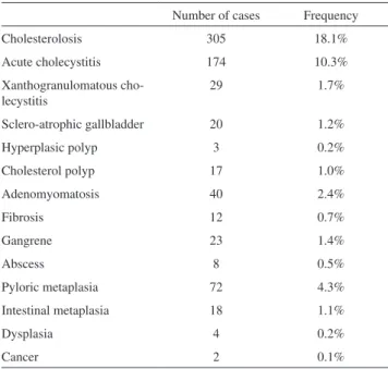
In 1,091 patients, no histological alteration was found, and the pathologist reported only chronic calculous cholecystitis. In the remaining patients, acute inlammation was observed in 174 patients (10.3%), gangrene in 23 (1.4%), abscess in 8 (0.5%), cholesterolosis in 305 (18.1%), adenomyomatosis in 40 (2.4%), xanthogranulomatous cholecystitis in 29 (1.7%), ibrosis in 12 (0.7%), sclero-atrophic gallbladder in 20 (1.2%), hyperplasic polyps in 3 (0.2%), cholesterol polyps in 17 (1.0%), pyloric metaplasia in 72 (4.3%), intestinal metaplasia in 18 (1.1%), dysplasia in 4 (0.2%) and cancer in 2 (0.1%). Adenomatous polyps were not observed in any patients (Table 1). More than one histological alteration was found in some patients.
In relation to gender, 1,238 patients were female (73.3%), and 451 patients were male (26.7%), making the female/male ratio was 2.7. The analysis of each
Table 1 - Frequency of the gallbladder histological altera-tions studied.
Number of cases Frequency
Cholesterolosis 305 18.1%
Acute cholecystitis 174 10.3%
Xanthogranulomatous cho-lecystitis
29 1.7%
Sclero-atrophic gallbladder 20 1.2%
Hyperplasic polyp 3 0.2%
Cholesterol polyp 17 1.0%
Adenomyomatosis 40 2.4%
Fibrosis 12 0.7%
Gangrene 23 1.4%
Abscess 8 0.5%
Pyloric metaplasia 72 4.3%
Intestinal metaplasia 18 1.1%
Dysplasia 4 0.2%
histological alteration studied and female or male gender, showed a significant statistical difference for acute cholecystitis, sclero-atrophic gallbladder, gangrene, abscess and cholesterol polyps, all of them more frequent among men (Table 2).
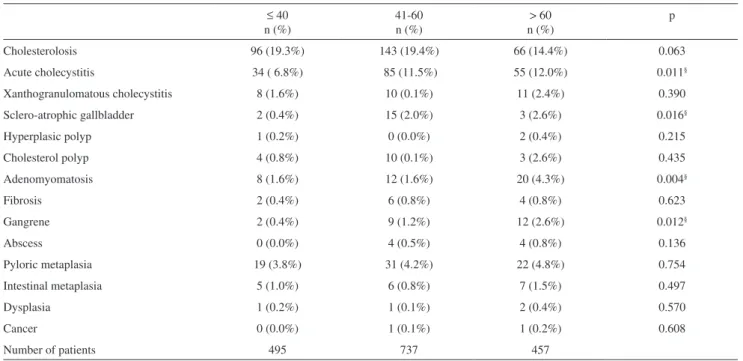
Patient ages varied from 11 to 90 years, with an average age of 50 years and median age of 51 years. Among the 1,689 patients, 495 (29.3%) were 40 years of age or younger, 737 (43.6%) were 41 to 60 years of age and 457 (27.1%) were over 60 years of age. The analysis of this three bands of age and each histological alteration studied demonstrated a signiicant statistical difference for acute cholecystitis, sclero-atrophic gallbladder, gangrene and adenomyomatosis. Sclero-atrophic gallbladders were observed more often in patients in the 41-60 age range, while all other gallbladder alterations with age-related signiicant statistical differences were observed more frequently in patients over 60 (Table 3). The study of the average age of each gallbladder histological alteration demonstrated, interestingly, a progressive increase in age for the pyloric metaplasia (50.6 ± 15 years), intestinal metaplasia (53.9 ± 14 years), dysplasia (56.8 ± 24 years) and cancer (60.0 ± 27 years) sequence; however, no statistical difference was observed (Table 4).
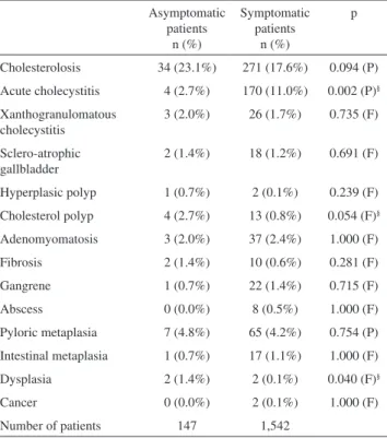
Regarding the presence or absence of symptoms, 147 (8.7%) patients were asymptomatic, and 1,542 (91.2%) presented symptoms before surgery. The statistical analysis of each histological alteration studied and the presence or absence of symptoms revealed a signiicant statistical difference for acute cholecystitis, cholesterol polyps and dysplasia. Acute cholecystitis had a bigger incidence among symptomatic patients, while cholesterol polyps and dysplasia had a bigger incidence among asymptomatic patients (Table 6).
Table 2 - Analysis of the gallbladder histological alterations according to gender.
Male n (%)
Female n (%)
p Cholesterolosis 76 (16.9%) 229 (18.5%) 0.437 (P) Acute cholecystitis 80 (17.7%) 94 (7.6%) 0.001 (P)§
Xanthogranulomatous cholecystitis
5 (1.1%) 24(1.9%) 0.245 (P) Sclero-atrophic gallbladder 13 (2.9%) 7 (0.6%) 0.001 (P)§
Hyperplasic polyp 0 (0%) 3 (0.2%) 0.569 (F)
Cholesterol polyp 12 (2.7%) 5 (0.4%) 0.001 (F)§
Adenomyomatosis 10 (2.2%) 30 (2.4%) 0.805 (P)
Fibrosis 6 (1.3%) 6 (0.5%) 0.096 (F)
Gangrene 14 (3.1%) 9 (0.7%) 0.001 (P)§
Abscess 5 (1.1%) 3 (0.2%) 0.036 (F)§
Pyloric metaplasia 15 (3.3%) 57(4.6%) 0.250 (P) Intestinal metaplasia 3 (0.7%) 15(1.2%) 0.430 (F)
Dysplasia 0 (0.0%) 4 (0.3%) 0.579 (F)
Cancer 0 (0.0%) 2 (0.2%) 1.000 (F)
Number of patients 451 1,238
(P) Pearson’s test; (F) Fisher’s test; § = statistically signiicant difference.
Table 3 - Analysis of the gallbladder histological alterations according to age.
≤ 40 n (%)
41-60 n (%)
> 60 n (%)
p
Cholesterolosis 96 (19.3%) 143 (19.4%) 66 (14.4%) 0.063
Acute cholecystitis 34 ( 6.8%) 85 (11.5%) 55 (12.0%) 0.011§
Xanthogranulomatous cholecystitis 8 (1.6%) 10 (0.1%) 11 (2.4%) 0.390
Sclero-atrophic gallbladder 2 (0.4%) 15 (2.0%) 3 (2.6%) 0.016§
Hyperplasic polyp 1 (0.2%) 0 (0.0%) 2 (0.4%) 0.215
Cholesterol polyp 4 (0.8%) 10 (0.1%) 3 (2.6%) 0.435
Adenomyomatosis 8 (1.6%) 12 (1.6%) 20 (4.3%) 0.004§
Fibrosis 2 (0.4%) 6 (0.8%) 4 (0.8%) 0.623
Gangrene 2 (0.4%) 9 (1.2%) 12 (2.6%) 0.012§
Abscess 0 (0.0%) 4 (0.5%) 4 (0.8%) 0.136
Pyloric metaplasia 19 (3.8%) 31 (4.2%) 22 (4.8%) 0.754
Intestinal metaplasia 5 (1.0%) 6 (0.8%) 7 (1.5%) 0.497
Dysplasia 1 (0.2%) 1 (0.1%) 2 (0.4%) 0.570
Cancer 0 (0.0%) 1 (0.1%) 1 (0.2%) 0.608
Number of patients 495 737 457
Metaplastic alterations, considered precancerous lesions, were analyzed separately from other histological alterations. The analysis of pyloric metaplasia and other gallbladder histological alterations studied showed a significant
statistical difference for adenomyomatosis, dysplasia, and cancer. In addition, the analysis of intestinal metaplasia and other gallbladder histological alterations studied also showed a signiicant statistical difference for pyloric metaplasia, adenomyomatosis, dysplasia and cancer (Table 5).
The only two patients with cancer were women, and they were diagnosed during the post-operative period by routine pathological examination. One patient was 41 years old and presented carcinoma in situ, with areas of pyloric metaplasia, intestinal metaplasia and dysplasia. The patient underwent a follow-up for ive years and remained disease-free during this period. The other patient, who was 79 year olds, presented with a papilliferous adenocarcinoma with invasion to the muscular layer and the presence of areas of pyloric metaplasia, intestinal metaplasia, dysplasia and adenomyomatosis. This patient was observed for one year and remained disease-free during the period.
DISCUSSION
In this study, there were low incidences of all studied histological alterations. All the gallbladders were submitted to a routine histopathological examination with only three histological segments (the fundus, body, neck) and any observed macroscopic alterations. According to
Albores-Table 4 - Average age of occurrence of each gallbladder histological alteration studied+
Average age with an alteration (yr)
Average age without an alteration (yr)
p
Cholesterolosis 48.5 ± 14 50.4 ± 15 0.050#
Acute cholecystitis 53.7 ± 14 49.6 ± 15 0.001#
Xanthogranulomatous cholecystitis
52.5 ± 17 50.0 ± 15 0.384
Sclero-atrophic gallbladder 53.7 ± 10 50.0 ± 15 0.117
Hyperplasic polyp 56.7 ± 25 50.0 ± 15 0.446
Cholesterol polyp 50.2 ± 13 50.0 ± 15 0.955
Adenomyomatosis 56.1 ± 15 49.9 ± 15 0.010#
Fibrosis 51.9 ± 15 50.0 ± 15 0.664
Gangrene 60.9 ± 14 49.9 ± 15 0.001#
Abscess 62.4 ± 12 50.0 ± 15 0.020#
Pyloric metaplasia 50.6 ± 15 50.0 ± 15 0.744
Intestinal metaplasia 53.9 ± 14 50.0 ± 15 0.275
Dysplasia 56.8 ± 24 50.0 ± 15 0.618
Cancer 60.0 ± 27 50.0 ± 15 0.350
+Continuous variables are described as mean ± SEM (standard error of
mean); the p value was derived using student’s T-test; # = statistically
sig-niicant difference.
Table 5 - Analysis of gallbladder histological alterations and metaplastic alterations
Pyloric metaplasia
n / p
Intestinal metaplasia
n / p Cholesterolosis 12 / 0.754 (P) 0 / 0.580 (F) Acute cholecystitis 2 / 0.320 (P) 5 / 0.310 (F) Xanthogranulomatous cholecystitis 1 /1.000 (F) 0 / 1.000 (F) Sclero-atrophic gallbladder 1 / 0.584 (F) 0 / 0.641 (P) Hyperplasic polyp 0 / 1.000 (F) 0 / 1.000 (F) Cholesterol polyp 0 / 1.000 (F) 0 / 1.000 (F) Adenomyomatosis 4 / 0.087 (F) 4 / 0.001 (F)§
Fibrosis 0 / 1.000 (F) 0 / 1.000 (F)
Gangrene 0 / 0.308 (P) 0 / 0.616 (P)
Abscess 0 / 1.000 (F) 0 / 1.000 (F)
Pyloric metaplasia --- 9 / 0.000 (F)§
Dysplasia 2 / 0.010 (F)§ 2 / 0.001 (F)§
Cancer 2 / 0.002 (F)§ 2 / 0.000 (F)§
(P) indicates Pearson’s test; (F) indicates Fisher’s test; § = statistically
signiicant difference
Table 6 - Analysis of the histological alterations according to the presence or absence of symptoms
Asymptomatic patients
n (%)
Symptomatic patients
n (%)
p
Cholesterolosis 34 (23.1%) 271 (17.6%) 0.094 (P) Acute cholecystitis 4 (2.7%) 170 (11.0%) 0.002 (P)§
Xanthogranulomatous cholecystitis
3 (2.0%) 26 (1.7%) 0.735 (F)
Sclero-atrophic gallbladder
2 (1.4%) 18 (1.2%) 0.691 (F) Hyperplasic polyp 1 (0.7%) 2 (0.1%) 0.239 (F) Cholesterol polyp 4 (2.7%) 13 (0.8%) 0.054 (F)§
Adenomyomatosis 3 (2.0%) 37 (2.4%) 1.000 (F)
Fibrosis 2 (1.4%) 10 (0.6%) 0.281 (F)
Gangrene 1 (0.7%) 22 (1.4%) 0.715 (F)
Abscess 0 (0.0%) 8 (0.5%) 1.000 (F)
Pyloric metaplasia 7 (4.8%) 65 (4.2%) 0.754 (P) Intestinal metaplasia 1 (0.7%) 17 (1.1%) 1.000 (F)
Dysplasia 2 (1.4%) 2 (0.1%) 0.040 (F)§
Cancer 0 (0.0%) 2 (0.1%) 1.000 (F)
Number of patients 147 1,542
(P) indicates Pearson’s test; (F) indicates Fisher’s test; § = statistically
Saavedra and Henson (1986), the incidence of histological alterations in chronic cholecystitis is generally a relection of the number of histological segments analyzed and the technique used. They report that when multiple sections of the gallbladder are examined, dysplasia and carcinoma
in situ are observed in 13.5% and 3.5% of patients,
respectively.11 In the study of Jukemura and associates with completed sampled gallbladders, the analysis of 475 gallbladder specimens from cholecystectomies demonstrated a cancer incidence of 1.68%.12
Additionally, a larger population sample would probably also affect the results of this study. Flum and associates, who used 1500,000 patients who underwent laparoscopic cholecystectomy to study the effect of intraoperative cholangiography per se in preventing common bile duct injury during cholecystectomy, argue that infrequent events can only be detected in large samples of population.13
The association between cholecystolithiasis and gallbladder cancer is well known, as cholecystolithiasis occurs in over 80% of all gallbladder cancer cases.2,4,5,8,14 Associations between cholecystolithiasis and other gallbladder alterations such as xanthogranulomatous cholecystitis, adenomyomatosis and pyloric and intestinal metaplasia have also been reported.15,16
In this study, cancer and dysplasia were only found in women, agreeing with the observations of other studies that gallbladder cancer occurs more often in women.1,17,18 Similarly, a greater incidence of metaplastic alterations would also be expected in women. This inding, reported by Duarte and associates,19 was also seen in this study, although no signiicant statistical difference was found.
The increased risk of gallbladder cancer in women is partially explained by the higher incidence of cholecystolithiasis in women when compared to men. Female hormones may play a role in the etiology of the disease. Higher and prolonged exposure to female sex hormones (such as estrogen and progesterone) may be a predisposing factor. Therefore, younger age at menarche, early age at irst pregnancy, multiple pregnancies and a prolonged reproductive period may increase the risk of biliary tract cancer.41
For males, a greater association with inflammatory gallbladder alterations, such as acute cholecystitis, gangrene and sclero-atrophic gallbladder, was seen, which has also been demonstrated in other trials.20-22 Acute cholecystitis has also been associated with cancer;2,23 however, in this study, the association between metaplastic lesions, dysplasia or cancer and inlammatory gallbladder alterations such as acute cholecystitis, sclero-atrophic gallbladder or ibrosis was not seen.
The prevalence of cholecystolithiasis and gallbladder
cancer increased with the age of the studied population. An estimated 1% of the patients over the age of 65 with cholecystolithiasis will develop gallbladder cancer, and the increased cancer risk among the elderly may be related to the duration of gallstone disease rather than the absolute age of the patient.24
Adenomyomatosis, a non-inflammatory gallbladder alteration, occurs in middle age patients, and the incidence increases with age. Originally depicted as a benign inding, it is currently identiied as a precancerous lesion, and cancer cases associated with areas of adenomyomatosis have been reported.17,25,26,43 As in other trials, this study revealed a higher incidence of adenomyomatosis within elderly patients.43
Xanthogranulomatous cholecystitis is an uncommon inlammatory and destructive gallbladder process that can spread to adjacent structures and be confused with cancer.27 This histological alteration occurs in approximately 2% of all cholecystectomies, affects men and women equally and is frequently associated with gallstones.42 The occurrence of cancer in gallbladders with xanthogranulomatous cholecystitis has been reported and has been observed in 9% to 12% of these cases.27,28 Similar to adenomyomatosis, xanthogranulomatous cholecystitis presented a higher incidence within elderly individuals in this study and, interestingly, occurred more often among women.
Metaplastic alterations (pyloric and/or intestinal metaplasia) are also associated with gallbladder cancer and are observed in the tumor area, although they are more frequently seen in the area surrounding the tumor. Dysplasia is also usually seen in the area surrounding the tumor, and this histological alteration may be a stage of progression from metaplasia to cancer.29-31 In this study, the association of intestinal metaplasia and adenomyomatosis was also conirmed.
Assuming that the metaplastic alterations and dysplasia are precancerous lesions, their incidence should also increase with age, a fact conirmed in this study. Yamagiwa and Tomiyama16 analyzed 1,000 resected gallbladders and observed that the number of cases of dysplasia and cancer increased with age, with the greatest incidence among patients over 60 years of age. In this same age group, they observed a greater number of cases of intestinal and pyloric metaplasia. Duarte and associates and Fernandes and associates also observed a bigger incidence of pyloric and intestinal metaplasia within patients over 50 years of age.19,32
Interestingly, the average age when the metaplastic alterations, dysplasia and cancer occur in this sequence demonstrated increases at three year intervals. The average age of patients with pyloric metaplasia was 10 years smaller than the average age of those with cancer. Roa and associates30 observed that the average age of patients with dysplasia and carcinoma in situ is 15 and 5 years younger, respectively, than patients with invasive cancer. This observation provides indirect evidence of the progression of these gallbladder lesions, as observed in other organs, such as the uterus and colon.
Adenomatous polyps were not observed in this study. The occurrence of polyps in the gallbladder is uncommon, estimated at approximately 1% of the cholecystectomies performed for cholecystolithiasis. Among all types of polyps, only adenomatous polyps are associated with the occurrence of cancer. The malignant transformation of an adenoma is an uncommon event, and as in other organs, its evolution is related to the size of the adenoma. Therefore, even though the adenoma-carcinoma sequence could exist in the gallbladder, it is probably less important than in other gastrointestinal tract tumors, such as colorectal cancer.30,33
Strangely enough, adenomyomatosis, pyloric metaplasia, intestinal metaplasia and dysplasia were observed in asymptomatic patients, which demonstrates that potentially serious lesions could be present in patients with few or no symptoms.
Ransohoff and Gracie34 performed follow-up on 123 patients with asymptomatic cholecystolithiasis for seven years with no occurrence of cancer in any of them.
However, the GREPCO Group35 in Italy performed follow-up on 118 patients with asymptomatic gallstones for 10 years, with one occurrence of cancer (0.8%) in a patient who died as a result of this disease at the age of 64.
Maringhini and associates36 performed follow-up on 2,583 patients for 13 years. During this period, ive patients (0.2%) developed cancer.
Despite the low incidence of gallbladder cancer in these trials, the fact that this disease progresses slowly must be taken into consideration. Therefore, a longer follow-up period may be required for the studied population to attain incidence rates similar to the general population. Additionally, it is very possible that cancer was not identiied among those patients with early stage lesions, which are often diagnosed by chance after a cholecystectomy.
In Chile, where the mortality for gallbladder cancer is the
leading cause of cancer deaths in the female population (15.6 death per 100,000 female habitants), the cholecystectomy is considered a crucial intervention for lowering the gallbladder cancer mortality rate. Chianale and associates37 and Andia and associates38 have correlated a reduced number of cholecystectomies with an increased gallbladder cancer mortality rate. In the USA, a country considered to have a moderate incidence of gallbladder cancer, Bartlett1 observed a reduction in gallbladder cancer mortality rates between 1965 and 1989, which coincides with an increased number of cholecystectomies.
K a p o o r3 9 i n I n d i a r e c o m m e n d s p r o p h y l a c t i c cholecystectomies for young patients with a long life expectancy who have asymptomatic gallstones; patients whose abdominal ultrasound examination shows a thickened gallbladder wall (greater than 3 mm) because 50% have xanthogranulomatous cholecystitis and the risk of cancer in these patients is higher; patients with large gallstones (greater than 3 cm); and patients with other conditions associated with gallbladder cancer, such as porcelain gallbladder, sessile polyps (greater than 1 cm), anomalous pancreaticobiliary junction, race and/or habitants of geographical locations with high incidence rates of gallbladder cancer.
Nevertheless, even though there are indications that performing prophylactic cholecystectomies in patients with asymptomatic gallstones may reduce the mortality rate of gallbladder cancer, this issue is still controversial. A systematic review published by Cochrane Library in 200740 compared the simple observation with the accomplishment of prophylactic cholecystectomy in patients with asymptomatic gallstones. Among 323 analyzed references, none congregated conditions for this revision. The authors (Gurusamy and Samraj) concluded that there is no evidence in literature to recommend cholecystectomy in patients with asymptomatic gallstones, and more studies are necessary.
1. Bartlett DL. Gallbladder cancer. Semin Surg Oncol. 2000;19:145-55. 2. Lam CM, Yuen AW, Wai AC, Leung RM, Lee AY, Ng KK, et sl.
Gallbladder cancer presenting with acute cholecystitis. A population-based study. Surg Endosc. 2005;19:697-701.
3. Shih SP, Schulick RD, Cameron JL, Lillemoe KD, Pitt HA, Choti MA, et al. Gallbladder cancer: the role of laparoscopy and radical resection. Ann Surg. 2007;245:893-901.
4. Donohue JH, Stewart AK, Menck HR. The National Cancer Data Base report on carcinoma of the gallbladder, 1989-1995. Cancer. 1998;83:2618-28.
5. Lazcano-Ponce EC, Miquel JF, Munoz N, Herrero R, Ferrecio C, Wistuba II, et al. Epidemiology and molecular pathology of gallbladder cancer. CA Cancer J Clin. 2001;51:349-64.
6. Akyürek N, Irkörücü O, Salman B, Erdem O, Sare M, Tatlicioglu E. Unexpected gallbladder cancer during laparoscopic cholecystectomy. J Hepatobiliary Pancreat Surg. 2004;11:357-61.
7. Misra S, Chaturvedi A, Misra NC. Gallbladder cancer. Curr Treat Options Gastroenterologist. 2006; 9:95-106.
8. Ziliotto Jr A, Kunzle JE, Sgarbi EC. Carcinoma primário de vesícula biliar. Rev Bras Cancerol. 1985;31:103-6.
9. Weinstein D, Herbert M, Bendet N, Sandbank J, Halevy A. Incidental inding of gallbladder carcinoma. Isr Med Assoc J. 2002;4:334-6. 10. Sun CD, Zhang BY, Wu LQ, Lee WJ. Laparoscopic cholecystectomy for
treatment of unexpected early-stage gallbladder cancer. J Surg Oncol. 2005; 91:253-7.
11. Albores-Saavedra J, Henson DE. Tumors of the gallbladder and extrahepatic ducts. In: Atlas of tumor pathology, 2nd series, fascicle 22.
Washington DC: Armed Forces Institute of Pathology, 1986. 12. Jukemura J, Leite KRM, Machado MCC, Montagnini AL, Penteado S,
Abdo EE, et al. Frequency of incidental gallbladder carcinoma in Brazil. ABCD Arq Bras Cir Dig. 1997;12:10-3.
13. Flum DR, Dellinger EP, Cheadle A, Chan L, Koepsell T. Intraoperative Cholangiography and Risk of Common Bile Duct Injury During Cholecystectomy. JAMA. 2003;289:1639-44.
14. Kapoor VK. Gallbladder cancer: a global perspective. J Surg Oncol. 2006; 93:607-9.
15. Martínez-Guzmán G, de la Rosa-Bayón J. Neoplasms and dysplasias of the gallbladder and their relationship with lithiasis. A case-control clinicopathological study. Rev Gastroenterol Mex. 1998;63:82-8. 16. Yamagiwa H, Tomiyama H. Intestinal metaplasia-dysplasia-carcinoma
sequence of the gallbladder. Acta Pathol Jpn. 1986;36:989-97. 17. Tazuma S, Kajiyama G. Carcinogenesis of malignant lesions of the
gallbladder. The impact of chronic inflammation and gallstones. Lagenbecks Arch Surg. 2001;386:224-9.
18. Kayhara M, Nagakawa T. Recent trends of gallbladder cancer in Japan: an analisis of 4770 patients. Cancer. 2007;110:572-80.
19. Duarte I, Llanos O, Domke H, Harz C, Valdivieso V. Metaplasia and precursor lesions of gallbladder carcinoma. Cancer. 1993;72:1878-84.
20. Botaitis S, Polychronidis A, Pitiakoudis M, Perente S, Simopoulos C. Does gender affect laparoscopic cholecystectomy? Surg Laparosc Endosc Percutan Tech. 2008;18:157-61.
21. Kanaan SA, Murayama KM, Merriam LT, Dawes LG, Prystowsky JB, Rege RV, ety al. Risk factors for conversion of laparoscopic to open cholecystectomy. J Surg Res. 2002;106:20-4.
22. Merriam LT, Kanaan SA, Dawes LG, Angelos P, Prystowsky JB, Rege RV, et sl. Gangrenous cholecystitis: analysis of risk factors and experience with laparoscopic cholecystectomy. Surgery. 1999;126:680-5; discussion 685-6.
23. Chao TC, Jeng LB, Jan YY, Hwang TL, Wang CS, Chen MF. Concurrent primary carcinoma of the gallbladder and acute cholecystitis. Hepatogastroenterology. 1998;45:921-6.
24. Sheth S, Bedford A, Chopra S. Primary gallbladder cancer: recognition of risk factors and the role of prophylactic cholecystectomy. Am J Gastroenterol. 2000; 95:1402-10.
25. Sermon A, Himpens J, Leman G. Symptomatic adenomyomatosis of the gallbladder-report of a case. Acta Chir Belg. 2003;103:225-9. 26. Aldridge MC, Gruffaz F, Bismuth H. Adenomyomatosis of the
gallbladder: a premalignant lesion? Surgery. 1991;109:107-10. 27. Spinelli A, Schumacher A, Pascher A, Lopez-Hanninen E, Al-abadi H,
Benckert C, et al. Extended surgical resection for xanthogranulomatous cholecystitis mimicking advanced gallbladder carcinoma: a case report and review of literature. World J Gastroenterol. 2006;12:2293-6. 28. Kwon A, Sakaida N. Simultaneous presence of xanthogranulomatous
cholecystitis and gallbladder cancer. J Gastroenterol. 2007;42:703-4. 29. Kijima H, Watanabe H, Iwafuchi M, Ishihara N. Histogenesis of
gallbladder carcinoma from investigation of early carcinoma and microcarcinoma. Acta Pathologica Japonica. 1989;39:235-44. 30. Roa I, de Aretxabala X, Araya JC, Roa J. Preneoplastic lesions in
gallbladder cancer. J Surg Oncol. 2006;93:615-23.
31. Black WC. The morphogenesis of gallbladder carcinoma. In: Fenoglio CM, Wolff M, editors. Progress in surgical pathology. New York: Masson; 1984, p. 207-33.
32. Fernandes JE, Franco MI, Suzuki RK, Tavares NM, Bromberg SH. Intestinal metaplasia in gallbladders: prevalence study. Sao Paulo Med J. 2008;126:220-2.
33. Roa I, de Aretxabala U, Morgan Fa R, Molina Ua R, Araya JC, Roa J,
et al. Pólipos e adenomas de la vesícula biliar: consideraciones clínico-patológicas. Rev Med Chil. 2004;132:673-9.
34. Ransohoff DF, Gracie WA. The natural history of silent gallstones: the innocent gallstone is not a myth. N Engl J Med. 1982;307:798-800. 35. Attili AF, De Santis A, Capri R, Repice AM, Maselli S. The natural
history of gallstones: the GREPCO experience. The GREPCO Group. Hepatology. 1995; 21:655-60.
36. Maringhini A, Moreau JA, Melton J, Hench VS, Zinsmeister AR, DiMagno EP. Gallstones, gallbladder cancer and other gastrointestinal malignancies. Ann Intern Med. 1987;107:30-5.
37. Chianale J, Valdivia G, Del Pino G, Nervi F. Mortalidad por cáncer vesicular en Chile y su relación con las tasas de colecistectomía. Análisis de la última década. Rev Méd Chile. 1990;118:1284-8.
38. Andia KM, Gederlini GA, Ferreccio RC. Gallbladder cancer: trend and risk distribution in Chile. Rev Med Chil. 2006;134:565-74.
39. Kapoor VK. Cholecystectomy in patients with asymptomatic gallstone to prevent gallbladder cancer - the case against. Indian J Gastroenterol. 2006;25:152-4.
40. Gurusamy KS, Samraj K. Colecistectomía versus no colecistectomía em pacientes com cálculos biliares asintomáticos. Systematic review in: La Biblioteca Cochrane Plus. 2007; Number 2.
41. Shukla V K, Chauhan V S, Mishra R N, Basu S. Lifestyle, reproductive factors and risk of gallbladder cancer. Singapore Med J. 2008;49:912-5. 42. Uchiyama K, Ozawa S, Ueno M, Hayami S, Hirono S, Ina S, et al.
Xanthogranulomatous cholecystitis: the use of preoperative CT indings to differentiate it from gallbladder carcinoma. J Hepatobiliary Pancreat Surg. 2009;16:333-8.