Severe Angle Class III skeletal malocclusion associated to
mandibular prognathism: orthodontic-surgical treatment
Marcelo Quiroga Souki1
The present case report describes the orthodontic treatment of a young adult patient (18y / 1m), Class III skeletal maloc-clusion, with mandibular prognathism and significant dental compensation. The canine relation was Class III, incisors with tendency to crossbite and open bite, moderate inferior crowding, and concave profile. Skeletal correction of malocclusion, facial profile harmony with satisfactory labial relationship, correction of tooth compensation and normal occlusal relation-ship were obtained with orthodontic treatment associated to orthognathic surgery. This case was presented to the Brazilian Board of Orthodontics and Facial Orthopedics (BBO), as part of the requirements to become a BBO diplomate.
Keywords: Angle Class III malocclusion. Orthodontic treatment. Orthognatic surgery.
How to cite this article: Souki MQ. Severe Angle Class III skeletal malocclu-sion associated to mandibular prognathism: orthodontic-surgical treatment. Den-tal Press J Orthod. 2016 Nov-Dec;21(6):103-14.
DOI: http://dx.doi.org/10.1590/2177-6709.21.6.103-114.bbo
» The author reports no commercial, proprietary or financial interest in the products or companies described in this article.
Contact address: Marcelo Quiroga Souki E-mail: [email protected]
» Patients displayed in this article previously approved the use of their facial and in-traoral photographs.
Submitted: September, 29 - Revised and accepted: October 06, 2016
1 Certificate and MSD in Orthodontics, PUC-Minas. Brazilian Board of
Orthodontic Diplomate.
INTRODUCTION
An 18-year-old patient, male, sought orthodontic treatment to correct the occlusion. His main complaint was related to his chin size and the lower teeth posi-tioned in front of the upper incisors. During the an-amnesis, he reported having had similar cases among his relatives. No systemic and/or medical abnormalities were described. His oral hygiene was unsatisfactory, however, with no previous history of caries and signii-cant periodontal disease.
DIAGNOSIS
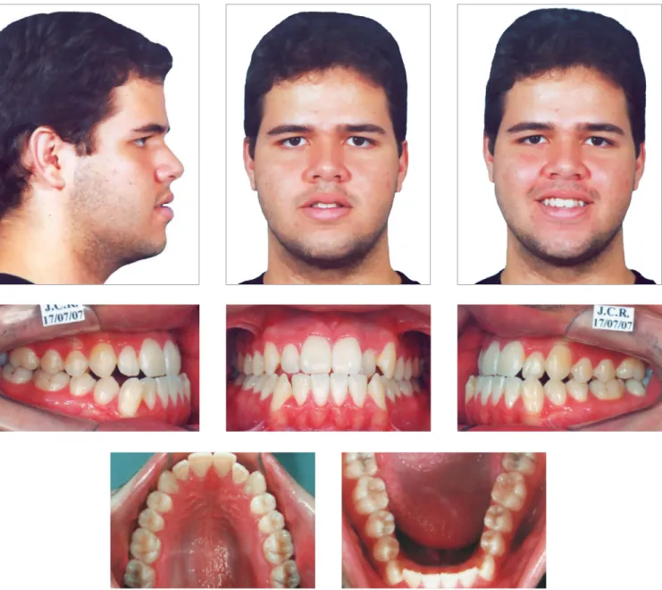
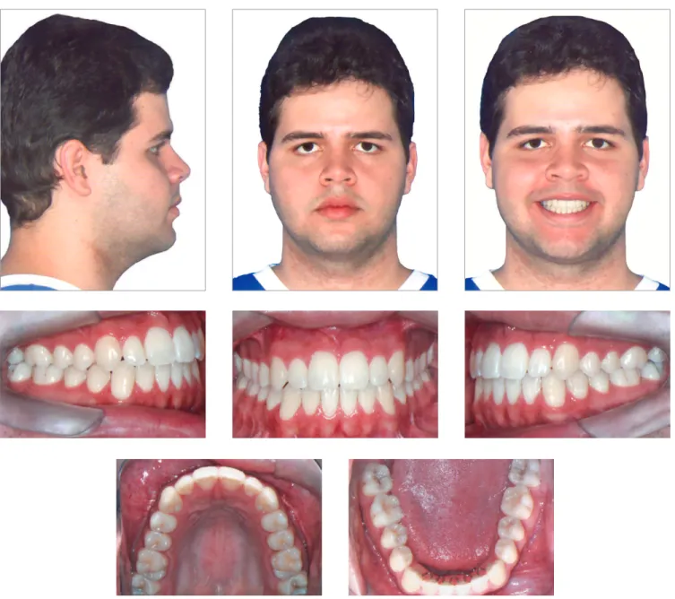
The clinical examination showed, in frontal view, a symmetrical face and a reduced exposure of the upper incisors upon smiling. In the lateral view, the proile was concave, associated to an incompe-tent lip seal at rest. The upper lip was well positioned (Upper lip – S-line = 0.5 mm), while the lower lip was protruded (Lower Lip – S-line = 4 mm) (Fig 1).
During the intraoral clinical examination, bilat-eral Angle Class III malocclusion was observed, as-DOI: http://dx.doi.org/10.1590/2177-6709.21.6.103-114.bbo
O presente caso clínico descreve o tratamento ortodôntico de um paciente com 18 anos e 1 mês de idade, portador de má oclusão esquelética de Classe III, com prognatismo mandibular e significativa compensação dentária. A relação entre caninos era de Classe III, incisivos com tendência à mordida cruzada e mordida aberta, moderado apinhamento inferior, além de perfil côncavo. A correção esquelética da má oclusão; a harmonia do perfil facial, com relação labial satisfatória; correção da compensação dentária e relação oclusal normal foram obtidas com o tratamento ortodôntico associado à cirurgia ortog-nática. Esse caso foi apresentado à Diretoria do Board Brasileiro de Ortodontia e Ortopedia Facial (BBO), como parte dos requisitos para a obtenção do título de Diplomado pelo BBO.
Severe Angle Class III skeletal malocclusion associated to mandibular prognathism: orthodontic-surgical treatment
BBOcase report
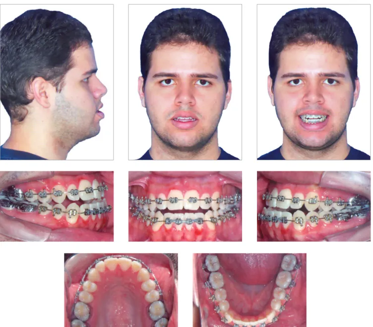
Figure 1 - Initial facial and intraoral photographs. sociated with significant dental compensation and moderate crowding in both arches. The upper
inci-sors were proclined (1.NA = 30o and 1-NA = 11mm),
while the lower incisors were vertical (1.NB = 22o
and IMPA = 83 o). Overbite and overjet were
re-duced, with tendency to open bite and crossbite in the anterior segment (Figs 1 and 2).
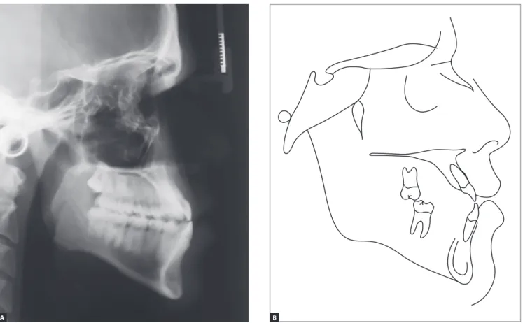
Panoramic radiograph indicated the presence of all permanent teeth, including the third molars.
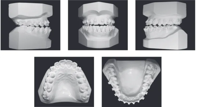
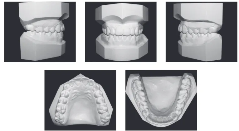
Figure 2 - Initial dental casts.
Severe Angle Class III skeletal malocclusion associated to mandibular prognathism: orthodontic-surgical treatment
BBOcase report
TREATMENT PLAN
With the aim of correcting skeletal malocclu-sion, achieve harmony of the facial profile, allow satisfactory labial relationship and passive lip seal, correct the dental compensation created by maloc-clusion and achieve a normal occlusal relationship, an orthodontic treatment associated with the surgi-cal correction of skeletal malocclusion through or-thognathic surgery was proposed. Initially, the pa-tient was referred to the buco-maxillofacial surgeon for surgical planning, preliminary orientations and decision about the upper and lower third molars. After his return, the fixed upper and lower appli-ances were installed, with Roth prescription brack-ets (0.022” x 0.028”), first molar bands with double convertible tube and second molars with single tubes in both arches. For dental leveling and align-ment, the following orthodontic archwire sequence was planned: 0.014” NiTi wire; 0.016”, 0.018” and
0.017” x 0.025” stainless steel archwire with ideal torques. After leveling and alignment conclusion and correction of dental inclinations, dental impressions were taken to help the presurgical preparation with the necessary wire bends. A surgical 0.019” x 0.025” archwire with welded hooks were programmed. Af-ter the orthognathic surgery, the need for the use of intermaxillary elastics and wire folds for the conclu-sion of the case was evaluated.
TREATMENT PROGRESS
The proposed treatment plan was followed as planned until the end of leveling and alignment. At this stage, it was perceived a great diiculty to obtain the ideal transverse arch positioning due to the occlusal interference created by the signiicant compensation presented by the patient original occlusion. Thus, an acetate plate was adapted in the upper arch, with oc-clusal bite-block, to eliminate the ococ-clusal locking and
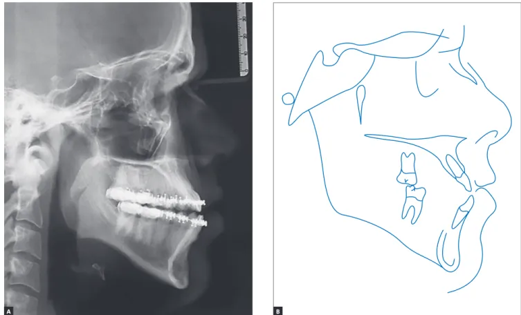
Figure 4 - Initial cephalogram (A) and cephalometric tracing (B).
facilitate the posterior segment torque movement in the lower arch, with greater eiciency. Subsequently, dental casts were performed and the inal preparation of the preoperative surgical preparation with the necessary bends was performed. Ater the pre-surgical orthodon-tics conclusion (Fig. 5 to 8), the inal surgical procedure plan was deined, along with the maxillofacial surgeon.
The surgical arches 0.019”X0.025” were installed with welded hooks, tied with steel wires in all the teeth. To achieve the best possible facial and skeletal results, Le Fort I maxillary surgery, with posterior impaction and clockwise rotation was performed, combined with sagittal mandibular osteotomy and an anterior and ver-tical mentoplasty.
Severe Angle Class III skeletal malocclusion associated to mandibular prognathism: orthodontic-surgical treatment
BBOcase report
Figure 6 - Intermediate: presurgical phase dental casts.
TREATMENT RESULTS
A significant improvement was seen in facial es-thetics. A pleasant facial smile was obtained, with an increase in the upper teeth exposure. The facial profile became straight, with improvement in the lip contour, as well as obtaining an appropriate lip seal due to reduction of the protrusion of the lower lip (reduced from 4 mm to 0 mm, to the Steiner’s S line) (Fig. 9 and Tab 1).
Intraoral records (Figs 9 and 10) demonstrated a satisfactory occlusal relationship in a lateral view, with good intercuspation and solid molar and canine Class I relationship. From the frontal view, it was possible to observe the dental midline coincidence and an adequate
overbite. Because of the improvement in occlusion, the patient presented anatomical conditions to obtain satis-factory occlusal guides, with lateral movement guided by the canines and protrusive by the incisors.
Cephalometric superimpositions showed a signii-cant improvement in the maxillary position, due to an advance and impaction of the posterior segment of the maxilla. A mandibular posterior displacement and an anterior and vertical repositioning of the chin were ob-served in the mandible. These modiications allowed an improvement in sagittal skeletal disharmony, with a reduction of 6 degrees in the ANB angle (Steiner) from -3 o to 3 o and in Wits (Jacobson) measurement from -9 mm to 0 mm (Table 1, Figs 11 to 14).
Figure 8 - Intermediate: presurgical phase cephalogram (A) and cephalometric tracing (B).
Severe Angle Class III skeletal malocclusion associated to mandibular prognathism: orthodontic-surgical treatment
BBOcase report
Figure 10 - Final dental casts.
Severe Angle Class III skeletal malocclusion associated to mandibular prognathism: orthodontic-surgical treatment
BBOcase report
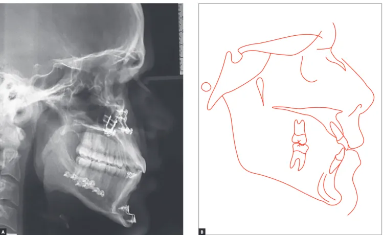
Figure 12 - Final cephalogram (A) and cephalometric tracing (B).
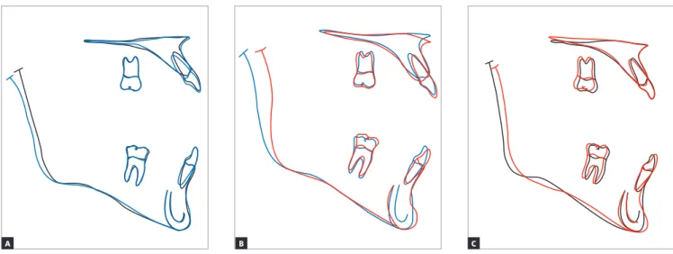
Figure 13 - Total superimpositions of cephalometric tracings: (A) initial (black) and pre-surgery (blue); (B) pre-surgery (blue) and final (red); (C) initial (black) and final (red).
B
B C
A
Figure 14 - Partial superimpositions of cephalometric tracings: (A) initial (black) and pre-surgery (blue); (B) pre-surgery (blue) and final (red); (C) initial (black) and final (red).
B C
A A
Table 1 - Cephalometric measurements: initial (A), pre-surgery (A1) and final (B).
Measurements Normal A A1 B Dif. A/B
Skeletal pattern
SNA (Steiner) 82° 84o 84,5o 88o 4
SNB (Steiner) 80° 87o 86,5o 85o 2
ANB (Steiner) 2° - 3o - 2o 3o 6
Wits (Jacobson) ♀ 0 ±2 mm
♂ 1 ±2 mm - 9mm - 4mm 0mm 9
Angle of convexity (Downs) 0° - 7o - 4o - 1o 6
Y-axis (Downs) 59° 54o 58o 56o 2
Facial angle (Downs) 87° 84o 92o 94o 10
SN-GoGn (Steiner) 32° 30o 32o 28o 2
FMA (Tweed) 25° 23o 29o 23o 0
Dental pattern
IMPA (Tweed) 90° 83o 89o 96o 13
1.NA (degrees) (Steiner) 22° 30o 34o 27o 3
1-NA (mm) (Steiner) 4 mm 11mm 11mm 9mm 2
1.NB (degrees) (Steiner) 25° 22o 30o 30o 8
1-NB (mm) (Steiner) 4 mm 8mm 10mm 10mm 2
1
1- Interincisal angle (Downs) 130° 131o 117o 121o 10
1-APo (Ricketts) 1 mm 9,5mm 10mm 5,5mm 4
Profile
Upper lip — S-line (Steiner) 0 mm 0,5mm - 1,5mm - 0,5mm 1
Severe Angle Class III skeletal malocclusion associated to mandibular prognathism: orthodontic-surgical treatment
BBOcase report
FINAL CONSIDERATIONS
The Angle Class III malocclusions can present a variable severity, with diferent levels of resolution complexity. In general, the greater the skeletal involve-ment, the more complex the orthodontic treatment be-comes1. When the patient presents skeletal and dental disharmonies, usually, a signiicant facial impairment, with a consequent psychosocial impact is expected2,3. During the growth period, it is possible to establish orthopedic therapy to harmonize maxillomandibular growth4,5. However, when the approach is too late, in post-pubertal stage, with no growth potential, the op-tions for treating these skeletal malocclusions become limited. Basically, dental compensation can be planned without skeletal disharmony correction or a complete orthodontic therapy correction associated with or-thognathic surgery6-9. In the case described, the patient and his relatives complained about, not only the den-tal aspect, but also the facial disharmony. For this rea-son, an ortho-surgical approach was proposed to cor-rect skeletal malocclusion and to harmonize the face.
REFERENCES
1. Baccetti T, McGill JS, Franchi L, McNamara JA Jr, Tollaro I. Skeletal efects of early treatment of Class III malocclusion with maxillary expansion and face-mask therapy. Am J Orthod Dentofacial Orthop. 1998 Mar;113(3):333-43.
2. Bernabé E, Sheiham A, de Oliveira CM. Condition-speciic impacts on
quality of life attributed to malocclusion by adolescents with normal occlusion and Class I, II and III malocclusion. Angle Orthod. 2008 Nov;78(6):977-82.
3. Nicodemo D, Pereira MD, Ferreira LM. Efect of orthognathic surgery
for class III correction on quality of life as measured by SF-36. Int J Oral Maxillofac Surg. 2008 Feb;37(2):131-4. Epub 2007 Oct 4.
4. Anne Mandall N, Cousley R, DiBiase A, Dyer F, Littlewood S, Mattick R, et al. Is early Class III protraction facemask treatment efective? A multicentre, randomized, controlled trial: 3-year follow-up. J Orthod. 2012 Sept;39(3):176-85.
5. Xu Y, Zhu P, Le L, Cai B. Conservative treatment for a growing patient with a severe, developing skeletal Class III malocclusion and open bite. Am J Orthod Dentofacial Orthop. 2014 June;145(6):807-16.
Ater the treatment plan presentation and explanation by the surgeon about the questions related to the surgi-cal procedure, orthodontic treatment was started. The preoperative phase was adequate, highlighting only the diiculty in the transverse arch coordination due to a bite interference during the torque movements in the lower arch. For that reason, it was necessary to use a removal bite-block in the upper arch to release lower tooth movement. The result achieved by the surgery was very favorable, making easy the post-surgical phase and orthodontic ending. In sequence, the patient was referred to speech pathology work to adapt the muscular structures and the oral functions. The final treatment results were highly positive, reaching all the proposed objectives: significant im-provement in facial profile and esthetics, in addition to functional and harmonic occlusion.
Acknowledgements
Thanks to Dr. Antônio de Albuquerque Brito for conducting the surgery of the presented case.
6. Javed O, Bernabé E. Oral Impacts on quality of life in adult patients with Class I, II and III malocclusion. Oral Health Prev Dent. 2016;14(1):27-32. 7. Silvola AS, Varimo M, Tolvanen M, Rusanen J, Lahti S, Pirttiniemi P. Dental
esthetics and quality of life in adults with severe malocclusion before and after treatment. Angle Orthod. 2014 July;84(4):594-9.
8. Pangrazio-Kulbersh V, Berger JL, Janisse FN, Bayirli B. Long-term stability of Class III treatment: rapid palatal expansion and protraction facemask vs LeFort I maxillary advancement osteotomy. Am J Orthod Dentofacial Orthop. 2007 Jan;131(1):7.e9-19.