1 7 8
Darzé et al
Pheochromocytoma-Induced segmental myocardial dysfunction mimicking an acute myocardial infarction in a patient with normal coronary arteries
Arq Bras Cardiol 2004; 82: 178-80.
Serviço de Cardiologia/Ecocardiografia - Hospital Aliança, Salvador, BA and Cardiology Division - John F. Kennedy Medical Center - Atlantis, FL. Mailing address: Eduardo S. Darzé - Serviço de Cardiologia/Ecocardiografia - Hospital Aliança - Av. Juracy Magalhães Jr., 2096 - Salvador, BA - Brazil - Cep 41920-000 E-mail: [email protected]
Received: 2/24/03 Accepted: 5/26/03
Arq Bras Cardiol, volume 82 (nº 2), 178-80, 2004
Eduardo S. Darzé, Roberto L. Von Sohsten
Salvador, BA, Brazil - Atlantis, FL, USA
Pheochromocytoma-Induced Segmental Myocardial
Dysfunction Mimicking an Acute Myocardial Infarction in a
Patient with Normal Coronary Arteries
Case Report
We report a case of pheochromocytoma-induced seg-mental myocardial dysfunction and electrocardiographic abnormalities mimicking an acute anterior myocardial infarction, probably due to coronary spasm. Coronary an-giography showed normal coronaries, and the electro-cardiographic and echoelectro-cardiographic changes resolved completely after therapy with an alpha-adrenergic blo-cker and tumor removal. Our case illustrates the importan-ce of maintaining a high index of suspicion in patients pre-senting with an unexpected myocardial event and a hyper-tensive crisis.
Pheochromocytoma is a catecholamine-secreting tu-mor that arises from chromaffin tissue of the sympathetic nervous system. The usual manifestations of this tumor in-clude palpitations, diaphoresis, headache, and paroxysmal hypertension. In addition to the classic symptoms, pheo-chromocytomas have been rarely associated with acute myocardial infarction and other cardiovascular complica-tions 1. We report the case of a patient with an adrenal
pheo-chromocytoma and normal coronary arteries who presen-ted with electrocardiographic and echocardiographic fdings consistent with an acute anterolateral myocardial in-farction (MI), with complete reversal of these abnormalities after alpha-adrenergic treatment.
Case Report
A 46-year-old female with history of hypertension was hospitalized with nausea, vomiting, and diaphoresis for 2 days. On physical examination, her initial blood
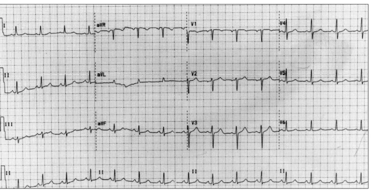
pres-sure was 230/130 mmHg; heart rate, 132 bpm; and her skin was mottled. The rest of the examination was unremarkable. The electrocardiogram (fig. 1) showed sinus tachycardia with ST segment elevation and deep, symmetric T wave in-version in leads V2 thru V6. The T waves were also inverted in leads I, aVL, II, and aVF. The QT interval was markedly prolonged and Q waves were present in leads II, III, aVF.
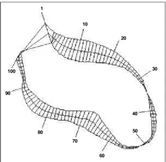
She was taken to the cardiac catheterization laboratory where a coronary angiogram showed no significant coro-nary disease. The left ventriculogram (fig. 2) showed severe anterolateral and apical hypokinesis. During the procedure, the patient required mechanical ventilation, and the arterial blood pressure became very labile, going from as high as 320/240 mmHg to as low as 70/30 mmHg, warranting the use of vasodilators and, at times, vasopressors. The diagnosis of pheochromocytoma was suspected, and therapy with an alpha-adrenergic blocker was started, which slowly control-led the blood pressure.
Laboratory data included 24-hour urinary vanillylman-delic acid, metanephrine, epinephrine, and norepinephrine levels that were, respectively, 313 mg (normal, 2-10), 76 (nor-mal, 0.3-0.9), 12339 ng (nor(nor-mal, 0-16), and 28316 ng (nor(nor-mal, 11-86). The blood urea nitrogen was 26 mg/dL and creatinine was 2.0 mg/dL. The peak CPK level was 951 IU/L with a nor-mal MB fraction. A computed tomography revealed a rounded mass in the left adrenal gland.
Arq Bras Cardiol 2004; 82: 178-80.
Darzé et al Pheochromocytoma-Induced segmental myocardial dysfunction mimicking an acute myocardial infarction in a patient with normal coronary arteries
1 7 9
Before discharge, the patient admitted that she had been having frequent bouts of nausea, vomiting, and severe headaches since she was diagnosed with hypertension 5 years before. She continues to do well 1 year after discharge, with good blood pressure control and no recurrence of symptoms.
Discussion
Pheochromocytoma-induced myocardial disease may take the form of ventricular hypertrophy due to long-stan-ding hypertension 2, dilated cardiomyopathy because of
persistent and prolonged exposure to high levels of
cate-cholamines 3, or, r arely, it may mimic an acute myocardial
infarction 4-9.
We report the case of a rare association between pheochromocytoma and reversible myocardial dysfunction in a patient with normal coronaries. Only a few cases of a pheochromocytoma crisis causing or mimicking an acute myocardial infarction have been reported in the literature. Their clinical presentations have varied significantly, from a silent myocardial infarction in one of the first cases reported in the literature 4, to frank cardiogenic shock 5. Striking
elec-trocardiographic changes suggesting myocardial ischemia are often present in patients with pheochromocytoma and include marked prolongation of the QT interval, deep and symmetric T wave inversion, and ST segment changes 10.
Although these are very suggestive of myocardial ische-mia, they have been described in other conditions such as subarachnoid hemorrhage and the use of certain drugs 10.
None of these situations is likely in the present case. Wall motion abnormalities have not been well characterized, but both segmental6 and global 7 myocardial dysfunction have
been reported. Although they are commonly associated, the ischemic ECG changes may not be accompanied by wall motion abnormalities 8. Additionally, despite striking ECG
changes and myocardial dysfunction, serum markers of myocyte necrosis may or may not be elevated 7. In our case,
the significant elevation in the CPK levels is probably of skeletal muscle origin and not myocardial injury because the MB fraction was normal.
The pathophysiology of myocardial dysfunction as-sociated with pheochromocytoma has been linked to either a direct toxic effect induced by catecholamines 3, or
myocar-dial stunning caused by coronary spasm 9. These changes
along with the ECG abnormalities are commonly reversible after treatment with alpha-adrenergic blockers and tumor re-moval 5,7. Although beta-blockers are certainly
recommen-ded in patients with an acute MI, they can worsen coronary spasm in cases of pheochromocytomas as a result of
unop-Fig. 1 - Electrocardiogram on admission showing ST segment elevation from V2-V6 and T wave inversion in V2-V6, I, II, aVL, and aVF. Note the marked prolongation in the QT interval.
1
10
20
30
40
50
60 70
80 90
100
1 8 0
Darzé et al
Pheochromocytoma-Induced segmental myocardial dysfunction mimicking an acute myocardial infarction in a patient with normal coronary arteries
Arq Bras Cardiol 2004; 82: 178-80.
posed alpha-adrenergic stimulation, and are, therefore, con-traindicated in this circumstance.
Our case illustrates an uncommon presentation of a pheochromocytoma crisis with ECG and wall motion abnor-malities mimicking myocardial infarction. The lack of eleva-tion of myocardial necrosis markers and complete reversal of segmental myocardial dysfunction suggest stunning due to coronary spasm as the underlying mechanism. Our report also reinforces the importance of maintaining a high index of
suspicion in patients who present with an unexpected myo-cardial infarction associated with a hypertensive crisis. The relevance of a thorough history cannot be overemphasized, because in our case and in a number of cases reported in the literature, symptoms suggestive of pheochromocytoma are found to have been present for years prior to the acute event, and are frequently missed on presentation 1. These patients
ought to be screened for pheochromocytoma, as early treat-ment may prevent serious morbidity and mortality.
Fig. 3 - Electrocardiogram 3 months after presentation, showing complete reversal of the previous abnormalities.
1. Cohen CD, Dent DM. Pheochromocytoma and acute cardiovascular death. Post Med J 1984; 60: 111-15.
2. Serfas D, Shoback DM, Lorell BH. Pheochromocytoma and hypertrophic cardio-myopahty. Lancet 1983; 2: 711-3.
3. Nanda AS, Feldman A, Liang CS. Acute reversal of pheochromocytoma-induced catecholamine cardiomyopathy. Clin. Cardiol 1995; 18: 421-3.
4. Boldt MH, Flexner M, Ortner AB. Pheochromocytoma associated with painless myocardial infarction. Ann Intern Med 1957; 46: 406-11.
5. Yamanaka O, Yasumasa F, Nakamura T, et al. “Myocardial stunning”-like pheno-menon during a crisis of pheochromocytoma. Jnp Circ J 1994; 58: 737-42.
References
6. Mauser M, Billmann P, Fleischmann D. Akuter myokardinfarkt bei der phaochro-mozytom-krise. Z Kardiol 2001; 90: 297-303.
7. Casazza F, Capozi A, Conconi B, et al. Danno miocardico acuto da feocromo-citoma. Ital Heart J 2000; 1: 686-9.
8. Costa J, Brandão A, Correia A, et al. Feocromocitoma extra-adrenal mimetizando enfarte agudo do miocárdio. Rev Port Cardiol 1999; 18: 1025-29.
9. Sheikhzadeh A, Fatourechi V, Paydar D, et al. Unusual cardiovascular mani-festation in a case of pheochromocytoma. Clin Cardiol 1983; 6: 136-42. 10. Cheng TO, Bashour TT. Striking electrocardiographic changes associated with