online | memorias.ioc.fiocruz.br
Inflammatory response of endothelial cells to hepatitis C virus
recombinant envelope glycoprotein 2
protein exposure
Ana Carolina Urbaczek1/+, Lívia Carolina de Abreu Ribeiro1, Valdecir Farias Ximenes2, Ana Afonso3,4,5, Camila Tita Nogueira1, Wesley Cardoso Generoso6, Juliana Vieira Alberice5,
Martina Rudnicki7, Renila Ferrer7, Luiz Marcos da Fonseca1, Paulo Inácio da Costa1
1Laboratório de Imunologia Clínica, Departamento de Análises Clínicas, Escola de Ciências Farmacêuticas 2Departamento de Química, Faculdade de Ciências, Universidade Estadual Paulista Julio de Mesquita Filho, Bauru, SP, Brasil 3Departamento de Parasitologia Médica, Unidade de Parasitologia Médica e Microbiologia, Instituto de Higiene e Medicina Tropcal,
Universidade Nova de Lisboa, Lisboa, Portugal 4Departamento de Morfologia e Patologia 6Departamento de Genética e Evolução,
Universidade Federal de São Carlos, São Carlos, SP, Brasil 5Grupo de Bioanalítica, Microfabricações e Separações, Departamento de
Química e Física Molecular, Instituto de Química de São Carlos, Universidade de São Paulo, São Carlos, SP, Brasil
7Escola de Ciências Farmacêuticas, Universidade de São Paulo, São Paulo, SP, Brasil
The hepatitis C virus (HCV) encodes approximately 10 different structural and non-structural proteins, includ-ing the envelope glycoprotein 2 (E2). HCV proteins, especially the envelope proteins, bind to cell receptors and can damage tissues. Endothelial inflammation is the most important determinant of fibrosis progression and, con-sequently, cirrhosis. The aim of this study was to evaluate and compare the inflammatory response of endothelial cells to two recombinant forms of the HCV E2 protein produced in different expression systems (Escherichia coli and Pichia pastoris). We observed the induction of cell death and the production of nitric oxide, hydrogen peroxide, interleukin-8 and vascular endothelial growth factor A in human umbilical vein endothelial cells (HUVECs) stimu-lated by the two recombinant E2 proteins. The E2-induced apoptosis of HUVECs was confirmed using the molecular marker PARP. The apoptosis rescue observed when the antioxidant N-acetylcysteine was used suggests that reac-tive oxygen species are involved in E2-induced apoptosis. We propose that these proteins are involved in the chronic inflammation caused by HCV.
Key words: HCV - E2 protein - inflammation - HUVEC
The hepatitis C virus (HCV), a member of the ge-nus Hepacivirus in the family Flaviviridae, is a small enveloped virus that possesses a positive-sense single-stranded RNA genome of approximately 9.6 kb (Hoof-nagle 2002, Penin et al. 2004, Kaukinen et al. 2013). The genome has a single open reading frame (ORF) (Taylor et al. 2000) encoding a polyprotein precursor of approxi-mately 3,000 amino acid residues that is cleaved by host and viral proteases to generate approximately 10 distinct structural and non-structural proteins (Encke et al. 1998, Penin et al. 2004). One of these proteins is envelope gly-coprotein 2 (E2), which undergoes post-translational modification after synthesis and possesses nine-11 po-tential glycosylation sites (Liu et al. 2001, Whidby et al. 2009). The E2 glycoprotein plays fundamental roles in the initiation of infection at different stages of the rep-lication cycle, including receptor binding, fusion with the host cell membrane and invasion (Bartenschlager & Lohmann 2000, Bartosch et al. 2003, Dubuisson et al. 2008, Lin et al. 2009).
doi: 10.1590/0074-0276140090
Financial support: FUNDECIF, FAPESP (2008/58957-0) + Corresponding author: [email protected] Received 11 March 2014
Accepted 29 July 2014
HCV infects approximately 170 million individu-als, representing 3% of the world’s population (Bian et al. 2009, Burke & Cox 2010, Ruggieri et al. 2013). The World Health Organization estimates that three-four million individuals are infected worldwide every year (Seeff & Hoofnagle 2003). The persistence of the infec-tion and the severity of the resultant inflammainfec-tion can lead to chronic hepatitis complicated by cirrhosis and/ or hepatocellular carcinoma (Ghany et al. 2003, Balasu-bramanian et al. 2005, Burke & Cox 2010, Kaukinen et al. 2013), making HCV infection one of the most preva-lent liver diseases in the world today. HCV infection is responsible for 60% of chronic liver diseases and is the major indication for liver transplants (Lauer & Walker 2001, Whidby et al. 2009).
al. 2007). Furthermore, it is believed that HCV proteins, especially the envelope proteins, can be toxic to cells in-dependent of direct viral infection by producing the “in-nocent bystander” effect (Balasubramanian et al. 2005). The vascular changes in the cirrhotic livers of pa-tients with chronic hepatitis C have attracted increasing interest because little is known about their relationship with major complications, such as portal hypertension, liver failure and hepatocellular carcinoma; thus, little is known about the prognostic implications of these vas-cular changes, highlighting the need for a more detailed characterisation of the inflammatory aspects in this sce-nario. Therefore, the aim of this study was to evaluate and compare the inflammatory response of endothe-lial cells [human umbilical vein endotheendothe-lial cells (HU-VECs)] to two recombinant forms of the HCV E2 protein produced in different expression systems.
SUBJECTS, MATERIALS AND METHODS
Strains, cell lines and media - Escherichia coli DH5α (Invitrogen, USA) was used for the general propagation of plasmids and E. coli BL21 (DE3) was used to express the E2 protein. Bacterial cells were grown under agita-tion at 37ºC in a low-salt Luria-Bertani medium contain-ing Zeocin at a final concentration of 25 µg/mL. Pichia pastoris KM71H(Muts) (Invitrogen) was used as the ex-pression host. Yeast cultures were maintained in a yeast extract-peptone-dextrose (YEPD) medium. The media for growth and induction were buffered complex glyc-erol medium (BMGY) and buffered complex methanol medium (BMMY), respectively, both at pH 4.0 HUVECs (ATCC® CRL-2873™) were grown in RPMI-1640 medi-um (Sigma Aldrich, USA) containing 10% foetal bovine serum and a mix of antibiotics and antifungals (Sigma Aldrich). The cultures were kept at 37ºC and 5% CO2 and disassociated from the culture dish using trypsin.
Cloning, expression and purification of E2 protein in E. coli - HCV cDNA was obtained from viral RNA ex-tracted with the QIAmp Viral RNA Mini Kit (QIAGEN, USA), according to the manufacturer’s protocol, using pooled sera from individuals with HCV genotype 1a provided by the Laboratory of Clinical Immunology of the Pharmaceutical Science School of Araraquara, São Paulo, Brazil. The HCV sequence was found by com-parison using the BLASTn local alignment program and its ORF was entirely sequenced. To express recombinant E2 protein, the soluble form of the protein without the transmembrane domain was selected (residues 384-661). The mature ORF was amplified with the forward primer 5’-GGCCATGGGGGA AACCCACGTCACCGG-3’ and reverse primer 5’-GCTCGAGGCTCGGACCT-GTCCCTGTC-3’ (the underlined bases indicate intro-duced restriction sites for NcoI and XhoI, respectively) (Rodríguez-Rodríguez et al. 2009). The pET42a plasmid was used to generate the mature E2 protein ORF flanked by glutathione S-transferase (GST) at the N-terminus and a 6x His tag at the C-terminus. The transformed E. coli BL21 were induced for 3 h with isopropylthio-β-galactoside (final concentration 0.4 mM) at 37ºC and 250 rpm when the optical density (OD) at 600 nm reached
0.5. The cells were pelleted, suspended in lysis buffer (10 mM Tris-HCl, 50 mM NaH2PO4 and 100 mM NaCl, pH 8.0) and subjected to sonication (5 pulses of 1 min each). The soluble phase was purified using Glutathione Sep-harose 4 Fast Flow (GE Healthcare, USA). The binding buffer employed was 10 mM Tris-base, 50 mM sodium phosphate and 100 mM sodium chloride at pH 8.0. The GST-tagged protein was eluted with a two-fold resin vol-ume of elution buffer (10 mM reduced glutathione and 50 mM Tris-HCl, pH 8.0). The fractions containing the pu-rified protein were dialysed against phosphate-buffered saline (PBS) (pH 8.0), quantified using the PierceTM BCA protein assay kit (Thermo Scientific, USA) and stored at -20ºC. This protein is referred to as E2B in this work.
Cloning, expression and purification of recombinant protein in P. pastoris - The E2 protein ORF was cloned into pPICZαA and the mature ORF was amplified with the forward primer AAGAATTCGAAACCCACGT-CACCGGGGGAA-3’ and the reverse primer 5’-AATCTAGATTCTCGGACCTGTCCCTGTCTTCC-3’ (the underlined bases indicate introduced EcoRI and XbaI restriction sites, respectively). The cloning was performed to create a recombinant plasmid contain-ing the E2 protein ORF flanked by the secretion signal peptide (α-factor) at the N-terminus and a 6x His tag at the C-terminus. Before P. pastoris transformation, the recombinant plasmid was linearised with PmeI endonu-clease and introduced into the yeast by electroporation (1.5 kV, 25 µF, 200 Ω) (Cregg 2007). Transformants were cultivated in solid YEPD with 1 M sorbitol and 100 µg/ mL Zeocin. The yeast transformants were screened for protein induction in 24-well plates (Boettner et al. 2002). One recombinant yeast colony was selected for protein production and purification. Expression induction for protein purification was performed as described in Gen-eroso et al. (2012), differing only in the use of BMGY and BMMY medium buffered with 100 mM McIlvaine’s buffer, pH 4.0. The supernatant was dialysed against PBS buffer (pH 8.0), concentrated using the Labscale TFF System (membrane Pellicon XL50, Millipore, USA) until 10-fold reduction and stored at -20ºC. This protein is referred to as E2Y in this work.
Cell death - HUVECs were seeded at 5 x 10⁴ cells/ mL and incubated for 24 h at 37ºC and 5% CO2 with recombinant E2B and E2Y using the concentrations and controls as described in the Cell viability section. The following controls were also added: cells without stimulation (negative) and annexin and propidium io-dide (PI) controls. The evaluation of cell death was per-formed using the Annexin V-FITC Annexin V Apop-tosis Detection Kit (BD Pharmingen, USA) according to the manufacturer’s protocol. The cells were analysed by flow cytometry (using a FACSCanto flow cytometer, BD Biosciences and FACSDiva software v.6.1.3). In each run, 30,000 cells were analysed and all experiments were performed in triplicate.
N-acetylcysteine (NAC) treatment - The effect of NAC on cells exposed to E2 recombinant proteins was studied with respect to apoptosis (PARP cleavage) and NO and hydrogen peroxide (H2O2) production. HUVECs were pre-incubated with 5 mM NAC for 1 h and treated with E2Y, E2B and controls as described above.
PARP cleavage - HUVECs at 5 x 10⁵ cells/mL were pre-incubated in the presence or absence of NAC (5 mM) for 1 h and incubated for 24 h at 37ºC and 5% CO2 with recombinant E2 proteins (E2B and E2Y) using the concentrations and control stimuli described in the Cell viability section. HUVECs were lysed in 10 mM Tris (pH 7.4), 1 mM EDTA, 0.5 mM EGTA, 150 mM NaCl, 1% Triton X-100, 50 mM NaF, 10 mM Na4P2O7·10H2O, 5 µg/mL aprotinin, 5 µg/mL leupeptin and 1 mM PMSF. To evaluate cell apoptosis, 20 µg of lysate protein was electrophoresed in 8% SDS-polyacrylamide gels and transferred onto nitrocellulose membranes (Hybond ECL). The membranes were blocked with 5% skim milk in Tris-buffered saline (TBS) containing 0.1% Tween-20 (TBS-T) and subsequently incubated with rabbit PARP antibody (1:2000 dilution, Santa Cruz Biotechnology, Inc, USA) overnight at 4ºC to detect full-length PARP (116 kDa) and cleaved PARP (carboxyl-terminal catalyt-ic fragment, 89 kDa). After washing with TBS-T for 1 h at room temperature (RT), the membranes were further incubated with a horseradish peroxidase-conjugated rab-bit polyclonal antibody (1:2000 dilution; Santa Cruz Bio-technology, Inc) for 2 h followed by 1 h washing (with 3-5 wash buffer changes). Protein bands were visualised with signal reagents. Actin levels were used to control for protein levels and were detected with an antibody against actin (Yang et al. 2004).
NO production - Total NO production was determined in the culture supernatant of HUVECs seeded at 5 x 10⁴ cells/mL incubated with E2B and E2Y and controls as described in the Cell viability section for 24 h at 37ºC and 5% CO2. The samples were measured in a NO analyser (Sievers Nitric Oxide Analyzer Overview, model NOA 280i, GE Analytical Instruments, USA), in which the ni-trites, nitrates and nitrosothiols present in the supernatant were converted into NO by a saturated solution of vana-dium trichloride in 0.8 M HCl at 90ºC. NO was detected by a chemiluminescent reaction in the gas phase between NO and ozone (Archer 1993, Jaiswal et al. 2000).
Arginase activity - Arginase activity was measured using urea. This reaction is based on L-arginine hydrol-ysis by arginase in cell lysates (Corraliza et al. 1994). Briefly, HUVECs were cultured with recombinant pro-teins, LPS, TNF-α and culture medium or culture super -natant of E. coli BL21 cells using the concentrations and stimuli described in the Cell viability section. The cells were lysed using 100 µL of 0.1% Triton X-100 for 30 min under agitation. Subsequently, 50 µL of cell lysate was added to 50 µL of 25 mM Tris-HCl and 25 µL of 100 mM MnCl2 and the final solution was incubated for 10 min at 56ºC for enzyme activation. Next, 50 µL of 0.5 M L-arginine (pH 9.7) was added and the test reaction was in-cubated at 37ºC for 60 min. The reaction was stopped by adding 400 µL of Stop Solution (96% H2SO4, 85% H3PO4 and water, at a proportion of 1:3:7 v/v/v). Twenty-five mi-crolitres of 9% α-isonitrosopropiophenone in 100% etha -nol was added and the reaction was incubated at 95ºC for 30 min. Finally, the cells were incubated at RT for 10 min and the absorbance was measured using a 540 nm fil-ter. The urea concentration was calculated using a linear equation generated by known quantities of urea. One unit of enzyme activity was defined as the amount of enzyme capable of producing 1 µmol of urea per minute.
H2O2 production - HUVECs at 5 x 10⁵ cells/mL were incubated for 2 h at 37ºC and 5% CO2 with recombinant E2B and E2Y using the concentrations and stimuli de-scribed in the Cell viability section. Approximately 600 ng/mL dihydrorhodamine 123 (DHR) (Sigma-Aldrich) was added and the cells were incubated at 37ºC for 10 min. The cells were washed with PBS (pH 7.2) and cen-trifuged for 5 min at 300 g. The supernatant was dis-carded and the cells were resuspended in 150 µL of PBS (pH 7.2). The samples were read in the FL1 channel us-ing a FACSCanto flow cytometer (BD Biosciences) and FACSDiva software v.6.1.3. The experiment included a control for spontaneous fluorescence (cells only) and a control for spontaneous production of H2O2 (DHR and cells without stimuli) (Walrand et al. 2003).
IL-8, TNF-α and vascular endothelial growth factor
A (VEGF-A) production - HUVECs at 5 x 10⁴ cells/mL were incubated for 24 h at 37ºC and 5% CO2 with recom-binant E2B and E2Y using the concentrations and con-trols described in the Cell viability section. An additional control using PMA (0.50 µM) was used in the TNF-α de -tection assay. The negative control consisted of 300 µL of culture medium and 300 µL of PBS (pH 7.2; medium of the protein dilution). Supernatants were collected and centrifuged at 2,860 g and 4ºC for 10 min. IL-8, TNF-α and VEGF-A production was measured by ELISA using the kit Human VEGF-A Platinum ELISA (eBioscience Inc, USA), according to the manufacturer’s instructions. Cytokine concentrations were calculated using a cytokine calibration curve. The results are expressed in pg/mL.
RESULTS
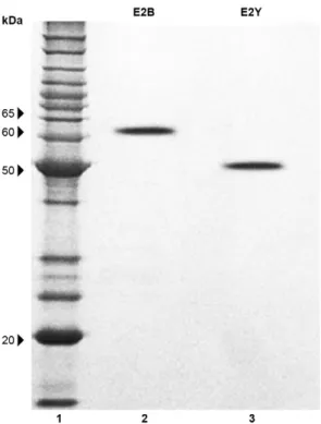
The recombinant E2 proteins were expressed in two different expression systems, the E. coli Rosetta strain (E2B) and the P. pastoris KM71H(Muts) strain (E2Y) (Fig. 1). The E2B protein exhibited a molecular weight of ap-proximately 63.5 kDa due to its expression as a fusion protein with GST (26 kDa) and the 6x His tag (1 kDa). The E2Y protein exhibited a molecular weight of ap-proximately 50.0 kDa due to its expression as a fusion protein with the 6x His tag (1 kDa). The N-glycosylation of the E2Y protein was confirmed by protein treatment with a peptide-N-glycosidase, PNGase F (New England Biolabs, USA), according to the manufacturer’s proto-col. The proteins exhibited different molecular weights (E2B = 36.5 kDa and E2Y = 49 kDa) due to the types of protein processing used in these two systems.
We observed that some concentrations of the recom-binant E2 proteins (E2Y: 62.5-250 µg/mL and E2B: 125-250 µg/mL) were slightly cytotoxic to HUVECs (Fig. 2). At 250 µg/mL, the decrease in viability was similar to that obtained when the cells were incubated with LPS or TNF-α. The cellular events provoked by the E2 proteins were evaluated using annexin V and PI assays, which indicated that early apoptosis was the main cause of cell death (Fig. 3). However, even at the highest concentra-tion of E2 protein used in this study, the proporconcentra-tion of apoptotic cells was always lower than 30%.
To further explore the mechanism of E2 protein-induced apoptosis, we investigated the degradation of PARP, which is thought to be one of the targets of acti-vated caspase-3 or 7 during apoptosis (Yang et al. 2004). Immunoblot analysis revealed that the recombinant E2 proteins induced the degradation of endogenous 116 kDa PARP, as shown by the appearance of 89 kDa fragments (Fig. 4A, B), which were clearly detected in all sam-ples treated with E2 protein or with the control stimuli, TNF-α (Fig. 4E, Line1) and LPS (Fig. 4E, Line 3). These results indicate that PARP cleavage is associated with E2-induced apoptosis in HUVECs. Moreover, the pre-treatment of the cells with the antioxidant NAC protect-ed against apoptosis by preventing PARP cleavage (Fig. 4C, D). The same effect was observed for the TNF-α (Fig. 4E, Line 2) and LPS (Fig. 4E, Line 4) controls.
The production of NO by HUVECs was stimulated by E2 protein treatments (Fig. 5). Statistical analysis of the results revealed that there was a statistically signifi-cant difference (p < 0.01) when compared with the spon-taneous control (negative control) or production stimu-Fig. 1: produced envelope glycoprotein 2 (E2) recombinant proteins
(sodium dodecyl sulfate polyacrylamide gel electrophoresis 12%). Channel 1: molecular weight marker [BenchMarkTM Protein Ladder
(10-220 kDa), Invitrogen]; 2: E2B (approximately 63.5 kDa); 3: E2Y (approximately 50 kDa).
Fig. 2: effect of envelope glycoprotein 2 (E2) recombinant proteins on human umbilical vein endothelial cells viability [3-(4, 5-dimeth-ylthiazol-2-yl)-2,5-diphenyltetrazolium bromide assay]. Results are expressed as mean and standard deviation of viable cells. The experi-ments were performed in triplicate. c-: negative control; c+: culture medium RPMI; LPS: lipopolysaccharide (1.0 µg/mL); Sup: culture supernatant Escherichia coli BL21; TNF-α: tumour necrosis factor
alpha (10 ng/mL); *: p < 0.05 compared to the negative control; ***: p < 0.001 compared to the negative control.
Fig. 3: cytotoxicity of envelope glycoprotein 2 (E2) proteins on hu-man umbilical vein endothelial cells. Results presented as mean and standard deviation of percentage obtained in the assay. In each run, 30,000 cells were analysed and all experiments were performed in triplicate. c-: negative control; early apoptosis: annexin V stained cells; late apoptosis: cells double-positive for annexin V and pro-pidium iodide (PI); LPS: lipopolysaccharide (1.0 µg/mL); necrosis: cells stained with PI; Sup: culture supernatant Escherichia coli BL21;
lated by the bacterial supernatant. The E2 proteins were as effective as the classical stimuli, LPS and TNF-α. One exception was the treatment with 7.81 µg/mL of E2 pro-tein, in which only a slight, but significant elevation in NO was observed relative to the more potent LPS and TNF-α stimuli. The higher production of NO was not the result of increased or decreased arginase activity rela-tive to the negarela-tive control (result not shown) because no
significant differences were observed when compared with the negative control. Our data also demonstrated that pre-treatment with NAC significantly decreased (p < 0.01) the E2 protein-induced NO production.
The production of H2O2 was evaluated in HUVECs after exposure to recombinant proteins at different con-centrations and control stimuli. The relative production of H2O2, calculated as the mean fluorescence intensity, is Fig. 8: vascular endothelial growth factor A (VEGF-A)production by envelope glycoprotein 2 (E2)-stimulated human umbilical vein endothelial cells. Results presented as mean and standard deviation. The experiments were performed in triplicate. LPS:
lipopolysaccha-ride (1.0 µg/mL); TNF-α: tumour necrosis factor alpha (10 ng/mL);
Sup: culture supernatant Escherichia coli BL21; c-: cells and medium and phosphate-buffered saline (pH 7.2); **: p < 0.01 compared to the negative control; ***: p < 0.001 compared to the negative control. Fig. 4: PARP cleavage in recombinant envelope glycoprotein 2
(E2)-induced apoptosis in human umbilical vein endothelial cells. Cells were pre-treated (C, D) or no (A, B) with N-acetylcysteine (NAC) for 1 h and incubated with recombinant E2 proteins, E2Y (A, C) and E2B (B, D) in different concentrations. The experiments were performed in triplicate. Line 2: 7.81 µg/mL; 3: 15.63 µg/mL; 4: 31.25 µg/mL; 5: 62.5 µg/mL; 6: 125 µg/mL; 7: 250 µg/mL; A-D1: lysate of untreated cells; E1: cells without pre-treatment with NAC and treated with
tu-mour necrosis factor alpha (TNF-α); E2: cells pre-treated with NAC and treated with TNF-α; E3: cells without pre-treatment with NAC
and treated with lipopolysaccharide (LPS); E4: cells pre-treated with NAC and treated with LPS; F1-4: actin.
Fig. 5: nitric oxide (NO) production by envelope glycoprotein 2 (E2)-stimulated human umbilical vein endothelial cells. Results are ex-pressed as mean and standard deviation of NO produced in µM. The experiments were performed in triplicate. c-: NO spontaneous pro-duction, cells and culture medium; LPS: lipopolysaccharide (1.0 µg/ mL); NAC: N-acetylcysteine; Sup: culture supernatant Escherichia coli BL21; TNF-α: tumour necrosis factor alpha (10 ng/mL); ***: p <
0.001 compared to the negative control.
Fig. 6: hydrogen peroxide production by envelope glycoprotein 2 (E2)-stimulated human umbilical vein endothelial cells. Results presented as mean and standard deviation of the mean fluorescence intensity (MFI). The experiments were performed in triplicate. c-: control fluorescence spontaneous, cells without stimulation; LPS: li-popolysaccharide (1.0 µg/mL); NAC: N-acetylcysteine; Sup: culture supernatant Escherichia coli BL21; TNF-α: tumour necrosis factor
alpha (10 ng/mL);***: p < 0.001 in relation to negative control.
Fig. 7: interleukin-8 (IL-8) production by envelope glycoprotein 2 (E2)-stimulated human umbilical vein endothelial cells. Results pre-sented as mean and standard deviation. The experiments were per-formed in triplicate. c-: cells and medium and phosphate-buffered saline (pH 7.2). LPS: lipopolysaccharide (1.0 µg/mL); Sup: culture supernatant Escherichia coli BL21; TNF-α: tumour necrosis factor
presented in Fig. 6. The E2 proteins were able to stimulate the production of H2O2 at all of the tested concentrations. Again, the production was similar to that obtained by stimulation with LPS and was inferior to that of TNF-α. Of the two E2 proteins, E2Y was more effective than E2B at concentrations of 7.81 µg/mL (p < 0.05). The results of pre-treatment with NAC revealed a significant decrease (p < 0.01) in the E2 protein-induced H2O2 production.
The E2 proteins were capable of inducing the pro-duction of IL-8 compared with non-stimulated cells. The detection of IL-8 production by HUVECs is presented in Fig. 7. There was a statistically significant difference (p < 0.05) between all of the stimuli tested compared with the negative control. Unlike the IL-8 results, the E2 proteins were not able to induce the production of TNF-α or LPS by HUVECs. However, 0.50 µM PMA induced HUVECs to produce 173.05 pg/mL TNF-α.
The detection of VEGF-A production by HUVECs in response to control stimuli and recombinant proteins is presented in Fig. 8. The E2 proteins significantly induced (p < 0.01) the production of VEGF-A by HUVECs.
The E2 protein-induced production of NO, H2O2, IL-8 and VEGF by HUVECs strongly supports the cy-totoxicity of these proteins.
DISCUSSION
There is evidence that endothelial cells are directly susceptible to infection by HCV (Fletcher et al. 2012) and that the damage caused by the infection leads to late complications, such as fibrosis, cirrhosis and hepatocel-lular carcinoma. These late complications are believed to be caused by numerous inflammatory molecules in response to viral infection of the liver (Ming-Ju et al. 2011). Consistent with this hypothesis, we found that E2 proteins were able to induce apoptosis and several inflammatory responses in HUVECs. The putative re-ceptors for E2 proteins in this cellular type have been described previously, including low-density lipoprotein receptor (Agnello et al. 1999), tetraspanin CD81 (Zhang et al. 2004), scavenger receptor class B type 1 (Scarsel-li et al. 2002), claudin-1 (Evans et al. 2007), occludin (OCLN) (Ploss et al. 2009) and transferrin receptor 1 (TfR1) (Martin & Uprichard 2013).
NO is an inorganic free radical molecule (Furchgott & Zawadzki 1980) that is highly diffusible and reactive (Bredt & Snyder 1992) and is involved in various physi-ological functions and pathphysi-ological conditions when produced in excess (Kaufman 1999, Benali-Furet et al. 2005, Deshpande et al. 2012). As a chronic inflammatory disease, hepatitis C induces an increase in NO produc-tion (Kane et al. 1997), which may play an important role in the pathogenesis of cirrhosis associated with infection (Hassan et al. 2002). Here, we have demonstrated for the first time that NO production by HUVECs was induced by both recombinant E2 proteins. This NO production may lead to later inflammation in the portal vein and subsequent fibrosis and cirrhosis.
The increased NO production could be the conse-quence of the increased expression of arginase in HU-VECs. HCV infection is associated with the develop-ment of hepatocellular carcinoma (Okuda 2007, Tan
et al. 2008) and can alter the expression of arginase, thereby stimulating tumourigenesis and hepatocellular carcinoma (Cao et al. 2009). However, this pathway does not appear to be relevant to endothelial cells because ar-ginase expression was not altered by the E2 proteins.
The E2 proteins were also able to induce the produc-tion of H2O2 by HUVECs. This is additional evidence of the role of E2 in the inflammatory response mediated by HCV. This result is consistent with the findings of Ming-Ju et al. (2011) and suggests the involvement of the E2 protein in H2O2 production and the development of in-flammation in the hepatic portal vein, with the increased expression of factors related to hepatic fibrosis.
Balasubramanian et al. (2003) reported that the HCV E2 protein was able to stimulate intracellular signalling pathways, leading to the induction of secretion of pro-inflammatory cytokine IL-8. The authors also reported that this production was dose-dependent. IL-8 is also observed in the serum of patients with chronic hepatitis C (Polyak et al. 2001, Akbar et al. 2011), demonstrating a correlation between inflammation, IL-8 serum levels and liver fibrosis (Kaplanski et al. 1997, Mahmood et al. 2002). These findings are evidence that IL-8 may play a role in HCV infection. Consistent with this hypothesis, recombinant E2 proteins stimulated the production of IL-8 in HUVECs. These results are consistent with the work of Balasubramanian et al. (2005). However, the E2 proteins were not able to induce the production of TNF-α by HUVECs. These results are also consistent with the work of Balasubramanian et al. (2005), who reported that HCV proteins can interact with the endothelium and that E2 protein did not induce the production of cytok-ines such as monocyte chemotactic protein-1, TNF-α and gamma interferon.
Analysis of peripheral blood mononuclear cells and liver biopsy samples of individuals chronically infected by the virus suggests that HCV infection may be able to induce apoptosis, causing damage to the liver while help-ing the virus to evade the immune system and facilitate viral dissemination (Hiramatsu et al. 1994, Pianko et al. 2001, Chiou et al. 2006). Here, we found that the E2 pro-teins were also able to induce apoptosis (early and late) as well as necrosis (fewer cells) in HUVECs. Similar results were also observed by Balasubramanian et al. (2005). The glycosylated protein expressed in P. pastoris (E2Y) was a more effective inducer of apoptosis as well as necro-sis relative to the non-glycosylated protein (E2B), dem-onstrating the influence of glycosylation on apoptosis. The E2-induced apoptosis of HUVECs was confirmed using the molecular marker PARP. Moreover, the apop-tosis induced by recombinant E2 protein was effectively rescued in cells pre-treated with NAC, suggesting that the generation of reactive oxygen species is involved in E2-induced apoptosis in HUVECs. We also suggest that the production of NO, H2O2, IL-8 and VEGF-A were not related to cell death induced by high concentrations of the recombinant protein, but are E2-specific effects.
and can be stimulated by HCV infection (Dvorak et al. 1992). Additionally, VEGF-A plays a role in the regu-lation of several cellular functions, including growth (Nasimuzzaman et al. 2007) and apoptosis (Höglinger et al. 2007). This effect was observed by Hassan et al. (2009) in liver biopsy samples of patients infected with HCV. Hepatic angiogenesis has been described in viral hepatitis, autoimmune liver cirrhosis, primary biliary cirrhosis and hepatocellular carcinoma (García-Monzón et al. 1995, Ker et al. 1999). HCV stimulates the synthe-sis and secretion of VEGF-A via virus-induced oxida-tive stress (Nasimuzzaman et al. 2007). In our study, the exposure of HUVECs to both recombinant E2 proteins induced the production of VEGF-A. We suggest that oxi-dative stress, as demonstrated by the production of NO and H2O2 in HUVECs in response to E2 proteins, may represent the stimulating factor of VEGF-A production. The literature reports that the HCV core protein is able to stimulate the production of VEGF-A, but there are no data regarding the E2 protein (Hassan et al. 2004, Abe et al. 2012). Therefore, this is the first demonstration that the E2 protein is also able to induce the production of VEGF-A and, consequently, angiogenesis.
HCV is a positive-stranded RNA virus that is un-able to integrate its genetic material into the host cell genome. The HCV genome does not contain oncogenes, suggesting that HCV induces hepatocellular carcinoma indirectly by causing chronic inflammation, cell death, proliferation and cirrhosis (Hassan et al. 2009). Here, we provide evidence that endothelial cells, such as HU-VECs, are susceptible to E2 HCV envelope proteins.
In conclusion, stimulation with E2 protein induced HUVECs to produce inflammatory and angiogenic fac-tors. Considering that endothelial inflammation is a deter-minant of fibrosis progression and cirrhosis, we propose that these cellular effects might be involved in the per-sistence and chronicity of HCV infection. These results may contribute to our understanding of the pathophysiol-ogy of hepatitis C and, consequently, to the development of new therapeutic strategies against the interaction of HCV structural proteins and the hepatic endothelium.
ACkNOWLEDGEMENTS
To Prof Flávio Henrique da Silva, for allowing the use of his laboratory in UFSCar and for providing equipment, re-agents and protocols for the production of recombinant pro-teins, to Prof Iracilda Zeppone Carlos, for providing many of the materials used in this research, as well as equipment from her laboratory (School of Pharmaceutical Sciences of Araraquara, UNESP), to Prof Dulcinéia Saes Parra Abdalla (School of Pharmaceutical Sciences, USP), for the donation of HUVEC, to Marisa Polezi Placeres (School of Pharmaceutical Sciences of Araraquara, UNESP), for technical assistance in cell culture, and to Prof Fernanda Anibal (UFSCar), for assis-tance in this paper revision.
REFERENCES
Abe M, Koga H, Yoshida T, Masuda H, Iwamoto H, Sakata M, Hanada S, Nakamura T, Taniguchi E, Kawaguchi T, Yano H, Torimura T, Ueno T, Sata M 2012. Hepatitis C virus core protein upregulates the expression of vascular endothelial growth factor via the
nu-clear factor-κB/hypoxia-inducible factor-1α axis under hypoxic
conditions. Hepatol Res 42: 591-600.
Agnello V, Abel G, Elfahal M, Knight GB, Zhang QX 1999. Hepatitis C virus and other Flaviviridae viruses enter cells via low density lipoprotein receptor. Proc Natl Acad Sci USA 96: 12766-12771.
Akbar H, Idrees M, Butt S, Awan Z, Sabar MF, Rehaman I, Hussain A, Saleem S 2011. High baseline interleukin-8 level is an indepen-dent risk factor for the achievement of sustained virological re-sponse in chronic HCV patients. Infect Genet Evol 11: 1301-1305.
Archer S 1993. Measurement of nitric oxide in biological models.
FASEB J 7: 349-360.
Balasubramanian A, Ganju RK, Groopman JE 2003. Hepatitis C vi-rus and HIV envelope proteins collaboratively mediate interleu-kin-8 secretion through activation of p38 MAP kinase and SHP2 in hepatocytes. J Biol Chem278: 35755-35766.
Balasubramanian A, Munshi N, Koziel MJ, Hu Z, Liang TJ, Groop-man JE, Ganju RK 2005. Structural proteins of hepatitis C virus induce interleukin 8 production and apoptosis in human endothe-lial cells. J Gen Virol 86: 3291-3301.
Bartenschlager R, Lohmann V 2000. Replication of hepatitis C virus.
J Gen Virol 81: 1631-1648.
Bartosch B, Dubuisson J, Cosset FL 2003. Infectious hepatitis C virus pseudo-particles containing functional E1-E2 envelope protein complexes. J Exp Med197: 633-642.
Benali-Furet NL, Chami M, Houel L, De Giorgi F, Vernejoul F, Lagorce D, Buscail L, Bartenschlager R, Ichas F, Rizzuto R, Pa-terlini-Bréchot P 2005. Hepatitis C virus core triggers apoptosis in liver cells by inducing ER stress and ER calcium depletion.
Oncogene24: 4921-4933.
Bian T, Zhou Y, Bi S, Tan W, Wang Y 2009. HCV envelope protein function is dependent on the peptides preceding the glycopro-teins. Biochem Biophys Res Commun378: 118-122.
Boettner M, Prinz B, Holz C, Stahl U, Lang C 2002. High-throughput screening for expression of heterologous proteins in the yeast Pi-chia pastoris. J Biotechnol99: 51-62.
Bredt DS, Snyder SH 1992. Nitric oxide, a novel neuronal messenger.
Neuron8: 3-11.
Burke KP, Cox AL 2010. Hepatitis C virus evasion of adaptive immune responses: a model for viral persistence. Immunol Res47: 216-227.
Cao W, Sun B, Feitelson MA, Wu T, Tur-Kaspa R, Fan Q 2009. Hepa-titis C virus targets over-expression of arginase I in hepatocar-cinogenesis. Int J Cancer124: 2886-2892.
Chiou HL, Hsieh YS, Hsieh MR, Chen TY 2006. HCV E2 may in-duce apoptosis of Huh-7 cells via a mitochondrial-related caspase pathway. Biochem Biophys Res Commun345: 453-458.
Corraliza IM, Campo ML, Soler G, Modolell M 1994. Determination of arginase activity in macrophages: a micromethod. J Immunol Methods 174: 231-235.
Cregg JM 2007. Pichia protocols, 2nd ed., Humana Press, Totowa, 268 pp.
Deshpande SR, Satyanarayana K, Rao MN, Pai KV 2012. Nitric ox-ide modulators: an emerging class of medicinal agents. Indian J Pharm Sci 74: 487-497.
Dubuisson J, Helle F, Cocquerel L 2008. Early steps of the hepatitis C virus life cycle. Cell Microbiol 10: 821-827.
Dvorak HF, Nagy JA, Berse B, Brown LF, Yeo KT, Yeo TK, Dvorak AM, van de Water L, Sioussat TM, Senger DR 1992. Vascular permeability factor, fibrin and the pathogenesis of tumor stroma formation. Ann N Y Acad Sci 667: 101-111.
Evans MJ, von Hahn T, Tscherne DM, Syder AJ, Panis M, Wölk B, Hatziioannou T, McKeating JA, Bieniasz PD, Rice CM 2007. Claudin-1 is a hepatitis C virus co-receptor required for a late step in entry. Nature446: 801-805.
Fletcher NF, Wilson GK, Murray J, Hu K, Lewis A, Reynolds GM, Stamataki Z, Meredith LW, Rowe IA, Luo G, Lopez-Ramirez MA, Baumert TF, Weksler B, Couraud PO, Kim KS, Romero IA, Jopling C, Morgello S, Balfe P, McKeating JA 2012. Hepatitis C virus infects the endothelial cells of the blood-brain barrier.
Gastroenterology 142: 634-643.
Furchgott RF, Zawadzki JV 1980. The obligatory role of endothelial cells in the relaxation of arterial smooth muscle by acetylcholine.
Nature288: 373-376.
García-Monzón C, Sánchez-Madrid F, García-Buey L, García-Arroyo A, García-Sánchez A, Moreno-Otero R 1995. Vascular adhesion molecule expression in viral chronic hepatitis: evidence of neoan-giogenesis in portal tracts. Gastroenterology108: 231-241.
Generoso WC, Malagó-Jr W, Pereira N, Henrique-Silva F 2012. Re-combinant expression and characterization of an endoglucanase III (cel12a) from Trichoderma harzianum (Hypocreaceae) in the yeast Pichia pastoris. Genet Mol Res 11: 1544-1557.
Ghany MG, Kleiner DE, Alter H, Doo E, Khokar F, Promrat K, He-rion D, Park Y, Liang TJ, Hoofnagle JH 2003. Progression of fi-brosis in chronic hepatitis C. Gastroenterology124: 97-104.
Hassan M, Ghozlan H, Abdel-Kader O 2004. Activation of RB/E2F signaling pathway is required for the modulation of hepatitis C virus core protein-induced cell growth in liver and non-liver cells. Cell Signal16: 1375-1385.
Hassan M, Selimovic D, Ghozlan H, Abdel-kader O 2009. Hepatitis C virus core protein triggers hepatic angiogenesis by a mechanism including multiple pathways. Hepatology49: 1469-1482.
Hassan MI, Kassim SK, Ali HS, Sayed el-DA, Khalifa A 2002. Evalu-ation of nitric oxide (NO) levels in hepatitis C virus (HCV) in-fection: relationship to schistosomiasis and liver cirrhosis among Egyptian patients. Dis Markers18: 137-142.
Hiramatsu N, Hayashi N, Katayama K, Mochizuki K, Kawanishi Y, Kasahara A, Fusamoto H, Kamada T 1994. Immunohistochemi-cal detection of Fas antigen in liver tissue of patients with chronic hepatitis C. Hepatology19: 1354-1359.
Höglinger GU, Breunig JJ, Depboylu C, Rouaux C, Michel PP, Al-varez-Fischer D, Boutillier AL, Degregori J, Oertel WH, Rakic P, Hirsch EC, Hunot S 2007. The pRb/E2F cell-cycle pathway mediates cell death in Parkinson’s disease. Proc Natl Acad Sci USA 104: 3585-3590.
Hoofnagle JH 2002. Course and outcome of hepatitis C. Hepatology 36 (Suppl. 1): S21-S29.
Jaiswal M, La Russo NF, Burgart LJ, Gores GJ 2000. Inflammatory cytokines induce DNA damage and inhibit DNA repair in cho-langiocarcinoma cells by a nitric oxide-dependent mechanism.
Cancer Res60: 184-190.
Kane JM, Shears LL, Hierholzer C, Ambs S, Billiar TR, Posner MC 1997. Chronic hepatitis C virus infection in humans: induction of hepatic nitric oxide synthase and proposed mechanisms for car-cinogenesis. J Surg Res69: 321-324.
Kaplanski G, Farnarier C, Payan MJ, Bongrand P, Durand JM 1997. Increased levels of soluble adhesion molecules in the serum of pa-tients with hepatitis C. Correlation with cytokine concentrations and liver inflammation and fibrosis. Dig Dis Sci42: 2277-2284.
Kaufman RJ 1999. Stress signaling from the lumen of the endoplas-mic reticulum: coordination of gene transcriptional and transla-tional controls. Genes Dev13: 1211-1233.
Kaukinen P, Sillanpää M, Nousiainen L, Melén K, Julkunen I 2013. Hepatitis C virus NS2 protease inhibits host cell antiviral response
by inhibiting IKKε and TBK1 functions. J Med Virol 85: 71-82. Ker CG, Chen HY, Juan CC, Lo HW, Shen YY, Chen JS, Lee KT,
Sheen PC 1999. Role of angiogenesis in hepatitis and hepatocel-lular carcinoma. Hepatogastroenterology46: 646-650.
Lauer GM, Walker BD 2001. Hepatitis C virus infection. N Engl J Med345: 41-52.
Lin X, Zhang Y, Bi S, Lu J, Zhao H, Tan W, Li D, Wang Y 2009. Hep-atitis C virus envelope glycoproteins complementation patterns and the role of the ecto and transmembrane domains. Biochem Biophys Res Commun385: 257-262.
Liu J, Zhu L, Zhang X, Lu M, Kong Y, Wang Y, Li G 2001. Expres-sion, purification, immunological characterization and applica-tion of Escherichia coli-derived hepatitis C virus E2 proteins.
Biotechnol Appl Biochem34: 109-119.
Mahmood S, Sho M, Yasuhara Y, Kawanaka M, Niiyama G, Togawa K, Ito T, Takahashi N, Kinoshita M, Yamada G 2002. Clinical significance of intrahepatic interleukin-8 in chronic hepatitis C patients. Hepatol Res 24: 413-419.
Martin DN, Uprichard SL 2013. Identification of transferrin receptor 1 as a hepatitis C virus entry factor. Proc Natl Acad Sci USA110: 10777-10782.
Ming-Ju H, Yih-Shou H, Tzy-Yen C, Hui-Ling C 2011. Hepatitis C virus E2 protein induce reactive oxygen species (ROS)-related fibrogenesis in the HSC-T6 hepatic stellate cell line. J Cell Bio-chem 112: 233-243.
Mosmann T 1983. Rapid colorimetric assay for cellular growth and survival: application to proliferation and cytotoxicity assays. J Immunol Methods 65: 55-63.
Nasimuzzaman M, Waris G, Mikolon D, Stupack DG, Siddiqui A 2007. Hepatitis C virus stabilizes hypoxia-inducible factor 1al-pha and stimulates the synthesis of vascular endothelial growth factor. J Virol81: 10249-10257.
Ng IO, Poon RT, Lee JM, Fan ST, Ng M, Tso WK 2001. Microvessel density, vascular endothelial growth factor and its receptors Flt-1 and Flk-1/KDR in hepatocellular carcinoma. Am J Clin Pathol 116: 838-845.
Okuda H 2007. Hepatocellular carcinoma development in cirrhosis.
Best Pract Res Clin Gastroenterol21: 161-173.
Penin F, Dubuisson J, Rey FA, Moradpour D, Pawlotsky JM 2004. Structural biology of hepatitis C virus. Hepatology39: 5-19.
Pianko S, Patella S, Ostapowicz G, Desmond P, Sievert W 2001. Fas-mediated hepatocyte apoptosis is increased by hepatitis C virus infection and alcohol consumption and may be associated with hepatic fibrosis: mechanisms of liver cell injury in chronic hepa-titis C virus infection. J Viral Hepat8: 406-413.
Ploss A, Evans MJ, Gaysinskaya VA, Panis M, You H, de Jong YP, Rice CM 2009. Human occludin is a hepatitis C virus entry factor required for infection of mouse cells. Nature457: 882-886.
Polyak SJ, Khabar KS, Rezeiq M, Gretch DR 2001. Elevated levels of interleukin-8 in serum are associated with hepatitis C virus infec-tion and resistance to interferon therapy. J Virol 75: 6209-6211.
Poon RT, Ho JW, Tong CS, Lau C, Ng IO, Fan ST 2004. Prognostic sig-nificance of serum vascular endothelial growth factor and endostatin in patients with hepatocellular carcinoma. Br J Surg91: 1354-1360.
Remick DG, Villarete L 1996. Regulation of cytokine gene expres-sion by reactive oxygen and reactive nitrogen intermediates. J Leukoc Biol59: 471-475.
2009. Structural properties of the ectodomain of hepatitis C virus E2 envelope protein. Virus Res 139: 91-99.
Ruggieri A, Anticoli S, Nencioni L, Sgarbanti R, Garaci E, Palamara AT 2013. Interplay between hepatitis C virus and redox cell sig-naling. Int J Mol Sci14: 4705-4721.
Scarselli E, Ansuini H, Cerino R, Roccasecca RM, Acali S, Filocamo G, Traboni C, Nicosia A, Cortese R, Vitelli A 2002. The human scavenger receptor class B type I is a novel candidate receptor for the hepatitis C virus. EMBO J21: 5017-5025.
Seeff LB, Hoofnagle JH 2003. Appendix: The National Institutes of Health Consensus Development Conference Management of Hepatitis C 2002. Clin Liver Dis7: 261-287.
Tan A, Yeh SH, Liu CJ, Cheung C, Chen PJ 2008. Viral hepatocar-cinogenesis: from infection to cancer. Liver Int 28: 175-188.
Taylor DR, Shi ST, Lai MM 2000. Hepatitis C virus and interferon resistance. Microbes Infect 2: 1743-1756.
Toi M, Bando H, Kuroi K 2000. The predictive value of angiogenesis for adjuvant therapy in breast cancer. Breast Cancer7: 311-314.
Wald O, Weiss ID, Galun E, Peled A 2007. Chemokines in hepati-tis C virus infection: pathogenesis, prognosis and therapeutics.
Cytokine 39: 50-62.
Walrand S, Valeix S, Rodriguez C, Ligot P, Chassagne J, Vasson MP 2003. Flow cytometry study of polymorphonuclear neutrophil oxidative burst: a comparison of three fluorescent probes. Clin Chim Acta331: 103-110.
Whidby J, Mateu G, Scarborough H, Demeler B, Grakoui A, Marcotri-giano J 2009. Blocking hepatitis C virus infection with recombinant form of envelope protein 2 ectodomain. J Virol83: 11078-11089.
Yang Y, Zhao S, Song J 2004. Caspase-dependent apoptosis and -inde-pendent poly(ADP-ribose) polymerase cleavage induced by trans-forming growth factor beta1. Int J Biochem Cell Biol 36: 223-234.
Zeremski M, Petrovic LM, Talal AH 2007. The role of chemokines as inflammatory mediators in chronic hepatitis C virus infection.
J Viral Hepat14: 675-687.