Disponible en castellano/Disponível em língua portuguesa SciELO Brasil www.scielo.br/rlae 1 RN, PhD, Assistant Professor at Juiz de Fora Federal University, Brazil, e-mail: [email protected]; 2 RN, PhD, Professor at University of São Paulo at Ribeirão Preto College of Nursing - WHO Collaborating Centre for Nursing Research Development, Brazil, e-mail: [email protected]
SUPERFICIAL PERIPHERAL VEIN TYPE CLASSIFICATION OF ADOLESCENTS, ADULTS AND
ELDERLY ACCORDING TO THE DELPHI TECHNIQUE
Cristina Arreguy-Sena1 Emilia Campos de Carvalho2
Arreguy-Sena C, Carvalho EC. Superficial peripheral vein type classification of adolescents, adults and elderly according to the Delphi technique. Rev Latino-am Enfermagem 2008 janeiro-fevereiro; 16(1):86-94.
This descriptive research attempts to cooperate with the standardization of communication about vein types in Nursing. Authors utilized the “Delphi” technique and aimed at elaborating and validating a peripheral vein type classification of adolescents/adults/elderly according to their vein characteristics. Initially, authors identified different vein types in literature. This was a preliminary classification that contained the titles and definitions, which were complemented during the expert (nurses, angiologists, anesthetists and biochemists) validation process. Twelve criteria and 27 vein types were validated (agreement level ≥ 90%). In addition, authors organized a set of pictures representing the different vein types. From these, 144 were used; 35 experts chose one picture to exemplify each classification title. The pictures representing 21 vein types were identified. This classification contributes to the clinical assessment of blood vessels and can be used in teaching, research and patient care management.
DESCRIPTORS: veins; classification; adolescent; adult; aged
CLASIFICACIÓN DE LAS VENAS PERIFÉRICAS SUPERFICIALES EN ADOLESCENTES,
ADULTOS Y ANCIANOS MEDIANTE LA TÉCNICA DELPHI
Esta investigación descriptiva tiene por objetivo colaborar en la estandarización de los tipos de venas para la práctica de enfermería. Se utilizó la técnica “Delphi”, para elaborar y validar una clasificación sobre los tipos de venas periféricas superficiales en adolescentes/adultos/ancianos, para ello se tuvo en consideración las características de los vasos. Inicialmente se identificó a través de la bibliografía los diferentes tipos de venas, los cuales conformaron la clasificación, usando títulos y definiciones que fueron complementados durante el proceso de validación por expertos (enfermeros, angiólogos, anestesiólogos y bioquímicos). Se validaron 12 criterios y 27 tipos de venas (nivel de concordancia ≥ 90%). Además, se elaboró un conjunto de fotos representativas para la población sobre los diferentes tipos de venas, de las cuales fueron usadas 144. De esta forma, 35 peritos escogieron una foto como ejemplo de cada título de clasificación, siendo identificadas las fotos representativas de 21 tipos de venas. Esta clasificación contribuyó en la evaluación clínica de los vasos sanguíneos, pudiendo ser utilizada para la enseñanza, investigación y gestión en el cuidado del paciente.
DESCRIPTORES: venas; classificación; adolescente; adulto; anciano
CLASSIFICAÇÃO DE VEIAS SUPERFICIAIS PERIFÉRICAS DE ADOLESCENTES, ADULTOS E
IDOSOS PELA TÉCNICA DELPHI
Esta pesquisa descritiva visa colaborar com a padronização da comunicação sobre os tipos de veias na prática de enfermagem. Utilizou a técnica “Delphi” e teve como objetivo elaborar e validar uma classificação sobre os tipos de veias superficiais periféricas de adolescentes/adultos/idosos, segundo características dos vasos. Inicialmente foram identificados pelos autores na literatura os diferentes tipos de veias, que constituíram a classificação, contendo títulos e definições que foram complementadas durante o processo de validação por peritos (enfermeiros, angiologistas, anestesistas e bioquímicos). Foram validados 12 critérios e 27 tipos de veias (ind. concordância ≥ 90%). Complementarmente foi constituído um conjunto de fotos, representacional na população, dos diferentes tipos de veias. Destas, 144 foram usadas; 35 peritos escolheram uma foto para exemplificar cada título da classificação. Foram identificadas fotos representativas de 21 tipos de veia. Esta classificação contribui para a avaliação clínica do vaso sanguíneo, podendo ser empregada no ensino, na pesquisa e na gestão do cuidado ao paciente.
INTRODUCTION
T
he historical evolution of the materials used for peripheral and/or central vein punctures is anexample of the rapid changes occurred in the health
sector, including the introduction of metallic needles(1);
the impact of the disinfection process of reusable
needles; the use of grindstones to whet or polish
needles until the 1980’s, in order to minimize the
trauma the roomba needles could cause to people’s
muscles and blood vessels; and the emergence of
the Aids pandemic, which created a new paradigm
about the self and heteroprotection when dealing with
biological materials and reinforced the need for the
adoption of disposable needles in health praxis(2-4). It
should be added that improvements in work legislation
and the increasing value the health workforce adds
to society also reinforced the replacement of
traditional activities (such as needle whetting, gauze
folding, among others) by new industrialized hospital
products. However, despite technological progress,
there are possibilities of damage to the vein network
during diagnosis, care and hemodynamic
assessment(5-7) .
Thus, professionals with good puncture skills
still may face challenges, especially considering the
wide range of characteristics and conditions of clients’
blood vessels. Equally, they may observe great
difficulty to name the vein types. Therefore, they
tended to use knowledge about the anatomic
nomenclature and evidence originated from the
morbid state(s) to name them. In this context, nurses
established a special communication process; a new
language that incorporated specific designations for
the veins that can be punctured. Sometimes, in group
communication, a new designation can be observed,
such as “challenging vein” for example.
Communication studies, including the use and
validation of nursing taxonomies, encouraged them
to elaborate and to validate a vein type classification.
The process of developing and using a classification
is not new to the profession; these classifications
contribute to the establishment of a common language,
providing comparable data or defining a precise
denomination for a fact or event, whether in teaching,
research or care.
A classification needs to reflect its purpose,
its conceptual domain; it needs to be logical, clear,
relevant and useful. This process involves continuous
development and refinement(8-9). Hence, this study is
relevant to enable an agreement about peripheral
puncture vein types through language standardization
among nursing professionals.
OBJECTIVE
Elaborate and validate a classification
containing a title, a definition and picture examples
about vein types.
METHODOLOGY
This descriptive study utilized the Delphi
technique(10). The research project was approved at
the UFJF Hospital Research Ethics Committee. It was
divided in two phases: 1) elaboration and validation
of a peripheral puncture vein type classification; 2)
assignment of a picture representing the vein type
listed in the classification.
In order to accomplish the first task, a group
of experts or judges evaluated the vein type
nomenclature and classification titles. The following
criteria were used for expert selection: 1) professional
category with a university degree, with practical
experience in peripheral blood vessels puncture for
diagnosis, hemodynamic or therapeutic ends, such as
nurses, angiologysts, anesthetists, biochemists; 2) the
selection of a representative of each professional
category was based on the time of experience in
teaching and/or research on blood vessel puncture,
the number of blood vessel puncture activities
performed, knowledge on the area and/or considering
peers recognition, as well as development of
professional activities in health institutions in the South
and Southeast regions of the country and 3) a minimum
sample of 16 experts(11), so as to assure the same or a
higher number of specialists when compared to the
possibilities for the five levels that constituted the Likert
scale we used. Therefore, the sample consisted of 35
participants: 14 nurses, seven angiologists, seven
anesthetists and seven biochemists. A 90% agreement
level was adopted for this phase to guarantee reliability.
In the preliminary version, the instrument presented
the different types of designations for the veins
(definitions and titles), formatted according to a
five-point Likert scale(11-16) (1 = completely irrelevant/
inadequate; 2 = moderately irrelevant/inadequate; 3
inadequacy level; 4 = moderately relevant/adequate
and 5 = completely relevant/adequate). Relevance and
adequacy were used as synonyms.The answers were
collected using statistical criteria. When experts did
not agree with the questions, their comments guided
subsequent discussions, with a view to: a) 1) offer new
elements for a new declaration and/or a new title; 2)
present the question for the experts’ discussion and
suggestions; 3) find out about individual opinions and
4) analyze the possibility/need to include new criteria.
These discussions were accompanied by new
approaches or led to the end of the questions, when
the expected level of agreement was obtained. This
cycle was repeated until they reached an agreement
with respect to all the diverging questions.
The preliminary content version of the
classification that was submitted to the experts’
analysis was a result of the authors’ previous
experience from clinical evidences about the blood
vessels that were localized in the client’s superior
members which they had the opportunity to see,
handle or evaluate, seeking to make it compatible
with a possible title and definition that are adequate
to the specificities of each situation. The classifications
were built according to a synergy structure for the
titles and declarations approach and according to
distinct and excluding criteria for elements of a same
criterion, being modified and expanded in accordance
with the emergent demand from the experts’ opinion.
Some pre-requisites were used as standardization/
reference strategies for the following criteria: width;
visibility; location having the joint as a reference;
location having the anatomic structure as a reference.
The nomenclature based on anatomy (Nomina
Anatomica) resulted from Latin and is considered
official at international level and highly mentioned in
text books, anatomy atlas as well as periodicals. It
should be added to the classification elaborated in
this research. In order to describe the blood vessels
“width” classification criterion, we sought to
approximate the blood vessel width to the
intravascular device itself, in which the rigid or flexible
needle devices were considered by means of ABNT
(Brazilian Association of Technical Norms)
measurement references. This institution is responsible
for the equalization of these devices in Brazil(17). For
the visibility criterion, we adopted the inspection
concepts, that is, resources characterized by a body
structure, systemized and identified by vision, while
equipped inspection referred to the use of any material
in the observation process. As the term articulation
refers to the connection between bone structures, an
articulation concept that was based on the external
body structure was elaborated and standardized
according to the participants’ suggestions. Next, they
were incorporated into the text. The criteria and types
of veins that were validated were:
Criterion: Mobility
1) Mobile vein - it is the vein that presents
position mobility or variation, no matter if it is in the
whole trajectory or in part of it. As a result, this type
of vein shows instability because it glides on deep plans
and adjoining areas when one tries to puncture it; 2)
Steady vein - it is the vein that presents mobility or
tendency to remain in the same position, no matter if
it is in its whole trajectory or in part of it, presenting
position stability because of the support of adjoining
deep and/or anatomy structures when one tries to
puncture it; 3) Vein that cannot be classified for
the proposed criterion - it is the vein that cannot
be classified according to the mobility criterion.
Criterion: Trajectory
1) Straight vein - it is the vein that presents
a trajectory that goes in the straight direction in the
whole or in part of its course, being perceived visually
or through touch as regular; 2) Tortuous vein - it is
the vein that presents a sinuous or bent trajectory,
no matter if it is in the whole or in part of its trajectory,
being perceived visually or through touch as irregular;
3) Vein that cannot be classified for the
proposed criterion - it is the vein that cannot be
classified according to the trajectory criterion.
Criterion: Insertion/Derivation
1) Oblique communicant vein is located
between the others, such as two vein drainage
territories. Its derivation or insertion angle is sharp
in one extremity and obtuse in the other; 2)
Horizontal Communicant Vein is located between
other two, such as two superficial vein drainage
territories. Its insertion or derivation angle is straight
in both extremities; 3) Vein that cannot be
classified for the proposed criterion - it is the
vein that cannot be classified according to the
Criterion: Width
1) Small width vein - adolescent/adult/
elderly veins whose external layer (adventitious) width is superior or close to the external width of number
27G (specify) or 25G (specify) scalp or similar to the correspondent width of other intravenous devices, and
its installation makes the internal layer (intimate) vessel width compatible with the mentioned width, without
causing vein expansion; 2) Medium width vein -adolescent/adult/elderly veins whose external layer
(adventitious) width is superior or close to the width of
a number 21G (specify) or 23G (specify) scalp or similar to the correspondent widths of other intravenous
devices, and its installation makes the internal layer (intimate) vessel width compatible with the mentioned
width, without causing vein expansion; 3) Large width vein - adolescent/adult/elderly veins whose external
layer (adventitious) width is superior to the width of a
number 19G (specify) scalp or similar to the correspondent widths of other intravenous devices, and
its installation makes the internal layer (intimate) vessel width compatible with the mentioned width, without
causing vein expansion; 4) Vein that cannot be classified for the proposed criterion - it is the vein
that cannot be classified according to the width criterion.
Criterion: Visibility
1) Visible vein is easily visualized, whether because of its large or expressive width, because of the
trajectory’s superficiality or because of its bluish or
greenish color observed through non equipped inspection; 2) Vein of difficult visualization is difficult to visualize
during non equipped inspection, whether because of its insignificant wall width (definitive or temporary), its
deepened insertion between other anatomy structures or the absence of a different color visualization in its
trajectory; 3) Vein that cannot be classified for the proposed criterion - it is the vein that, during non
equipped or equipped inspection (for example, when the body structure tourniquet is employed), shows an
impossible visualization, no matter whether because of
its insignificant wall width (definitive or temporary), deepened insertion in other anatomy structures or the
absence of different colors in its trajectory.
Criterion: Palpability
1) Palpable vein - it is the vein that, when
inspected, appears to be engorged, visible and/or
prominent, and easily identified through palpation;
2) Non palpable vein - it is the vein that during an
inspection does not seem to be engorged and is
difficult to perceive through touch; 3) Vein that
cannot be classified for the proposed criterion
- it is the vein that cannot be classified according to
the palpability criterion.
Criterion: Location with reference to the joint
1) Vein located in the joint - it is the vein
located in the superior limb joint (ginglymus, ellipsoid
and simple), i.e., in the region between the 10% space
of the value of the circumference of the articulation
structure, proximally or distally when we take the line
of the joint flexion analyzed as a reference; 2) Vein
located out of the joint - it is the vein located out
of the superior limb joint, i.e., out of the region
between the 10% space of the value of the
circumference of the articulation structure, proximally
or distally when we take the line of the joint flexion
as a reference; 3) Vein that cannot be classified
for the proposed criterion - it is the vein that
cannot be classified according to the location with
reference to the joint criterion.
Criterion: Location with reference to the anatomy
structure
1) Arm’s vein - it is the vein located in
the superior right or left arm, in the anterior, medium
or lateral member’s face (veins: axillary, basilic,
cephalic, its derivations or anatomy variations); 2)
Forearm’s vein - it is the vein located in the right
or left superior forearm member, in the anterior,
medium or lateral’s member’s face (veins: basilic,
intermediate basilic, cephalic, intermediate cephalic,
intermediate brachial, radial, ulnar), its derivations
or anatomy variations; 3) Hand’s vein - it is the
vein located in the right or in the left hand in the
d o r s a l f a c e ( s u p e r f i c i a l a r c h p a l m v e i n , i t s
derivations or its anatomy variations); 4) Finger’s
vein - it is the located in the index finger phalanx
on the right or on the left in the dorsal, medium or
lateral (lateral and medium superficial fingers
veins), its derivations or anatomy variations; 5)
Vein that cannot be classified for the proposed
criterion - it is the vein that cannot be classified
according to the location with reference to the
Criterion: Width regularity of the vein trajectory
1) Regular or homogenous trajectory
width vein - it is the vein that presents integer and
non expanded valves, as the regularity appearance
and width constancy (without stenosis areas in the
vein trajectory) have been detected during the
inspection and touch; 2) Vein with irregular
trajectory width of the valve or nodded type
-it is the vein that presents small phlebectasias or
dilatations located in the valves insertion, causing,
during the insertion and the palpation, some
irregularities in the vessel width and resulting in the
appearance of some nodes in the vein trajectory. 3)
Vein that cannot be classified for the proposed
criterion - it is the vein that cannot be classified
according to the location with reference to the
anatomy structure and the international nomenclature
criterion.
Criterion: The vein trajectory elasticity
1) Hardened consistency trajectory
vein - it is the vein that, when palpated, presents
some circumscribed areas with lessened elasticity
a n d d i s t e n s i o n , g i v i n g t o t h e p e r s o n w h o i s
performing this procedure the sensation of palpating
a consistent and hardened structure (that appears
t o b e a c o r d ) ; 2) F l e x i b l e c o n s i s t e n c y
trajectory vein - it is the vein that, when palpated,
keeps its elasticity and distension properties, giving
the person who is performing this procedure the
sensation of flexibility and softness of the vessel
walls; 3) Vein that cannot be classified for the
proposed criterion - it is the vein that cannot be
classified according to the consistency of the vein
trajectory.
Criterion: Continuity Solution of the Vessel Walls
1 ) V e i n w i t h b l o o d v e s s e l w a l l s
continuity solution - it is the vein that presents
circumscribed discontinuity of all vessel layers.
They are presented with some ruptures, which are
c a u s e d b y a c c i d e n t s ( t r a u m a s , s t r o k e s ) ,
pathological situations (coagulation disturbs), the
use of the indicated or self determined medicine
a n d / o r i t s u n d e s i r a b l e e f f e c t s ( p u n c t u r e s ,
t r a n s f i x i o n s ) . W h e n i n s p e c t e d a n d m e a s u r e d ,
evidences appear of content extravasation and the
site where the device needle was inserted can be
identified; 2) Vein without blood vessel walls
continuity solution - it is the vein that does not
present discontinuity in its walls. They do not have
ruptures caused by accidents (traumas, strokes)
or the use of indicated or self determined medicine
a n d / o r i t s u n d e s i r a b l e e f f e c t s ( p u n c t u r e s ,
transfixions). Thus, they do not present inspection,
palpation or measurement detectable evidences of
content extravasation, nor is it possible to identify
a likely intravenous insertion place in its trajectory;
3 ) V e i n t h a t c a n n o t b e c l a s s i f i e d f o r t h e
proposed criterion - it is the vein that cannot be
classified according to the continuity solution of the
vessel walls criterion.
Criterion: Puncture Easiness
1) Easily punctured vein - it is the
straight, steady vein, of large or medium width, which
is palpable and homogenous, with a flexible
consistency trajectory, without continuity solution.
According to the characteristics, this propitiates the
vein puncture process; 2) Difficultly punctured
vein - it is the mobile and tortuous vein, of small
width, which is difficult or impossible to visualize and
non palpable. It presents nods and hardened vein
trajectory, with continuity solution and, according to
the vein characteristics, this makes the vein puncture
process difficult; 3) Vein that cannot be
classified for the proposed criterion - it is the
vein that cannot be classified according to the
puncture possibility criterion.
Criterion: Other criteria to be included
The agreement levels obtained in the
validation are described in the results section.
I n t h e s e c o n d s t a g e, a p i c t u r e
r e p r e s e n t i n g e a c h v e i n t y p e w a s i d e n t i f i e d ,
although the photographic resource is- according
to the methodological point of view- incompatible
with criteria that could be understood through the
use of a semiotic non-visual technique, which
constitutes a restriction of the present study.
I n i t i a l l y, a s e t o f p i c t u r e s o f t h e d i f f e r e n t
adolescents’, adults’ and elderly persons’ forearm
order to take the pictures, we selected a sample
with six representatives of each race, both male
and female and living in the states of Minas Gerais,
Paraná and São Paulo (Brazil). The pictures were
standardized according to participants’ age group,
position in which the pictures were taken (frontal
o r l a t e r a l c a m e r a p o s i t i o n ) , e n v i r o n m e n t
temperature when pictures were taken (30ºC to
38ºC or 86ºF to 100.4ºF), tourniquet use in the
photographed body structure or not, presence of
a numeric rank that allowed to dimension the
picture size in relation to the body structure and
inclusion of different races and male and female
participants. Eight photos of each participant were
taken. Nine of them were outpatients and belonged
to three races, totaling 216 photos. Photos of three
of the participants of each race were excluded,
because of a lack of age group compatibility,
although authors adopted a 2-year variation (above
or under) in the participant age group. This study
considered 144 photos. Afterwards, at least one
photo identifying each vein type and representing
the adopted criteria was selected. Four expert
nurses were responsible for the preliminary trial;
two of them were academics and the other two
clinicians, and all of them had more than ten years
of professional experience in teaching and/or care.
Three photos of each sample were made available
to three experts, who selected the number of
p h o t o s f o r e a c h t i t l e w h i c h t h e y c o n s i d e r e d
appropriate. Next, they worked together to reduce
their size. The fourth expert excluded one of the
three photos. Thus, only two photos were presented
to the participants of the first stage. Therefore,
the agreement level presented in table 1 about the
picture selection refers to the choice of the best
picture from two images that had previously been
considered representative of the criterion.
RESULTS AND DATA ANALYSIS
T h e 3 5 e x p e r t s w h o e l a b o r a t e d a n d
validated the vein type classification had different
backgrounds and professions. Seven of them were
male anesthetics, 32 to 50 years old, and their
professional experience varied from two to 28
years. All of them had medical residence and a
specialist title in the area. The other seven experts
were angiologists and, because this is a new
s p e c i a l i z a t i o n , s o m e v a s c u l a r s u r g e o n s w e r e
included among the experts of this professional
category. All of them were male, who had done
their medical residences and had a specialist title,
with ages ranging from 34 to 70 years (five of them
w e r e 4 3 t o 5 8 y e a r s o l d ) , a n d p r o f e s s i o n a l
experience varying from 2 to 40 years; among the
seven biochemists, five were men and two women,
with ages varying from 30 to 58 years, five of them
had completed their medical residences and two
of them possessed only the university degree, and
work experience in the area varied from 7 to 30
years. Among the nurses, there were 13 women
and one man. Their ages varied from 35 to 58
years, and their work experience from 13 to 25
years, showing an early start of their professional
career. Among the nurses, three of them had a
Doctoral degree, seven a Master’s degree, three
were specialists and one of them an Undergraduate
d e g r e e . A l l p a r t i c i p a n t s w e r e i n v o l v e d w i t h
p e r i p h e r a l b l o o d v e s s e l a s s e s s m e n t a n d / o r
handling.
This study resulted in a classification about
the superficial vein types of adolescent, adult and/
o r e l d e r l y, c o m p o s e d o f 1 2 d i f f e r e n t c r i t e r i a
(mobility, trajectory, insertion/derivation, width,
visibility, palpability, location with reference to the
joint, location with reference to the anatomic
structure, width regularity of the vein trajectory;
vein trajectory elasticity, continuity solution of the
vessel walls, puncture easiness). The agreement
index about the titles and declarations among the
experts can be analyzed in four groups, such as:
A) criteria that obtained unanimous agreement
a m o n g e x p e r t s f o r t i t l e s a n d d e c l a r a t i o n s
s i m u l t a n e o u s l y ; B ) c r i t e r i a t h a t o b t a i n e d
unanimous title concordance among experts and
d e c l a r a t i o n c o n c o r d a n c e s u p e r i o r t o 9 0 % : C )
c r i t e r i a t h a t o b t a i n e d u n a n i m o u s d e c l a r a t i o n
concordance among experts and title concordance
s u p e r i o r t o 9 0 % ; D ) c r i t e r i a t h a t o b t a i n e d
concordance within the adopted score level, but
inferior to 100% among experts both for title and
declaration. The vein types called “cannot be
classified for any of the proposed criteria” were
unanimously considered as such on all criteria; the
l e v e l s o b t a i n e d b y t h e o t h e r c a t e g o r i e s a r e
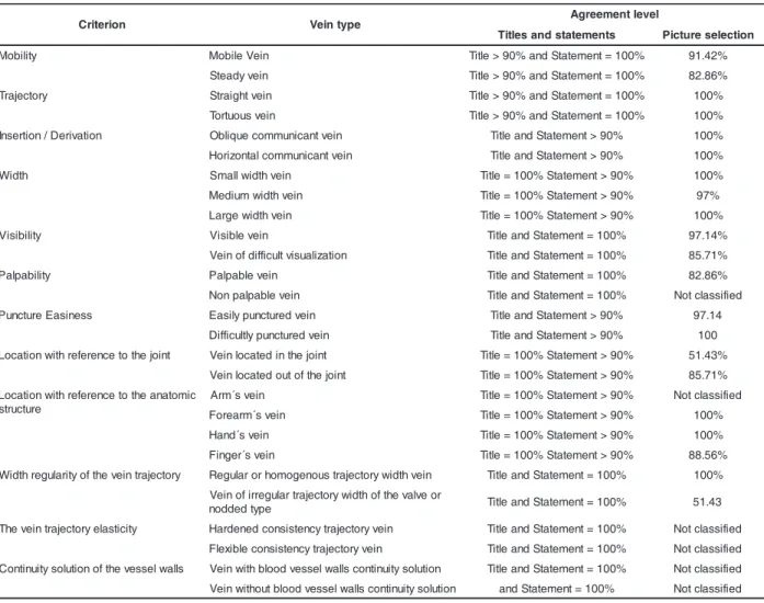
Table 1 - Agreement level obtained in criteria validation, vein types and selection of pictures to classify peripheral
veins
The 144 color photos were analyzed by
experts, who preserved two good examples of photos
for a better selection. All pictures that obtained the
major part should correspond to the title for which they
were selected. This fact was confirmed by the comments
of more than 95% of the 35 experts who participated
in the first work stage, since they stated that all
pictures they had seen were excellent. The experts’
concordance index for the best pair of available photos
was distributed like this: 1) unanimity; 2) 97,14%; 3)
91,42%; 4) 88,56% ; 5) 85,71%; 6) 82,86% ; 7)
51,43%. Figures 1 to 4 show black and white pictures
of these situations. It is also important to consider that
the experts have plural labor practice for the use of
the vein network according to the developed/evolved
action point of view or its use purpose. Due to this
fact, some items have obtained differentiated
assessment with evaluation and agreement.
Figure 1 - Vein types. Examples of vein pictures classified according to the Mobility (1, 2), Trajectory (3,4) and
Insertion / Derivation (5, 6) criteria
/QDKNG8GKP 5VGCF[8GKP 5VTCKIJV8GKP 6QTVWQWU8GKP 1DNKSWG %QOOWPKECPV8GKP *QTK\QPVCN %QOOWPKECPV8GKP n o i r e t i r
C Veintype Agreementlevel
s t n e m e t a t s d n a s e l t i
T Pictureselection
y t il i b o
M MoblieVein Tilte>90%andStatement=100% 91.42%
n i e v y d a e t
S Tilte>90%andStatement=100% 82.86%
y r o t c e j a r
T Straightvein Tilte>90%andStatement=100% 100%
n i e v s u o u t r o
T Tilte>90%andStatement=100% 100%
n o it a v i r e D / n o it r e s n
I Obilquecommunicantvein TilteandStatement>90% 100%
n i e v t n a c i n u m m o c l a t n o z i r o
H TilteandStatement>90% 100%
h t d i
W Smallwidthvein Tilte=100%Statement>90% 100%
n i e v h t d i w m u i d e
M Tilte=100%Statement>90% 97%
n i e v h t d i w e g r a
L Tilte=100%Statement>90% 100%
y t il i b i s i
V Visiblevein TilteandStatement=100% 97.14%
n o it a z il a u s i v tl u c if f i d f o n i e
V TilteandStatement=100% 85.71%
y t il i b a p l a
P Palpablevein TilteandStatement=100% 82.86%
n i e v e l b a p l a p n o
N TilteandStatement=100% Notclassiifed
s s e n i s a E e r u t c n u
P Easliypuncturedvein TilteandStatement>90% 97.14
n i e v d e r u t c n u p y lt l u c if f i
D TilteandStatement>90% 100
t n i o j e h t o t e c n e r e f e r h t i w n o it a c o
L Veinlocatedinthejoint Tilte=100%Statement>90% 51.43%
t n i o j e h t f o t u o d e t a c o l n i e
V Tilte=100%Statement>90% 85.71%
c i m o t a n a e h t o t e c n e r e f e r h t i w n o it a c o L e r u t c u r t s n i e v s ´ m r
A Tilte=100%Statement>90% Notclassiifed
n i e v s ´ m r a e r o
F Tilte=100%Statement>90% 100%
n i e v s ´ d n a
H Tilte=100%Statement>90% 100%
n i e v s ´ r e g n i
F Tilte=100%Statement>90% 88.56%
y r o t c e j a r t n i e v e h t f o y t i r a l u g e r h t d i
W Regularorhomogenoustrajectorywidthvein TilteandStatement=100% 100%
r o e v l a v e h t f o h t d i w y r o t c e j a r t r a l u g e r r i f o n i e V e p y t d e d d o
n TilteandStatement=100% 51.43
y t i c it s a l e y r o t c e j a r t n i e v e h
T Hardenedconsistencytrajectoryvein TilteandStatement=100% Notclassiifed
n i e v y r o t c e j a r t y c n e t s i s n o c e l b i x e l
F TilteandStatement=100% Notclassiifed
s ll a w l e s s e v e h t f o n o it u l o s y t i u n it n o
C Veinwithbloodvesselwallsconitnuitysoluiton TilteandStatement=100% Notclassiifed
n o it u l o s y t i u n it n o c s ll a w l e s s e v d o o l b t u o h t i w n i e
Figure 2 - Vein types. Examples of vein pictures classified according to the Width (1, 2, 3); Visibility (4,5)
Palpability (6)
5OCNN9KFVJ8GKP
Ō/GFKWO9KFVJ
8GKP
Ō.CTIG9KFVJ8GKP
Ō8KUKDNG8GKP
8GKPQH&KHHKEWNV
8KUWCNK\CVKQP
Ō2CNRCDNG8GKP
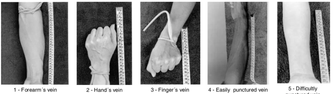
Figure 3 - Vein types. Examples of vein pictures classified according to the criteria: Location with reference to
the anatomy structure (1, 2, 3); Puncture Easiness (4, 5)
(QTGCTOyUXGKP
*CPFyUXGKP
(KPIGTyUXGKP
'CUKN[RWPEVWTGFXGKP
&KHHKEWNVN[ RWPEVWTGFXGKP
Figure 4 - Vein types. Examples of vein pictures classified according to the criteria: Location with reference to
the joint (1,2); Width regularity of the vein trajectory (3,4) and cannot be classified for any of the proposed
criteria (5)
8GKPNQECVGFKPVJG
LQKPV
8GKPNQECVGFQWVQH
VJGLQKPV
4GIWNCTQT JQOQIGPQWUVTCLGEVQT[
YKFVJXGKP
8GKPQHKTTGIWNCT VTCLGEVQT[YKFVJQHVJG
XCNXGQTPQFFGFV[RG
8GKPVJCVECPPQVDG ENCUUKHKGFHQTVJG RTQRQUGFETKVGTKQP
With respect to the process of choosing the
set of pictures to represent the different vein types, it
is important to emphasize that some titles could not
be exemplified, given the methodological trajectory,
which used visual assessment to select the pictures:
1) “hardened consistency trajectory vein” and “flexible
consistency trajectory vein”, which belong to the “vein
trajectory elasticity” criterion; 2) “non palpable vein”,
included in the palpability criterion;
For other titles, pictures could not be validated
either, due to the population selection criteria: “vein
without blood vessel walls continuity solution” and
“vein with blood vessel walls continuity solution”,
included in the “continuity solution of the vessel walls”
criterion. These vein conditions are usually observed
in hospitalized persons, after multiple punctures.
However, according to the selection criteria, all
photographed subjects were healthy and possessed
intact veins.
Finally, one other title could not be exemplified
either, as pictures in this study represented forearm
for this criterion. This was the case for “arm’s vein”,
included in the “location with reference to the anatomic
structure”(11) criterion.
Despite the construction and validation of this
classification, including the types and criteria described
above, this study is limited by the lack of picture
examples for the above mentioned titles. However,
the description of these veins contributes to their
naming / labeling in clinical practice.
FINAL CONSIDERATIONS
The classification of adolescents, adults and
elderly persons’ superficial peripheral vein types
developed in this research is composed of 12
consensus criteria (agreement level of more than 90%
for completely adequate/relevant and not inferior to
10% for moderately adequate/relevant): mobility,
trajectory, insertion/derivation, width, visibility,
palpability, location with reference to the joint, location
with reference to the anatomy structure, width
regularity of the vein trajectory, the vein trajectory
elasticity continuity solution of the vessel walls and
puncture easiness; the “other criterion to be included”
was equally approved. This aspect adds to the
consideration that a classification should foresee the
inclusion of new terms or their refinement.
The consensus among the 35 experts
concerning the titles and declaration concordance index
of each criterion was facilitated by the use of the Delphi
technique. In order to measure the participants’ opinion,
the use of a Likert scale in all stages favored an
agreement on the evaluation parameter. When
participants chose the pictures, they had already
accomplished a title and declaration approximation
about what each photo should represent.
First, four experts selected the pictures,
avoiding exhaustive meetings with all 35 participants
and facilitating the operational conduction of this stage.
Pictures exemplifying 21 titles were obtained and the
other titles could not be exemplified through photos,
because of the incompatibility of the visual criterion
with the variables defined in this work. However, other
pictures can be added, considering the criterion
“location with reference to the anatomy structure” for
example. The classification, in spite of being simple,
covers a diversity of criteria through which a vein
can be analyzed. Some experts manifested the utility
of this classification for undergraduate teaching,
research on the theme and professional recycling on
vein puncture, either verbally or in writing.
REFERENCES
1. Meyers L. Intravenous catheterization. Am J Nurs 1945; 45(2):930-1.
2. Maeda ST. Escalpe, equipo de soro e seringa descartável: critérios qualitativos para subsidiar a compra. [dissertação]. São Paulo (SP): Escola de Enfermagem/USP; 1998. 3. Arreguy-Sena C. A relação entre o preconceito social e o comportamento de infectados pelo HIV numa instituição hospitalar, segundo a percepção desses. [dissertação] Rio de Janeiro (RJ): EEAN/UFRJ; 1991.
4. Ministério da Saúde (BR). Manual de normas técnicas para prevenção da transmissão da AIDS. Brasília (DF): Ministério da Saúde, 1989.
5. Arreguy-Sena C. A trajetória de construção e validação dos diagnósticos de enfermagem: Trauma vascular e risco para trauma vascular. [Tese ] Ribeirão Preto (SP): EERP/USP; 2002. 6. Lastória S. Tromboflebite superficial. In: Maffei FHA, Lastória S, Yoshida WB, Rollo HA. Doenças vasculares periféricas. 2ª ed. São Paulo (SP): MEDSI; 1995, p.831-40. 7. Phillips LD. Manual de terapia intravenosa. 2ª ed. Porto Alegre (RGS): Artmed; 2001.
8. Clark KJ. How nurses can participate in the development of. an ICNP. Int Nurs Rev 1996 November-December; 43(6):171-4.
9. Nielsen GH, Mortensen RA. The architecture for an international classification for nursing practice (ICNP). Int Nurs Rev 1996 November-December; 43(6):175-82. 10. Lindeman CA. Delphi survey of priorities in clinical nursing research. Nurs Res 1975; 24(6):434-41.
11. Duffield C. The Delphi Techinique: a comparison of results obtained using two expert panels. Lut J Nurs Stud. 1993; 30: 2 7 7 - 3 7 .
12. Faro ACM Técnica Delphi na Validação das intervenções de Enfermagem. Rev Esc Enfermagem USP 1997 agosto; 31(1):259-73.
13. Guilford JP. Psychometric methods. Bombay (EUA): Mc Graw Hill; 1971.
14. Rodrigues A. Psicologia Social. Petrópolis (RJ): Vozes; 1993. 15. Procter S, Hunt M. Using the Delphi survey technique to develop a professional definition of nursing for analysing nursing workload. J Adv Nurs 1994; 19:1003-14.
16. Salmond SW. Orthopaedic nursing research priorities: a Delphi study. Orthop Nurs 1994; (2):31-45.
17. Associação Brasileira de Normas Técnicas - ABNT. NBR 9753. Escalpe estéril e de uso único: requisitos e métodos de ensaio. Rio de Janeiro (RJ): ABNT: abril 1997.