238
R
ELATO DEC
ASOReceived for publication 11/09/2014 - Accepted for publication 26/05/2015 The authors declare no conflicts of interests.
1 Orbit Sector, Department of Ophthalmology and Visual Science - Universidade Federal de São Paulo, São Paulo, SP, Brazil.
Atypical presentation of pilomatrixoma
in the tarsal conjunctiva
Apresentação atípica de pilomatricoma na conjuntiva tarsal
Mirtha Ramirez Dittrich
1, Jacqueline Martins de Sousa
1, Paulo Gois Manso
1A
BSTRACTPilomatrixoma is a rare benign tumor, which usually affects young women in the head and neck region. The eyelid is a common site of the disease, though it is very rare in the tarsal conjunctiva. The pilomatrixoma has clinical pleomorphism, which confuses this disease with other similar conditions. The diagnosis is made by pathological examination in most cases. The treatment is surgical, performed by total excision of the lesion with clear margins and recurrence is very uncommon. We present an unusual case of probable recurrent pilomatrixoma in the left upper tarsal conjunctiva.
Keywords: Pilomatrixoma/diagnosis; Conjunctiva/pathology; Conjunctival diseases/diagnosis; Case reports
R
ESUMOO pilomatricoma é uma neoplasia benigna rara, geralmente acomete mulheres jovens, ocorrendo na região da cabeça e pescoço. A pálpebra é um local comum de aparecimento do pilomatricoma, contudo, seu aparecimento na conjuntiva tarsal é muito raro. O diagnóstico é feito pelo exame anátomo-patológico na maioria dos casos, pois seu pleomorfismo clínico o confunde com outras alterações. O tratamento é cirúrgico, realizado por meio da exérese total da lesão com margens livres. São raros os casos de recidiva. Apresentamos um caso atípico de provável recidiva de pilomatricoma, localizado na conjuntiva tarsal superior à esquerda.
Descritores: Pilomatricoma/diagnóstico; Túnica conjuntiva/patologia; Doença da túnica conjuntiva/diagnóstico; Relatos de casos
Rev Bras Oftalmol. 2016; 75 (3): 238-40
239
I
NTRODUCTIONP
ilomatrixoma was first described by Malherbe in 1880, named calcifying epithelioma of sebaceous glands.1 In1961, Forbis and Helwig suggested the term pilomatrixoma for this lesion and reserve epithelioma for malignancies.1,2 It is characterized by a benign tumor originating
from a hair follicle, and is more common in young women. 1 The
most affected regions are the head and neck, although the tarsal conjunctiva is a very uncommon location. There are several clinical forms of presentation, which can hinder the diagnosis and may cause confusion with other, more common lesions. 1,3 A specific
malignant presentation called pilomatrixoma carcinoma should be remembered, being more frequent in older men, as it is aggressive and has a tendency to recur and metastasize. 1,4
We report clinical findings, the histopathological exam and management of a rare case of tarsal conjunctival pilomatrixoma.
C
ASER
EPORTA 53 year-old, white female patient, who is a housekeeper, from the city of Sao Paulo in Brazil, complaining of a painless tumor with progressive growth in the left upper eyelid for four months was referred to our service. She reported a similar lesion ten years ago in the same region, which was resected two years ago, and confirmed as squamous papilloma by histological exam. She had exotropia since childhood, and no other important ophthalmic or personal history.
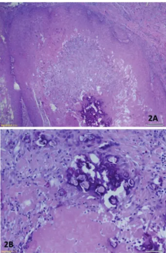
An ophthalmological examination revealed visual acuity of 20/20 in both eyes. There was a whitish nodular lesion with soft-elastic consistency, regular edges and not adhered to deep planes in the nasal region of the tarsal conjunctiva of the left upper eyelid, measuring 1 cm in diameter at its widest and better seen by means of the eversion maneuver. (Figure 1) She presented alternating exotropia, without other changes in the ophthalmologic evaluation. A total thickness excisional biopsy was performed with to-tal tumor ablation with no intraoperative or postoperative complications. The histological examination showed epithelial neoplasm with areas of calcification and ghost cells, which confirmed the diagnosis of pilomatrixoma with free margins. (Fi-gure 2) The patient has been accompanied for one year without signs of recurrence, and with good aesthetics.
Rev Bras Oftalmol. 2016; 75 (3): 238-40
D
ISCUSSIONAlthough our patient was aged 53 at the time of diagnosis, she reported a similar lesion 10 years ago. Therefore it is possible that the lesion began when the patient was near 40 years of age. Pilomatrixomas have a bimodal incidence, occurring mainly in the first three decades of life (some reports showed an 80% rate of incidence under the age of ten) and some cases between 50 and 65 years old. 1,2,4-7 According to others, pilomatrixoma often affects
patients in the first three decades of life. 1,3,7 Thus, our patient
seems to be have presented the lesion at an unusual age of onset. Most case reports have been described in caucasians, as was the case of our patient. However, there was no correlation between ethnicity and the disease.1,2 Pilomatrixoma is a rare
disease, with a prevalence of 0.15 to 1.25% and affects mostly women 1-5,8 and the case we described here was also a female.
Our patient had a nodular sessile whitish lesion in the medial region of the left upper conjunctival tarsus, emphasizing that this entity may have several clinical presentations.2 Four
cases of pilomatrixoma were diagnosed with histological confirmation in a total of 621 eyelid tumors within 30 years, with clinical pleomorphism: subcutaneous nodule, sessile tumor with necrotic surface, pedicle tumor causing ectropion and nodule with a cystic appearance. 3 All these patients had their lesion in
the left eyelid and three of them in the lower left eyelid. According to others, 69% of head pilomatrixomas occur in the upper eyelid or eyebrow and only 17% of the periorbital lesion affects the tarsal conjunctiva. 1 In a study with nine patients with
Figure 1: A- Preoperative photo showing lesion in the left upper
eyelid. B- Patient looking down, showing the lesion in the left upper eyelid. C- Eversion of the upper left eyelid observing high, whitish, irregular lesion measuring 1cm in diameter.
Figure 2: A - Epithelial neoplasm with areas of calcification (HE
40x). B - Details showing areas of calcification and ghost cells (HE 200x).
240
Rev Bras Oftalmol. 2016; 75 (3): 238-40
pilomatrixoma in a total of 17 cases of tumors derived from hair follicles within eight years, the majority was female without previous systemic disease, 82% affected the upper eyelid and the majority in the medial region. 9 Pilomatrixomas can appear
on any area of the body, though about 50% occur in the head and neck region, mainly on the neck, frontal, periorbital and pre-auricular regions. 5 The most common periorbital areas affected
are: eyebrow, eyelid and canthal areas. Some authors suggest that the distribution of pilomatrixoma is related to the density of hair follicles. 1,6,7,9
The disease is not hereditary and there are no reports of predisposing factors. 6 Its pathogenesis is associated with a mutation
in the exon 3 of the CTNNB1 gene, located on chromosome 3p22-p21.3, which encodes the beta-catenin protein. This mutation also occurs in cases of follicular carcinoma. 1,4,6,9 There are case reports
of multiple and familiar pilomatrixomas, which are rarer than the single lesions and were associated with various syndromes, such as Steinertmyotonic dystrophy, Gardner syndrome, Turner syndrome, trisomy 9, spina bifida, Soto syndrome, Rubinstein-Taybi syndrome, xerodermapigmentosum, and Churg-Strauss syndrome. 1,4,6 However, our patient had no other systemic changes.
The growth of the lesion is usually slow and progressive. When in the eyelid, the lesion size is usually up to 1cm, as we noted in our patient. 1,9 There are several eyelid pathologies with
similar characteristics and they should be remembered as differential diagnosis, such as dermoid cyst, inclusion cyst, Moll cyst, sebaceous cyst, foreign body reaction, pyogenic granuloma, papilloma, chalazion and even sarcoidosis.1,3,6 Malignancies may
not be excluded in the first evaluation. In our case, we initially suspected malignancy, because the aspect and the report of lesion recurrence after excision in the same location, although the first histologic result revealed a benign lesion. As a differential diagnosis, it is important to think about the malignant tumors sebaceous gland carcinoma and pilomatrixoma carcinoma, although they are much rarer. 3,10 The pilomatrixoma carcinoma
is a malignant variant of pilomatrixoma, whose differential diagnosis is performed by histology, showing abundant proliferation of basaloid cells, infiltrative growth pattern and extends to deep planes. There are no specific molecular and immunohistochemical markers for it. 4 It affects mainly older
men, is very rare in the eyelid, may have recurrence in about 50-60% of the cases and can metastasize in 10-12% of cases to the lungs, bone, brain, lymph nodes, skin and retroperitoneum, with poor prognosis.1,4 There were only 101 cases of pilomatrixoma
carcinoma described in the English scientific literature (Pubmed database) up until 2013. 4
The definitive diagnosis of pilomatrixoma is histological. Two populations of cells can be predominant namely: basaloid cells (basophilic nucleus and scant cytoplasm) and ghost cells (enucleated cells eosinophils pale). 1,6,7,9 Ghost cells are more
common in older tumors and they represent necrotic areas where there were living basaloid cells. 6 The presence of ghost cells is
suggestive, but not pathognomonic for pilomatrixoma, because they can be observed in epidermoid cysts, chronic inflammation
Corresponding author:
Mirtha Ramirez Dittrich
R Botucatu, 821 – Vila Clementino, São Paulo, SP, Brazil. Zip code: 04023-062phone: 55 – 11 – 5085-2010
E-mail: [email protected]
of the hair follicle with hyperkeratosis and chronic dermatoses.1
Pilomatrixoma lesion has precise limits and areas of calcification, foreign body granulomatous reaction, ossification and pseudocapsule formation may be present. 3,6
There are no reports of spontaneous regression of pilomatrixoma. The treatment of choice is total excision of the lesion with clear margins. 1,5,7,9 The tumor is never attached to the
subcutaneous tissue, but may be attached to the skin, making its removal difficult, as in our case. In this situation, the best approach is total excision of the lesion with the adjacent skin. 1,5 Incomplete
resection of the lesion can cause recurrence, described in up to 3% of cases. 1,5 In our case, it is very likely that the lesion found
was a recurrence of a previously undiagnosed pilomatrixoma. Although histological diagnosis is not particularly difficult, it may not have been noticed at the time of first evaluation due to the rarity of the disease.
R
EFERENCES1. Levy J, Ilsar M, Deckel Y, Maly A, Anteby I, Pe’er J. Eyelid Pilomatrixoma: a description of 16 cases and a review of the literature. Surv Ophthalmol. 2008;53(5):526-35.
2. Mendes MH, Souza LP. Recurrent pilomatricoma on the left eye-brow: case report. Arq Bras Oftalmol. 2009;72(3):380-3. 3. Marback EF, Cardoso C, Moitinho LM, Marback RL.
Clinico-pathologic study of eyelid pilomatrixoma: the experience of the ‘Hospital Universitário Prof. Edgard Santos’. Arq Bras Oftalmol. 2007;70(3):501-3.
4. Cornejo KM, Deng A. Pilomatrix carcinoma: A case report and review of the literature. Am J Dermatopathol. 2013;35(3):389-94. 5. Mendes Neto JA, Raposo RM, Segalla DK, Leonhard FD. Pilomatrixoma in the head and neck. Braz J Otorhinolaryngol. 2009;75(4):618.
6. Passi N, Chawla U, Jyoti, Khurana AK. Pilomatrixoma of eyelid in a middle aged. Nepal J Ophthalmol. 2011;3(5):88-90. 7. Rodríguez CI, Mencía EG, Gutiérrez ED, Suárez AG. An unusual
presentation of a pilomatrixoma in the eyelid. Arch Soc Esp Oftalmol. 2006;81(8):483-6.
8. Doxanas MT, Green WR, Arentsen JJ, Elsas. Lid lesions in child-hood: A histopathologic survey at the Wilmer Institute (1923-1974). J Pediatr Ophthalmol. 1976;13(1):7-39.
9. Mencia GE, Gutiérrez DE, Ricoy J, Gómez LI, Monescillo J, García TJ. Benign hair-follicle derived eyelid tumors in adults. Arch Soc Esp Oftalmol. 2002;77(11):605-10.
10. Vianna LM, Cariello AJ, Lowen MS, Sant’Anna AE, Hofling-Lima AL. Sebaceous carcinoma of the eyelid – different diagnos-tic times, different outcomes: case reports. Arq Bras Oftalmol. 2011;74(6):444-6.