Open Access
Research article
Mitochondrial haplogroup H1 is protective for ischemic stroke in
Portuguese patients
Alexandra Rosa
1, Benedita V Fonseca
1, Tiago Krug
1, Helena Manso
1,2,
Liliana Gouveia
3, Isabel Albergaria
2, Gisela Gaspar
2, Manuel Correia
4,
Miguel Viana-Baptista
5, Rita Moiron Simões
6, Amélia Nogueira Pinto
6,
Ricardo Taipa
4, Carla Ferreira
7, João Ramalho Fontes
7, Mário Rui Silva
8,
João Paulo Gabriel
8, Ilda Matos
9, Gabriela Lopes
4, José M Ferro
3,
Astrid M Vicente
1,2and Sofia A Oliveira*
1Address: 1Instituto Gulbenkian de Ciência, Oeiras, Portugal, 2Instituto Nacional de Saúde Dr. Ricardo Jorge, Lisboa, Portugal, 3Serviço de
Neurologia, Hospital de Santa Maria, Lisboa, Portugal, 4Serviço de Neurologia, Hospital Geral de Santo António, Porto, Portugal, 5Serviço de
Neurologia, Hospital Garcia de Orta, Almada, Portugal, 6Serviço de Neurologia, Hospital Fernando Fonseca, Amadora, Portugal, 7Serviço de
Neurologia, Hospital São Marcos, Braga, Portugal, 8Serviço de Neurologia, Hospital de São Pedro, Vila Real, Portugal and 9Serviço de Neurologia,
Hospital Distrital de Mirandela, Mirandela, Portugal
Email: Alexandra Rosa - arosa@uma.pt; Benedita V Fonseca - mfonseca@igc.gulbenkian.pt; Tiago Krug - tkrug@igc.gulbenkian.pt;
Helena Manso - hmanso@igc.gulbenkian.pt; Liliana Gouveia - lilianafog@gmail.com; Isabel Albergaria - isabel.albergaria@insa.min-saude.pt; Gisela Gaspar - gisela.gaspar@insa.min-saude.pt; Manuel Correia - mmcorreia@mail.telepac.pt; Miguel
Viana-Baptista - mbatista.neuro@fcm.unl.pt; Rita Moiron Simões - rita_moiron_simoes@hotmail.com;
Amélia Nogueira Pinto - ameliapinto@hotmail.com; Ricardo Taipa - ricardotaipa@gmail.com; Carla Ferreira - carla.m.c.ferreira@gmail.com; João Ramalho Fontes - fontes@hsmbraga.min-saude.pt; Mário Rui Silva - uavc@chvrpr.min-saude.pt; João Paulo Gabriel - Jp.sequeira@iol.pt; Ilda Matos - Ildamariasilvamatos@hotmail.com; Gabriela Lopes - gab.lopes@clix.pt; José M Ferro - jmferro@fm.ul.pt;
Astrid M Vicente - avicente@igc.gulbenkian.pt; Sofia A Oliveira* - soliveira@igc.gulbenkian.pt * Corresponding author
Abstract
Background: The genetic contribution to stroke is well established but it has proven difficult to identify the genes and the disease-associated alleles mediating this effect, possibly because only nuclear genes have been intensely investigated so far. Mitochondrial DNA (mtDNA) has been implicated in several disorders having stroke as one of its clinical manifestations. The aim of this case-control study was to assess the contribution of mtDNA polymorphisms and haplogroups to ischemic stroke risk.
Methods: We genotyped 19 mtDNA single nucleotide polymorphisms (SNPs) defining the major European haplogroups in 534 ischemic stroke patients and 499 controls collected in Portugal, and tested their allelic and haplogroup association with ischemic stroke risk.
Results: Haplogroup H1 was found to be significantly less frequent in stroke patients than in controls (OR = 0.61, 95% CI = 0.45–0.83, p = 0.001), when comparing each clade against all other haplogroups pooled together. Conversely, the pre-HV/HV and U mtDNA lineages emerge as potential genetic factors conferring risk for stroke (OR = 3.14, 95% CI = 1.41–7.01, p = 0.003, and OR = 2.87, 95% CI = 1.13–7.28, p = 0.021, respectively). SNPs m.3010G>A, m.7028C>T and m.11719G>A strongly influence ischemic stroke risk, their allelic state in haplogroup H1 corroborating its protective effect.
Conclusion: Our data suggests that mitochondrial haplogroup H1 has an impact on ischemic stroke risk in a Portuguese sample.
Published: 1 July 2008
BMC Medical Genetics 2008, 9:57 doi:10.1186/1471-2350-9-57
Received: 3 March 2008 Accepted: 1 July 2008
This article is available from: http://www.biomedcentral.com/1471-2350/9/57
© 2008 Rosa et al; licensee BioMed Central Ltd.
Background
Stroke is a complex disorder resulting from the interplay of genetics and environment, and genes potentially hav-ing an impact on disease pathogenesis (e.g. genes involved in hemostasis), intermediate phenotypes (e.g. atherosclerosis) or clinical risk factors (e.g. blood pressure regulation) have been tested for association with stroke risk [1]. Mostly nuclear genes have been intensively inves-tigated thus far, while the role of the mitochondrial genome has been neglected. Mitochondria are extranu-clear organelles whose primary function is the production of ATP through the oxidative phosphorylation (OXPHOS) respiratory chain. They also play a decisive role in intracellular signaling, metabolic pathways such as Krebs' or tricarboxylic acid cycle and the metabolism of amino acids, lipids, cholesterol and steroids. Mitochon-drial function is required for normal vascular cell growth and function, and its dysfunction can result in apoptosis, favoring atherosclerotic plaque rupture. mtDNA is mater-nally inherited, does not recombine and exhibits high mutation and fixation rates, therefore making it an impor-tant tool in phylogenetics. Human mtDNA is a haploid, circular molecule of approximately 16,600 nucleotides encoding for thirteen OXPHOS polypeptides, twenty-two transfer RNAs and two ribosomal RNAs (Figure 1) [2]. Particular combinations of certain polymorphisms define mitochondrial haplogroups and subclades (Table 1), which tend to be associated to broad geographic areas and/or populations [3].
Particular variants of the mitochondrial genome have been linked to aging [4,5], the strongest risk factor for stroke, and to several neurological and vascular disorders. Among the best-known examples of a mitochondrial dis-order is that of MELAS (MIM: 540000), a mitochondrial
encephalopathy characterized by lactic acidosis and stroke-like episodes. This syndrome is caused by the m.3243A>G mutation, an A to G transition at mtDNA nucleotide position 3243 [6,7]. Leber's hereditary optic neuropathy (LHON, MIM: 535000), a vascular disease of the optic disc, is also caused by mtDNA mutations that lead to respiratory chain dysfunction [8]. Interestingly, the phylogenetic background of haplogroup J influences the clinical penetrance and expression of the m.11778G>A and m.14484T>C primary LHON mutations [9,10]. This exemplifies how, although defined on the basis of evolu-tionarily neutral polymorphisms, common mtDNA varia-tion of phylogenetic relevance assumes a funcvaria-tional role on the expression of particular complex traits. mtDNA variation has been associated with non-Mendelian and non-maternally inherited complex disorders such as Par-kinson's disease [11], Alzheimer's disease [12], myocar-dial infarction [13], obesity [14], occipital stroke in migraine [15,16], and mean intima-media thickness of bilateral carotid arteries [17]. Increased mitochondrial oxidative stress and dysfunction has been linked to many ischemic stroke risk factors, including hypertension [18], diabetes [19], inflammation [20], plaque rupture [20], tobacco smoke and alcohol exposure [21]. The goal of the present study was to determine whether mtDNA SNPs or haplogroups predispose to ischemic stroke in a large cohort of Portuguese patients and controls.
Methods
Study subjectsFive hundred thirty four unrelated patients with a clinical diagnosis of ischemic stroke, who were under the age of 65 at stroke onset, were recruited through Neurology and Internal Medicine Departments throughout Portugal. Stroke was defined by the presence of a new focal
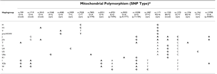
neuro-Table 1: Type of investigated mitochondrial markers and haplogroup determination.
Mitochondrial Polymorphism (SNP Type)*
Haplogroup m.709 G>A (ncod) m.1719 G>A (ncod) m.3010 G>A (ncod) m.3348 A>G (syn) m.4580 G>A (syn) m.5999 T>C (syn) m.7028 C>T (syn) m.7805 G>A (p.V74I) m.8251 G>A (syn) m.8701 A>G (p.T59A) m.9055 G>A (p.A177T) m.10398 A>G (p.T114A) m.1087 3T>C (syn) m.1171 9G>A (syn) m.1230 8A>G (ncod) m.1270 5C>T (syn) m.1336 8G>A (syn) m.1361 7T>C (syn) m.13708 G>A (p.A458T)
H C G
H1 A C G
V A T G
pre-HV/HV G T G
J G G A A C A
J1b G A G A A C A
T A A A C A
U A G C
U4 C A G C
U5 A G C C
U6a G A A G C
K1 A G G
I G A A A G T T
X2b G A G A A T T A
W A G A A A T T
L G G C T
Each haplogroup was determined by the combination of bolded alleles, and the alleles not bolded aided in the phylogenetic assignment. The polymorphisms are named after their base pair position and alleles.
Genomic localization of the investigated markers within the human mitochondrial DNA molecule Figure 1
logical deficit, with an acute onset and symptoms and signs persisting for more than 24 hours, and was con-firmed by Computed Tomography Scan (97% of cases) and/or Magnetic Resonance Imaging (in 25% of patients) [22]. All patients were seen and all neuroradiology tests were reviewed by study neurologists. Trauma, tumors, infection and other causes of neurological deficit were excluded.
Data collection forms were developed for this study that included extensive clinical information such as stroke characteristics, general clinical observation, neurological symptoms and signs, complications and interventions during hospitalization and situation at discharge. Data was also collected on relevant lifestyle aspects and previ-ous clinical risk factors.
Four hundred ninety nine unrelated healthy individuals were included in this study as a control sample popula-tion. Since stroke is a late-onset disease, the control group was selected from a group of healthy volunteers with a higher mean age than the case group, thus minimizing the chances for mis-classification as "stroke-free". Control individuals were verified to be free of stroke by direct interview before recruitment, but no brain imaging stud-ies were performed. The interview also included questions on established clinical and life-style risk factors for stroke.
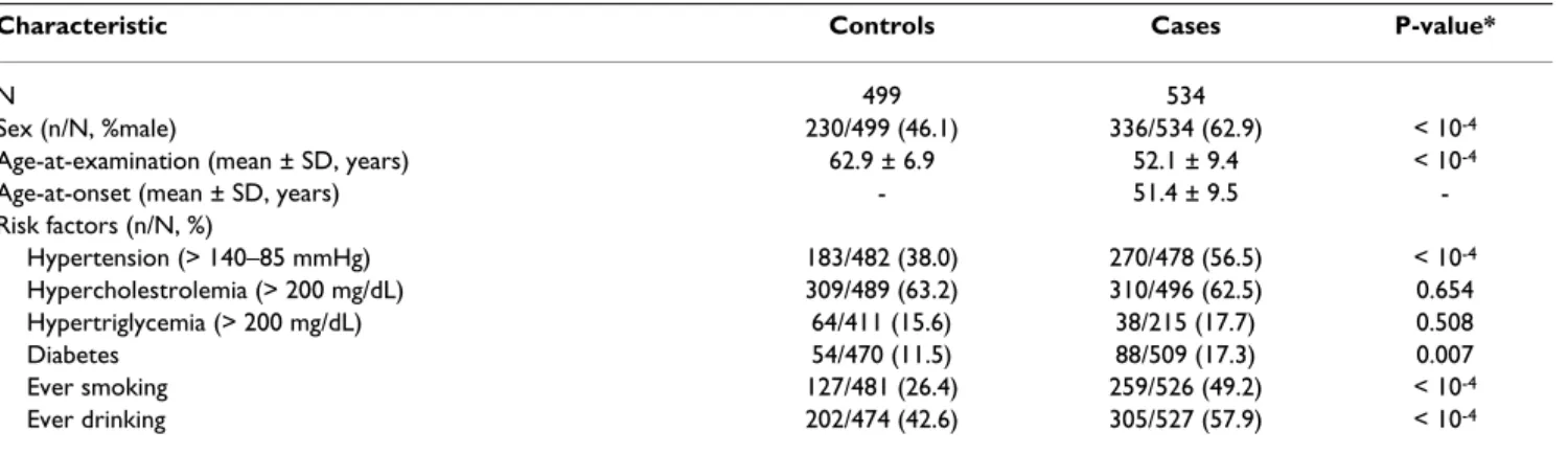
The principal demographic and clinical characteristics and frequency of risk factors of this study sample are shown in Table 2. The research protocol was approved by the ethics committees of participating institutions, and participants were informed of the study and provided informed consent.
SNP selection and haplogroup definition
We studied nineteen mtDNA SNPs (Figure 1 and Table 1) of phylogenetic relevance for classifying the Portuguese mitochondrial haplogroup variation which includes the
most prevalent West Eurasian haplogroups (H, H1, V, pre-HV/HV, J, J1b, T, U, U4, U5, K1, I, X2b, and W), as well as some African haplogroups (U6a and L) more frequent in Portugal and in the Iberian Peninsula than in other Euro-pean countries [23,24]. Haplogroups and their subclades, which show different frequencies and distributions in human populations, are defined by the combination of multiple markers (Table 1), embracing the information from the whole set of branches of the mtDNA tree rather than the status at any single point mutation. The nomen-clature of clades follows Torroni et al. [25], Richards et al. [26], and Macaulay et al. [27]. The "Other" haplogroup category in Table 3 (19 controls and 20 patients) includes individuals whose haplogroups could not be assigned to the clades in Table 1.
Genotyping
Genomic DNA was extracted from whole blood samples using the NucleoSpin Blood XL kit (Macherey-Nagel; Düren, Germany) or a salting out procedure. SNPs were genotyped using Sequenom's iPlex assay (primer exten-sion of multiplex products with detection by matrix-assisted laser desorption/ionization time-of-flight mass spectrometry) following manufacturer's protocol and detected in a Sequenom MassArray K2 platform. The primer sequences are available upon request and were designed using Sequenom's (San Diego, USA) MassAR-RAY® Assay Design 3.0 software according to the
Cam-bridge reference sequence [2]. Extensive quality control was performed using eight HapMap controls of diverse ethnic affiliation, sample duplication within and across plates, non-Mendelian maternal inheritance check in three large pedigrees, and a minimum of 90% call rate. Genotype determinations were performed blinded to affection status. 0.2% of all calls were heterozygous, most likely due to mtDNA heteroplasmy, and these were not included in the analyses.
Table 2: General characteristics of the ischemic stroke case-control study sample
Characteristic Controls Cases P-value*
N 499 534
Sex (n/N, %male) 230/499 (46.1) 336/534 (62.9) < 10-4
Age-at-examination (mean ± SD, years) 62.9 ± 6.9 52.1 ± 9.4 < 10-4
Age-at-onset (mean ± SD, years) - 51.4 ± 9.5
-Risk factors (n/N, %)
Hypertension (> 140–85 mmHg) 183/482 (38.0) 270/478 (56.5) < 10-4
Hypercholestrolemia (> 200 mg/dL) 309/489 (63.2) 310/496 (62.5) 0.654
Hypertriglycemia (> 200 mg/dL) 64/411 (15.6) 38/215 (17.7) 0.508
Diabetes 54/470 (11.5) 88/509 (17.3) 0.007
Ever smoking 127/481 (26.4) 259/526 (49.2) < 10-4
Ever drinking 202/474 (42.6) 305/527 (57.9) < 10-4
Statistical analysis
An unpaired Student's t test and a χ2 test were used to
compare quantitative and qualitative clinical and demo-graphic data, respectively, between cases and controls. χ2
tests were performed to explore the association of each mtDNA SNP and haplogroup with stroke risk. For haplo-group analyses, we compared each haplohaplo-group with all other haplogroups pooled together. To adjust the associa-tion analysis for confounding factors, age-at-examinaassocia-tion, hypertension, diabetes and ever smoking were included as covariates in multivariate logistic regression with back-ward elimination of risk factors. The interaction i among
covariates in regression models was not strong (-0.5 <i <
0.5). Logistic regressions were performed using the R free-ware [28]. Odds ratios (ORs) and their associated 95% confidence intervals (CIs) were uncorrected for confound-ing variables in the χ2 tests and corrected for covariates in
regression models. Results were considered significant below the conventional level of 0.05. Since some of the markers are in linkage disequilibrium and the haplogroup comparisons are not independent, we did not perform corrections for multiple testing and uncorrected p-values are reported.
Results
Table 2 shows the general characteristics of our dataset. Since stroke is a very common late-onset disorder, we chose to have the control group significantly older than
the case group to minimize misclassification biases of control individuals. Male to female ratio, hypertension, diabetes, ever smoking, and ever drinking were signifi-cantly higher in ischemic stroke patients than in controls, and the effects of these potentially confounding variables were accounted for in the multivariate logistic regressions with backward elimination of risk factors. Our group of patients has a similar risk factor profile than previously described older groups of ischemic stroke cases with sim-ilar male to female ratios [29,30], and therefore can be considered representative of the general ischemic stroke population.
The mtDNA haplogroup distribution in the control group (Table 3) was in agreement with previously published data on a similar Portuguese normal population [23,24], with 8.3–9.9% of the individuals having mtDNA haplo-groups characteristic of African populations (L and U6). With the genotyped SNPs, a haplogroup could not be assigned to an almost equal percentage of individuals in the control and patient groups (3.8 and 3.7%, respec-tively, classified as "Others" in Table 3), which again is in concordance with other studies using equivalent approaches [11,31]. These individuals have either an ambiguous SNP-profile or belong to rare Eurasian haplo-groups (e.g. R, Z, M). The fact that L, U6, and "Others" haplogroup categories are present in equivalent propor-tions in cases (12.0%) and controls (10.8%) (Table 3)
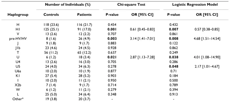
fur-Table 3: Results of mitochondrial haplogroup association testing with ischemic stroke risk.
Number of Individuals (%) Chi-square Test Logistic Regression Model
Haplogroup Controls Patients P-value OR [95% CI] P-value OR [95% CI]
H 118 (23.6) 116 (21.7) 0.454 0.432
H1 125 (25.1) 91 (17.0) 0.001 0.61 [0.45–0.83] 0.007 0.57 [0.38–0.85]
V 13 (2.6) 12 (2.2) 0.707 0.861
pre-HV/HV 8 (1.6) 26 (4.9) 0.003 3.14 [1.41–7.01] 0.008 4.68 [1.51–14.54]
J 9 (1.8) 9 (1.7) 0.883 0.122
J1b 23 (4.6) 24 (4.5) 0.928 0.862
T 56 (11.2) 65 (12.2) 0.637 0.249
U 6 (1.2) 18 (3.4) 0.021 2.87 [1.13–7.28] 0.038 4.01 [1.08–14.90]
U4 13 (2.6) 16 (3.0) 0.705 0.286
U5 24 (4.0) 34 (6.3) 0.278 0.048 2.17 [1.01–4.67]
U6a 10 (2.0) 10 (1.9) 0.877 0.71
K1 27 (5.4) 28 (5.2) 0.903 0.184
I 10 (2.0) 11 (2.1) 0.950 0.500
X2b 7 (1.4) 9 (1.7) 0.714 0.789
W 6 (1.2) 11 (2.1) 0.279 0.394
L 25 (5.0) 34 (6.4) 0.348 0.913
Other* 19 (3.8) 20 (3.7) -
-Significant uncorrected P-values (< 0.05) are highlighted in bold. Crude and adjusted odds ratios (OR) and 95% confidence intervals (CI) are shown only for significantly associated haplogroups.
ther suggests that our dataset was well matched for ethnicity and lacks significant substructure.
Results of crude and adjusted association analyses of mitochondrial haplogroups are shown in Figure 2 and Table 3, and those of single-markers are presented in Fig-ure 3 and Table 4. Sub-haplogroup H1 was found to be significantly less frequent in ischemic stroke patients than in controls when comparing each clade against all others pooled together (χ2 test OR = 0.61, 95%CI = 0.45–0.83, p
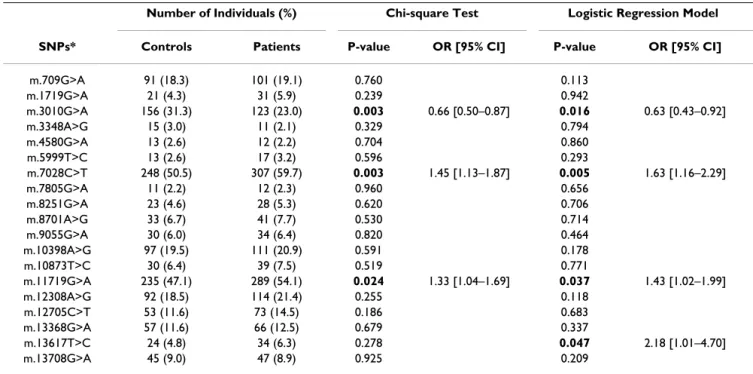
= 0.001); when significant risk factors were included in the model as covariates (age-at-examination, hyperten-sion, diabetes and ever smoking), the association remained significant (logistic regression OR = 0.57, 95%CI = 0.38–0.85, p = 0.007). The sub-haplogroup H1 is thus protective for ischemic stroke in this dataset. SNPs m.3010G>A, m.7028C>T and m.11719G>A, which together define H1, were found to consistently influence the risk for ischemic stroke in both the uncorrected χ2 test
and the logistic regression model, their allelic state in H1 corroborating its protective effect. Stratification by sex revealed that the crude association of haplogroup H1 is quite consistent among males (OR = 0.65, 95%CI = 0.43– 0.98, p = 0.038) and females (OR = 0.58, 95%CI = 0.37– 0.93, p = 0.021), even though it did not reach significance in females when adjusted for co-variates (Figure 2), most likely due to the small sample size.
Conversely, the pre-HV/HV, also known as R0 [32] (χ2
test OR = 3.14, 95%CI = 1.41–7.01, p = 0.003; logistic regression OR = 4.68; 95%CI = 1.51–14.54, p = 0.008), and U (χ2 test OR = 2.87, 95%CI = 1.13–7.28, p = 0.021;
logistic regression OR = 4.01, 95%CI = 1.08–14.90, p = 0.038) mtDNA lineages emerge as potential genetic fac-tors conferring risk for stroke (Figure 2 and Table 3). The relatively rare U5 sub-clade and its defining polymor-phism m.13617T>C showed a trend for association with stroke risk only with the logistic regression test (OR = 2.17, 95%CI = 1.01–4.67, p = 0.048, and OR = 2.18; 95%CI = 1.01–4.70, p = 0.047, respectively).
Discussion
To the best of our knowledge, this is the first comprehen-sive association study of mtDNA variation with ischemic stroke risk in an European population. In a large popula-tion sample of ethnically-matched cases and controls, we found that haplogroup H1 is protective while haplo-groups pre-HV/HV and U increase risk for ischemic stroke. Since these haplogroups are defined by the combination of several polymorphisms also present in other clades (e.g. allele A of m.3010G>A is a phylogenetic marker of subclades H1 and J1b), the observed haplogroup associa-tions cannot be attributed to particular SNPs, but instead to their precise arrangement. To exclude the possibility that the observed associations are due to population strat-ification with study participants of African or non-West
Table 4: Results of mitochondrial SNP association testing with ischemic stroke risk. Significant uncorrected P-values (< 0.05) are highlighted in bold. Crude and adjusted odds ratios (OR) and 95% confidence intervals (CI) are shown only for significantly associated polymorphisms.
Number of Individuals (%) Chi-square Test Logistic Regression Model
SNPs* Controls Patients P-value OR [95% CI] P-value OR [95% CI]
m.709G>A 91 (18.3) 101 (19.1) 0.760 0.113
m.1719G>A 21 (4.3) 31 (5.9) 0.239 0.942
m.3010G>A 156 (31.3) 123 (23.0) 0.003 0.66 [0.50–0.87] 0.016 0.63 [0.43–0.92]
m.3348A>G 15 (3.0) 11 (2.1) 0.329 0.794
m.4580G>A 13 (2.6) 12 (2.2) 0.704 0.860
m.5999T>C 13 (2.6) 17 (3.2) 0.596 0.293
m.7028C>T 248 (50.5) 307 (59.7) 0.003 1.45 [1.13–1.87] 0.005 1.63 [1.16–2.29]
m.7805G>A 11 (2.2) 12 (2.3) 0.960 0.656
m.8251G>A 23 (4.6) 28 (5.3) 0.620 0.706
m.8701A>G 33 (6.7) 41 (7.7) 0.530 0.714
m.9055G>A 30 (6.0) 34 (6.4) 0.820 0.464
m.10398A>G 97 (19.5) 111 (20.9) 0.591 0.178
m.10873T>C 30 (6.4) 39 (7.5) 0.519 0.771
m.11719G>A 235 (47.1) 289 (54.1) 0.024 1.33 [1.04–1.69] 0.037 1.43 [1.02–1.99]
m.12308A>G 92 (18.5) 114 (21.4) 0.255 0.118
m.12705C>T 53 (11.6) 73 (14.5) 0.186 0.683
m.13368G>A 57 (11.6) 66 (12.5) 0.679 0.337
m.13617T>C 24 (4.8) 34 (6.3) 0.278 0.047 2.18 [1.01–4.70]
m.13708G>A 45 (9.0) 47 (8.9) 0.925 0.209
Eurasian ancestries, we performed the statistical analyses in the overall dataset excluding the individuals with hap-logroups U6a, L, and "Others" (54 controls and 64 patients), and obtained the same associations (data not shown). Unlike H1, the pre-HV/HV, U and U5
haplo-groups were found in a small number of individuals, and therefore their association with stroke risk is only sugges-tive. Low counts tend to inflate the qui-square values and lead to false-positive results. We did not study the associ-ation of mtDNA with stroke subtypes since a much larger
Logistic regression odds ratios and confidence intervals (CIs) for mtDNA haplogroup association with ischemic stroke risk Figure 2
Logistic regression odds ratios and confidence intervals (CIs) for mtDNA haplogroup association with ischemic stroke risk. Bars indicate 95% CIs and are shown as dotted lines when the upper confidence limit (CL) is over 7. The upper CL is indicated when it is over 7 and the respective lower CL is greater than 1.
● ●
● ●
● ●
● ●
● ●
● ●
● ●
● ●
01234567
Haplogroups
OR and 95% CI
H H1 V pre−HV.HV J J1b T U U4 U5 U6a K1 I X2b W L
●
All Male Female 14.54 32.19 14.91
Logistic regression odds ratios and confidence intervals (CIs) for mtDNA SNPs association with ischemic stroke risk Figure 3
Logistic regression odds ratios and confidence intervals (CIs) for mtDNA SNPs association with ischemic stroke risk. Bars indicate 95% CIs and are shown as dotted lines when the upper confidence limit (CL) is over 6. The upper CL is indicated when it is greater than 6.
●
● ●
● ●
● ●
● ●
●
● ●
● ●
●
● ●
●
●
0123456
OR and 95% CI
m.709G>A m.1719G>A m.3010G>A m.3348A>G m.4580G>A m.5999T>C m.7028C>T m.7805G>A m.8251G>A m.8701A>G m.9055G>A
m.10398A>G m.10873T>C m.11719G>A m.12308A>G m.12705C>T m.13368G>A m.13617T>C m.13708G>A
● All Male Female
SNP
sample size would be required to have a representative number of individuals in each subtype and haplogroup category. Stratification by sex was performed as there are clear differences between male and female ischemic patients [33], and some of the associations (e.g. adjusted association of H1 in females) most likely did not reach statistical significance due to the relatively small sample sizes.
Earlier studies have addressed the contribution of mtDNA variation to stroke susceptibility. The m.12308A>G poly-morphism defining haplogroups U and K, previously associated with occipital stroke in migraine [15,16] and suggested to increase the risk of developing stroke in MELAS patients with the m.3243A>G mutation [34], was not associated with ischemic stroke in our dataset. How-ever, an association of the U5 subcluster with migrainous stroke has been reported [16] and is consistent with our tentative association of the U5 haplogroup and its defin-ing m.13617T>C polymorphism with ischemic stroke. m.5178C>A, associated with aging [4] and cerebrovascu-lar disorders (cerebral hemorrhage or infarction) in a small Japanese case-control sample [35] and with intima-media thickness in carotid arteries of Japanese type 2 dia-betic individuals [17], could not be investigated here as it is Asian-specific [36]. Haplogroup A, unlike its defining polymorphisms m.663A>G in the 12S rRNA gene and m.8794C>T in the ATPase 6 gene, was recently found associated with atherothrombotic cerebral infarction in 440 Japanese females after adjustement for significant co-variates [37]. None of the three SNPs we studied in the 12S rRNA and ATPase 6 genes (m.709G>A, m.8701A>G, and m.9055G>A) were associated with ischemic stroke, suggesting that haplogroup A, but not its defining SNPs individually or other SNPs in the same genes, may consti-tute a risk factor for stroke in Japanese. Finally, we did not try to replicate the reported association with lacunar cere-bral infarction of the m.16189T>C variant in the mtDNA hypervariable region [38] as we only investigated SNPs in the coding region and this polymorphism is not restricted to any particular haplogroup [39,40]. These discrepancies among reports highlight: i) the difficulty of finding repro-ducible mitochondrial genome associations with disease due to the continent-specificity of some mtDNA SNPs and clades, and ii) the necessity of performing association studies in very large samples so that even uncommon hap-logroups are represented by a sufficient number of indi-viduals. A power analysis of mitochondrial haplogroup association studies such as the present one (investigating 17 haplotypes) reveals that a sample of size similar to ours (515 cases and 515 controls) only provides 50% power to detect a change in haplogroup frequency from 0.251 in controls to 0.17 in cases (as observed here for H1) at a sig-nificance level of 0.05 [41]. Even though we only had 50% power, we detected an association of H1 at a significance
level of 0.001, and this association would survive a Bon-ferroni correction for the seventeen crude or adjusted association tests performed for haplogroups, suggesting that it is an important association. Much larger cohorts are required for less common clades or finer changes in hap-logroup frequency, and therefore the present study pro-vides preliminary evidence of association that requires further validation in independent cohorts.
Although the polymorphisms that characterize the phyl-ogeny are thought to be evolutionarily neutral, they may cause subtle alterations in the encoded transcripts or pro-teins, which collectively and over time, influence the risk of a stroke event. Given that stroke is mostly a late-onset disorder, it does not affect the successful transmission of mtDNA alleles and their fixation in the population. Addi-tionally, several reports have documented the tissue-spe-cific accumulation of mitochondrial deletions with aging [42,43], and it is conceivable that mtDNA polymor-phisms or haplogroups which are neutral under normal circumstances become advantageous in post-mitotic tis-sues in the presence of acquired mutations.
The associated m.3010G>A non-coding polymorphism, located in the conserved 3' end of the 16S rRNA gene, lies near non-coding point mutations known to confer resist-ance to chloramphenicol, a prokaryotic and mitochon-drial protein synthesis inhibitor [44]. The synonymous m.7028C>T transition is located in the cytochrome c oxi-dase (COX) subunit I gene (COI) of complex IV. This pro-tein complex is the terminal enzyme of the respiratory chain, which collects electrons from reduced cytochrome c and catalyzes the reduction of oxygen to water, and con-sists of 13 polypeptide subunits, 3 of which are mtDNA-encoded. m.11719G>A is a synonymous SNP in the ND4 gene. ND4 gene product is a subunit of the respiratory complex I which accepts electrons from NADH, transfers them to ubiquinone and uses the energy released to pump protons across the mitochondrial inner membrane. A mutation in ND4 (m.11778G>A) causing an arginine to histidine change at amino acid 340 [MIM 516003.0001] accounts for over 50% and 90% of all LHON cases among Caucasians and Asians, respectively. Interestingly, the penetrance of this mutation is higher within a J haplo-group background, but its effect is most prominent on the J2 subclade [8,9]. The physical proximity of the associated polymorphism in ND4 to known mutations suggests that it lies in or close to important functional domains and has the potential to alter the protein's function.
patients display complex I and IV deficits relative to matched controls, prior to their first stroke, and to identify phenotypic differences among haplogroups using trans-mitochondrial hybrid cell (cybrid) technology [45]. In rats, a reduction in the aerobic capacity is concomitant with a decrease in the amount of proteins required for mitochondrial biogenesis and oxidative function in skele-tal muscle, and with an increase in cardiovascular risk fac-tors [46].
The ethiopathogenic complexity of stroke is paralleled by that of mitochondrial disorders, probably in part due to their dual genetic control (mitochondrial and nuclear) and interplay with the environment. A small minority of complex I to IV subunits are mtDNA-encoded and pro-duced, while the majority of subunits are nuclear-encoded and transported into the organelle. It is likely that mtDNA polymorphisms and haplogroups act synergistically with nuclear genetic factors and environmental components, and therefore mtDNA-encoded gene/nuclear-encoded gene and mtDNA-encoded gene/environment epistatic interactions may explain a larger fraction of the ischemic stroke heritability.
Conclusion
We found suggestive evidence for association of the mito-chondrial haplogroup H1 with ischemic stroke. For a deeper insight of the role of mtDNA variants in ischemic stroke, the full-sequencing of the molecule and the repli-cation of the same polymorphisms in a large, well-matched, independent dataset are mandatory. If repli-cated in other populations, these influences on ischemic stroke risk are a relevant matter of public health given that haplogroups H1, pre-HV/HV, U, and U5 represent about 20% of the European population.
Competing interests
The authors declare that they have no competing interests.
Authors' contributions
MC, JMF, AMV, and SAO participated in study design. BVF, IA, GG, LG, MC, MVB, RMS, ANP, RT, CF, JRF, MRS, JPG, IM, GL, and JMF contributed to sample collection. AR, BVF, TK, HM, IA and GG generated genotyping data. AR, TK, HM, AMV and SAO assisted in statistical analysis. AR, TK, HM, JMF, AMV and SAO interpreted the results. All authors intervened in manuscript preparation and/or critical revision, read and approved the final manuscript.
Acknowledgements
We are deeply grateful to all study participants and to the genotyping unit at the Instituto Gulbenkian de Ciência. This work was supported in part by the Marie Curie International Reintegration Grant 513760 (SAO), the Marie Curie Intra-European Fellowship 024563 (SAO), the grant PTDC/ SAU-GMG/64426/2006 from the Portuguese Fundação para a Ciência e a
Tecnologia (FCT), FCT fellowships (AR, TK, HM), and fellowships from the Portuguese Instituto do Emprego e Formação Profissional (BVF, TK).
References
1. Casas JP, Hingorani AD, Bautista LE, Sharma P: Meta-analysis of genetic studies in ischemic stroke: thirty-two genes involving approximately 18,000 cases and 58,000 controls. Arch Neurol
2004, 61:1652-1661.
2. MITOMAP: A Human Mitochondrial Genome Database
[http://www.mitomap.org]
3. Kivisild T, Shen P, Wall DP, Do B, Sung R, Davis K, Passarino G, Underhill PA, Scharfe C, Torroni A, Scozzari R, Modiano D, Coppa A, de Knijff P, Feldman M, Cavalli-Sforza LL, Oefner PJ: The role of selection in the evolution of human mitochondrial genomes.
Genetics 2006, 172:373-387.
4. Tanaka M, Gong JS, Zhang J, Yoneda M, Yagi K: Mitochondrial gen-otype associated with longevity. Lancet 1998, 351:185-186. 5. Ivanova R, Lepage V, Charron D, Schächter F: Mitochondrial
gen-otype associated with French Caucasian centenarians. Geron-tology 1998, 44:349.
6. Goto Y, Nonaka I, Horai S: A mutation in the tRNA(Leu)(UUR) gene associated with the MELAS subgroup of mitochondrial encephalomyopathies. Nature 1990, 348:651-653.
7. Kobayashi Y, Momoi MY, Tominaga K, Momoi T, Nihei K, Yanagisawa M, Kagawa Y, Ohta S: A point mutation in the mitochondrial tRNA-leu (UUR) gene in MELAS (mitochondrial myopathy, encephalopathy, lactic acidosis and stroke-like episodes). Bio-chem Biophys Res Commun 1990, 173:816-822.
8. Howell N, Kubacka I, Halvorson S, Howell B, McCullough DA, Mackey D: Phylogenetic analysis of the mitochondrial genomes from Leber hereditary optic neuropathy pedi-grees. Genetics 1995, 140:285-302.
9. Torroni A, Petrozzi M, D'Urbano L, Sellitto D, Zeviani M, Carrara F, Carducci C, Leuzzi V, Carelli V, Barboni P, De Negri A, Scozzari R:
Haplotype and phylogenetic analyses suggest that one Euro-pean-specific mtDNA background plays a role in the expres-sion of Leber hereditary optic neuropathy by increasing the penetrance of the primary mutations 11778 and 14484. Am J Hum Genet 1997, 60:1107-1121.
10. Hudson G, Carelli V, Spruijt L, Gerards M, Mowbray C, Achilli A, Pyle A, Elson J, Howell N, La Morgia C, Valentino ML, Huoponen K, Savon-taus ML, Nikoskelainen E, Sadun AA, Salomao SR, Belfort R Jr, Grif-fiths P, Man PY, de Coo RF, Horvath R, Zeviani M, Smeets HJ, Torroni A, Chinnery PF: Clinical expression of Leber hereditary optic neuropathy is affected by the mitochondrial DNA-haplo-group background. Am J Hum Genet 2007, 81:228-233.
11. Walt JM van der, Nicodemus KK, Martin ER, Scott WK, Nance MA, Watts RL, Hubble JP, Haines JL, Koller WC, Lyons K, Pahwa R, Stern MB, Colcher A, Hiner BC, Jankovic J, Ondo WG, Allen FH Jr, Goetz CG, Small GW, Mastaglia F, Stajich JM, McLaurin AC, Middleton LT, Scott BL, Schmechel DE, Pericak-Vance MA, Vance JM: Mitochon-drial polymorphisms significantly reduce the risk of Parkin-son disease. Am J Hum Genet 2003, 72:804-811.
12. Carrieri G, Bonafè M, De Luca M, Rose G, Varcasia O, Bruni A, Mal-etta R, Nacmias B, Sorbi S, Corsonello F, Feraco E, Andreev KF, Yashin AI, Franceschi C, De Benedictis G: Mitochondrial DNA haplogroups and APOE4 allele are non-independent varia-bles in sporadic Alzheimer's disease. Hum Genet 2001,
108:194-198.
13. Nishigaki Y, Yamada Y, Fuku N, Matsuo H, Segawa T, Watanabe S, Kato K, Yokoi K, Yamaguchi S, Nozawa Y, Tanaka M: Mitochondrial haplogroup N9b is protective against myocardial infarction in Japanese males. Hum Genet 2007, 120:827-836.
14. Okura T, Koda M, Ando F, Niino N, Tanaka M, Shimokata H: Asso-ciation of the mitochondrial DNA 15497G/A polymorphism with obesity in a middle-aged and elderly Japanese popula-tion. Hum Genet 2003, 113:432-436.
15. Majamaa K, Finnilä S, Turkka J, Hassinen IE: Mitochondrial DNA haplogroup U as a risk factor for occipital stroke in migraine.
Lancet 1998, 352:455-456.
Publish with BioMed Central and every scientist can read your work free of charge
"BioMed Central will be the most significant development for disseminating the results of biomedical researc h in our lifetime."
Sir Paul Nurse, Cancer Research UK
Your research papers will be:
available free of charge to the entire biomedical community
peer reviewed and published immediately upon acceptance
cited in PubMed and archived on PubMed Central
yours — you keep the copyright
Submit your manuscript here:
http://www.biomedcentral.com/info/publishing_adv.asp
BioMedcentral 17. Matsunaga H, Tanaka Y, Tanaka M, Gong JS, Zhang J, Nomiyama T,
Ogawa O, Ogihara T, Yamada Y, Yagi K, Kawamori R: Antiathero-genic mitochondrial genotype in patients with type 2 diabe-tes. Diabetes Care 2001, 24:500-503.
18. Puddu P, Puddu GM, Cravero E, De Pascalis S, Muscari A: The puta-tive role of mitochondrial dysfunction in hypertension. Clin Exp Hypertens 2007, 29:427-434.
19. Mootha VK, Lindgren CM, Eriksson KF, Subramanian A, Sihag S, Lehar J, Puigserver P, Carlsson E, Ridderstråle M, Laurila E, Houstis N, Daly MJ, Patterson N, Mesirov JP, Golub TR, Tamayo P, Spiegelman B, Lander ES, Hirschhorn JN, Altshuler D, Groop LC: PGC-1alpha-responsive genes involved in oxidative phosphorylation are coordinately downregulated in human diabetes. Nat Genet
2003, 34:267-273.
20. Kobayashi S, Inoue N, Ohashi Y, Terashima M, Matsui K, Mori T, Fujita H, Awano K, Kobayashi K, Azumi H, Ejiri J, Hirata K, Kawashima S, Hayashi Y, Yokozaki H, Itoh H, Yokoyama M: Interaction of oxi-dative stress and inflammatory response in coronary plaque instability: important role of C-reactive protein. Arterioscler Thromb Vasc Biol 2003, 23:1398-1404.
21. Cakir Y, Yang Z, Knight CA, Pompilius M, Westbrook D, Bailey SM, Pinkerton KE, Ballinger SW: Effect of alcohol and tobacco smoke on mtDNA damage and atherogenesis. Free Radic Biol Med
2007, 43:1279-1288.
22. Adams HP Jr, Bendixen BH, Kappelle LJ, Biller J, Love BB, Gordon DL, Marsh EE 3rd: Classification of subtype of acute ischemic stroke. Definitions for use in a multicenter clinical trial. TOAST. Trial of Org 10172 in Acute Stroke Treatment.
Stroke 1993, 24:35-41.
23. Pereira L, Prata MJ, Amorim A: Diversity of mtDNA lineages in Portugal: not a genetic edge of European variation. Ann Hum Genet 2000, 64:491-506.
24. González AM, Brehm A, Pérez JA, Maca-Meyer N, Flores C, Cabrera VM: Mitochondrial DNA affinities at the Atlantic fringe of Europe. Am J Phys Anthropol 2003, 120:391-404.
25. Torroni A, Huoponen K, Francalacci P, Petrozzi M, Morelli L, Scozzari R, Obinu D, Savontaus ML, Wallace DC: Classification of Euro-pean mtDNAs from an analysis of three EuroEuro-pean popula-tions. Genetics 1996, 144:1835-1850.
26. Richards MB, Macaulay VA, Bandelt HJ, Sykes BC: Phylogeography of mitochondrial DNA in western Europe. Ann Hum Genet
1998, 62:241-260.
27. Macaulay V, Richards M, Hickey E, Vega E, Cruciani F, Guida V, Scoz-zari R, Bonné-Tamir B, Sykes B, Torroni A: The emerging tree of West Eurasian mtDNAs: a synthesis of control-region sequences and RFLPs. Am J Hum Genet 1999, 64:232-249. 28. The Comprehensive R Archive Network
[http://cran.r-project.org/]
29. Helgadottir A, Gretarsdottir S, St Clair D, Manolescu A, Cheung J, Thorleifsson G, Pasdar A, Grant SF, Whalley LJ, Hakonarson H, Thorsteinsdottir U, Kong A, Gulcher J, Stefansson K, MacLeod MJ:
Association between the gene encoding 5-lipoxygenase-acti-vating protein and stroke replicated in a Scottish population.
Am J Hum Genet 2005, 76:505-509.
30. Nakayama T, Asai S, Sato N, Soma M: Genotype and haplotype association study of the STRK1 region on 5q12 among Japa-nese: a case-control study. Stroke 2006, 37:69-76.
31. Ruiz-Pesini E, Lapeña A-C, Díez-Sánchez C, Pérez-Martos A, Montoya J, Alvarez E, Díaz M, Urriés A, Montoro L, López-Pérez MJ, Enriquez JA: Human mtDNA haplogroups associated with a high or reduced spermatozoa motility. Am J Hum Genet 2000,
67:682-696.
32. Torroni A, Achilli A, Macaulay V, Richards M, Bandelt HJ: Harvesting the fruit of the human mtDNA tree. Trends Genet 2006,
22:339-345.
33. Roquer J, Campello AR, Gomis M: Sex differences in first-ever acute stroke. Stroke 2003, 34:1581-1585.
34. Pulkes T, Sweeney MG, Hanna MG: Increased risk of stroke in patients with the A12308G polymorphism in mitochondria.
Lancet 2000, 356:2068-2069.
35. Ohkubo R, Nakagawa M, Ikeda K, Kodama T, Arimura K, Akiba S, Saito M, Ookatsu Y, Atsuchi Y, Yamano Y, Osame M: Cerebrovas-cular disorders and genetic polymorphisms: mitochondrial DNA5178C is predominant in cerebrovascular disorders. J Neurol Sci 2002, 198:31-35.
36. Attimonelli M, Accetturo M, Santamaria M, Lascaro D, Scioscia G, Pappada G, Russo L, Zanchetta L, Tommaseo-Ponzetta M: HmtDB, a Human Mitochondrial Genomic Resource Based on Varia-bility Studies Supporting Population Genetics and Biomedi-cal Research. BMC Bioinformatics 2005, 6(Suppl 4):S4.
37. Nishigaki Y, Yamada Y, Fuku N, Matsuo H, Segawa T, Watanabe S, Kato K, Yokoi K, Yamaguchi S, Nozawa Y, Tanaka M: Mitochondrial haplogroup A is a genetic risk factor for atherothrombotic cerebral infarction in Japanese females. Mitochondrion 2007,
7:72-79.
38. Liou CW, Lin TK, Huang FM, Chen TL, Lee CF, Chuang YC, Tan TY, Chang KC, Wei YH: Association of the mitochondrial DNA 16189 T to C variant with lacunar cerebral infarction: evi-dence from a hospital-based case-control study. Ann N Y Acad Sci 2004, 1011:317-324.
39. Brehm A, Pereira L, Kivisild T, Amorim A: Mitochondrial portraits of the Madeira and Açores archipelagos witness different genetic pools of its settlers. Hum Genet 2003, 114:77-86. 40. Maca-Meyer N, González AM, Larruga JM, Flores C, Cabrera VM:
Major genomic mitochondrial lineages delineate early human expansions. BMC Genet 2001, 2:13.
41. Samuels DC, Carothers AD, Horton R, Chinnery PF: The power to detect disease associations with mitochondrial DNA haplo-groups. Am J Hum Genet 2006, 78:713-720.
42. Corral-Debrinski M, Horton T, Lott MT, Shoffner JM, Beal MF, Wal-lace DC: Mitochondrial DNA deletions in human brain: regional variability and increase with advanced age. Nat Genet
1992, 2:324-329.
43. Meissner C, Bruse P, Oehmichen M: Tissue-specific deletion pat-terns of the mitochondrial genome with advancing age. Exp Gerontol 2006, 41:518-524.
44. Kearsey SE, Craig IW: Altered ribosomal RNA genes in mito-chondria from mammalian cells with chloramphenicol resistance. Nature 1981, 290:607-608.
45. King MP, Attardi G: Human cells lacking mtDNA: repopulation with exogenous mitochondria by complementation. Science
1989, 246:500-503.
46. Wisløff U, Najjar SM, Ellingsen O, Haram PM, Swoap S, Al-Share Q, Fernström M, Rezaei K, Lee SJ, Koch LG, Britton SL: Cardiovascu-lar risk factors emerge after artificial selection for low aero-bic capacity. Science 2005, 307:418-420.
Pre-publication history
The pre-publication history for this paper can be accessed here: