Improved Muscle Function in Duchenne
Muscular Dystrophy through L-Arginine and
Metformin: An Investigator-Initiated,
Open-Label, Single-Center, Proof-Of-Concept-Study
Patricia Hafner1,2*, Ulrike Bonati1, Beat Erne3, Maurice Schmid1, Daniela Rubino1, Urs Pohlman1, Thomas Peters4, Erich Rutz5, Stephan Frank6, Cornelia Neuhaus7, Stefanie Deuster8, Monika Gloor9, Oliver Bieri9, Arne Fischmann10, Michael Sinnreich2,3, Nuri Gueven11, Dirk Fischer1,2
1Division of Neuropaediatrics, University of Basel Children's Hospital, Basel, Switzerland,2Department of Neurology, University of Basel Hospital, Basel, Switzerland,3Department of Biomedicine, University of Basel, Basel, Switzerland,4Interdisciplinary Center of Nutritional and Metabolic Diseases, St. Claraspital, Basel, Basel, Switzerland,5Paediatric Orthopaedic Department, University of Basel Children's Hospital, Basel, Switzerland,6Division of Neuropathology, Institute of Pathology, University of Basel Hospital, Basel, Switzerland,7Therapy Department, University of Basel Children's Hospital, Basel, Switzerland,8Hospital Pharmacy, University of Basel Hospital, Basel, Switzerland,9Department of Radiology, Division of Radiological Physics, University of Basel Hospital, Basel, Switzerland,10Division of Neuroradiology, University of Basel Hospital, Basel, Switzerland,11 Pharmacy, School of Medicine, University of Tasmania, Hobart, TAS, Australia
*patricia.hafner@ukbb.ch
Abstract
Altered neuronal nitric oxide synthase function in Duchenne muscular dystrophy leads to impaired mitochondrial function which is thought to be one cause of muscle damage in this disease. The study tested if increased intramuscular nitric oxide concentration can improve mitochondrial energy metabolism in Duchenne muscular dystrophy using a novel therapeu-tic approach through the combination of L-arginine with metformin. Five ambulatory, geneti-cally confirmed Duchenne muscular dystrophy patients aged between 7–10 years were
treated with L-arginine (3 x 2.5 g/d) and metformin (2 x 250 mg/d) for 16 weeks. Treatment effects were assessed using mitochondrial protein expression analysis in muscular biop-sies, indirect calorimetry, Dual-Energy X-Ray Absorptiometry, quantitative thigh muscle MRI, and clinical scores of muscle performance. There were no serious side effects and no patient dropped out. Muscle biopsy results showed pre-treatment a significantly reduced mitochondrial protein expression and increased oxidative stress in Duchenne muscular dystrophy patients compared to controls. Post-treatment a significant elevation of proteins of the mitochondrial electron transport chain was observed as well as a reduction in oxida-tive stress. Treatment also decreased resting energy expenditure rates and energy sub-strate use shifted from carbohydrates to fatty acids. These changes were associated with improved clinical scores. In conclusion pharmacological stimulation of the nitric oxide path-way leads to improved mitochondria function and clinically a slowing of disease progression
OPEN ACCESS
Citation:Hafner P, Bonati U, Erne B, Schmid M, Rubino D, Pohlman U, et al. (2016) Improved Muscle Function in Duchenne Muscular Dystrophy through L-Arginine and Metformin: An Investigator-Initiated, Open-Label, Single-Center, Proof-Of-Concept-Study. PLoS ONE 11(1): e0147634. doi:10.1371/journal. pone.0147634
Editor:Mario D. Cordero, University of Sevilla, SPAIN
Received:August 24, 2015
Accepted:January 5, 2016
Published:January 22, 2016
Copyright:© 2016 Hafner et al. This is an open access article distributed under the terms of the
Creative Commons Attribution License, which permits unrestricted use, distribution, and reproduction in any medium, provided the original author and source are credited.
Data Availability Statement:All relevant data are within the paper.
in Duchenne muscular dystrophy. This study shall lead to further development of this novel therapeutic approach into a real alternative for Duchenne muscular dystrophy patients.
Trial Registration
ClinicalTrials.govNCT02516085
Introduction
Duchenne muscular dystrophy (DMD) is an X-linked recessive neuromuscular disorder that affects 1 in 3.500–6.000 male births. DMD is characterized by rapid and irreversible replacement of normal muscle by connective tissue and fat. Although the disease causing gene product, dys-trophin, is present in many different tissues throughout the body, disease pathology is predomi-nantly restricted to muscle tissue. In the muscle, dystrophin is located close to the inner surface of the plasmalemma and interacts as a structural protein both with a number of membrane pro-teins that form the dystrophin-associated glycoprotein complex (DGC), and cytoskeletal propro-teins [1,2]. Therefore, loss of dystrophin in DMD is associated with loss of cytoskeletal and sarcolem-mal integrity. It is believed that this structural defect gives rise to dysregulated calcium homeosta-sis through mechano-sensitive Ca++-channels, activation of proteases, such as calpain, and increased production of reactive oxygen species (ROS), which cause protein and membrane dam-age. One of the major sources of cellular ROS are mitochondria, implying altered mitochondrial function in DMD. However, while patients with mitochondrial dysfunction disorders frequently display impaired muscle function [3], mitochondrial dysfunction as a feature of DMD is not gen-erally accepted despite numerous reports. One of the first publications that described impaired oxidative phosphorylation as a feature of DMD was reported in 1985 [4]. Later, using31P mag-netic resonance spectroscopy, increased ADP and Pi levels relative to ATP and reduced phospho-creatine levels were found in muscle of DMD patients [5]. Sperl et al. [6] also reported decreased oxidation rates in muscle biopsies from DMD patients and some indication of loose coupling of oxidative phosphorylation in mitochondria from those patients. These findings were also sup-ported by later observations of reduced rates of cellular respiration and lower activities of enzymes of the mitochondrial respiratory chain in biopsy samples of a DMD patient.
Some of this mitochondrial dysfunction is recapitulated in themdx-mouse model of DMD. Analysis of skeletalmdxmuscle showed a 50% decrease in the activity of all respiratory chain linked enzymes compared to control animals[7]. The authors also reported that isolated mito-chondria frommdxmuscles had only 60% of maximal respiration rates compared to control and attributed this impairment to a Ca++-overload of dystrophin-deficient muscle fibers. Inter-estingly, this study identified no deficiencies in cardiac muscle. Contrary to that, Braun et al. [8] reported that irrespective of muscle type, the absence of dystrophin had no effect on the maximal capacity of oxidative phosphorylation, or on coupling between oxidation and phos-phorylation. Finally, Millay et al. [9] reported a strong link between mitochondrial-dependent necrosis and muscular dystrophy in several mouse models (incl. themdx-model), which strongly suggests that mitochondria play a major role in the pathology of DMD. Consistent with impaired mitochondrial function in DMD, low fat utilization as energy substrate during early stages of the disease has been suggested [10,11]. This hypothesis is supported by observa-tions that muscle tissue is being increasingly replaced by adipose tissue in DMD patients [12].
In DMD, loss of dystrophin also results in a severe reduction of neuronal nitric oxide (NO) synthase (nNOS) activity [13], which under normal conditions converts intramuscular L-argi-nine to NO [14]. NO stimulates mitochondrial biogenesis by increasing SIRT1 and PGC-1α
of Diagnostic and Interventional Radiology (AF) is supported by a grant from Bracco Suisse SA (http:// imaging.bracco.com/ch-en/bracco-suisse-sa). The funders had no role in study design, data collection and analysis, decision to publish, or preparation of the manuscript.
concentrations [15], and is also critical for regulating muscular energy balance by activating AMP-activated protein kinase (AMPK) [16]. It is thought that NO and AMPK synergistically increase mitochondrial function and biogenesis through independent mechanisms. Therefore, impaired nNOS function could contribute to the observed mitochondrial dysfunction in DMD. Children with DMD have elevated synthesis of asymmetric dimethylarginine (ADMA), diminished Homoarginine (hArg) synthesis and reduced NO bioavailability compared to healthy children [17]. Increasing NO levels to stimulate mitochondrial function, to reduce oxi-dative stress, and to improve fat utilization for energy production appears promising to amelio-rate the pathology of DMD. Skeletal muscle nNOS activation is AMPK dependent [18] and there is broad evidence for beneficial effects of AMPK activation in the mdx mouse model. 5-Aminoimidazole-4-carboxamide ribonucleotide (AICAR), an AMPK inducer, reduces mus-cle fatigability and improves performance of musmus-cles from mdx mice by increasing PGC-1α
and mitochondrial biogenesis [19]. Chronic AMPK stimulation triggers beneficial adaptations [20] and ameliorates the dystrophic phenotype in the mdx mouse model [21]. One of the best known pharmacologically AMPK activators is metformin that can elevate AMPK concentra-tions in human skeletal muscle [22]. In accordance, metformin stimulates PGC-1αexpression
in the mdx mouse [23] and protects skeletal muscle from toxic degeneration [24]. Taken together, there is evidence the metformin possibly could ameliorate the dystrophic phenotype via augmenting the AMPK dependent nNOS stimulation.
To test this hypothesis of a synergistic effect of NO and AMPK to stimulate mitochondrial function, this study aimed to evaluate the subclinical and clinical benefits of the combined ther-apy with the NO precursor L-arginine, and the pharmacological AMPK activator and indirect nNOS stimulator metformin, in DMD patients. This approach should potentiate the effect of either a solely therapy with metformin or L-Arginine.
Material and Methods
Ethics statement
An investigator-initiated, open-label, single-center, proof-of-concept-study approved by the local Ethics Committee (EKBB EK209/11) and National Swiss Drug Agency (Swissmedic) (2012DR2001) was conducted. Approval was obtained October 25th2011. Participant recruit-ment started in January 2012; last follow up visit was performed in October 2012. Patients and parents were informed on preclinical data, alternative treatments, risks, and possible benefits of the study. Oral informed assent from affected children and written informed consent from parents was obtained.
This study was registered after enrolment of participants started because registration was not mandatory at that time. The authors confirm that all ongoing and related trials for this drug/intervention are registered.
Patients
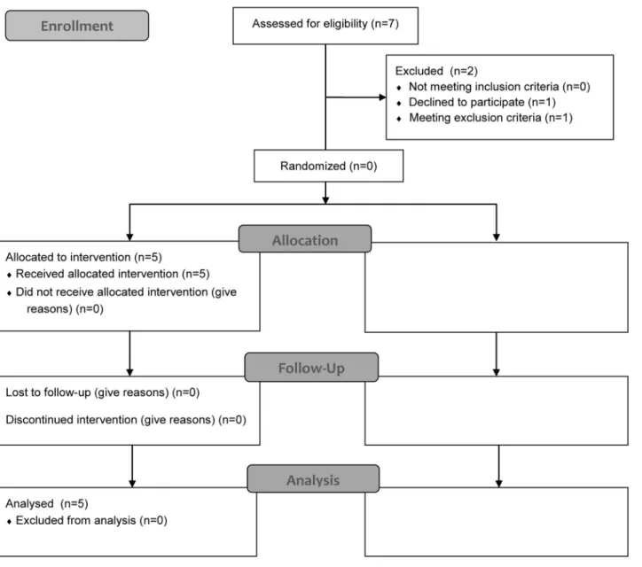
Patient number was low because of the intention of this pilot study to proof the concept of pharmacological stimulation of the nitric oxide pathway by multiple and also invasive assess-ments. The patient disposition of this pilot- trial is illustrated inFig 1.
Treatment and dose selection
L-argininehydrochloride drinking solution (L-argininehydrochloride, Selectchemie Zürich, Switzerland) was administered daily as an oral dose of 3 x 2.5g (t.i.d.) over the study period of 16 weeks. The arginine dose is in line with previous studies that reported a doubling of L-arginine serum concentration in children aged 7 to 17 years [25,26].
Metformin is an oral biguanide antidiabetic drug approved by Swissmedic for insulin resis-tance and type 2 diabetes. Metformin-associated lactic acidosis is a very rare but relevant side
Fig 1. Study profile according CONSORT flow chart.
effect only occurring in patients with impaired renal function. Metformin may cause mild and transient gastrointestinal side effects but no severe side effects in otherwise healthy subjects. Metformin (Sandoz Pharmaceuticals AG, Rotkreuz, Switzerland) was given twice daily at a dose of 2x250 mg (b.i.d) over the study period of 16 weeks.
Medication was administered at home by the parents. To assess compliance of medication intake empty and full vials and blisters were returned by the patients at the end of the 16 week study period.
Study design and safety
At screening, patients fulfilling all inclusion criteria and no exclusion criteria and giving informed consent were enrolled into the study. At baseline and after completion of the trial (week 16), muscle metabolism was assessed in all patients by a punch biopsy of vastus lateralis muscle and indirect calorimetry. Furthermore, quantitative thigh muscle MRI, whole-body Dual-Energy X-Ray absorptiometry (DEXA), clinical assessments, and laboratory parameters were performed. Treatment was started one day after baseline visit. For safety reasons selected laboratory parameters and vital signs were assessed subsequently (at week 2, 4, and 8) and at the final visit at week 16. All parameters at baseline and at week 16 were performed in the same order and time schedule to minimize measuring errors.
Clinical assessment of muscle function
Clinical response to treatment was assessed by the motor function measure (MFM), which was performed by certified physiotherapists and is a validated clinical score assessing both ambula-tory and non-ambulaambula-tory patients [27]. This measure was chosen to facilitate the assessment of patients who might lose independent ambulation during the trial. The MFM is recommended in the guidelines on the clinical investigation of medicinal products for the treatment of DMD published by the European Medicines Agency [28]. In addition, timed motor performance tests (2 min walking distance, timed Gower`s maneuver and 10 meter walking time) were employed [29].
Indirect calorimetry and DEXA
After intake of a standardized meal and a fasting period of at least 12 hours indirect calorimetry was performed (Deltatrac II, MBM-200 Metabolic Monitor) [30]. DEXA was performed to determine body’s content of bone, fat, and muscle. This technique uses two slightly diverging sources of X radiation for tissue differentiation. The irradiation dose was 1–2μSv (Hologic
QDR 4500 A (S/N 45959) [31].
Magnetic resonance imaging (MRI)
To evaluate the muscle fat fraction (MFF) and healthy muscle fraction (HMF), a two-point Dixon method (2PD) was used [32]. MRI was performed on a 1.5 Tesla scanner (Magnetom Avanto, Siemens Healthcare, Erlangen, Germany) as described [33].
Muscle biopsy analysis
Tissue was frozen and stored at -80°C until processing. To assess markers of nitric oxide levels, mitochondrial biogenesis, and ROS-induced damage we measured the levels of cGMP, nitrotyrosine, carbonylated proteins, and the levels of different mitochondrial respiratory complexes by ELISA or Western blot respectively. Each analysis was repeated three times for each muscle biopsy sample. Two different protein extractions were performed: one proce-dure without detergent to be used for the ELISA format and a detergent-based one adapted for muscle tissues to be used in western blot detection. Protein content of extracts was mea-sured using the Lowry based DC Protein Assay (Bio-Rad Laboratories, U.S.). For measuring the levels of cGMP in muscle extracts a commercial cGMP ELISA Kit (Cell Biolabs Inc., San Diego, Ref: STA-505) was used following the manufacturer’s instructions. The nitrotyrosine content was quantified using a colorimetric OxiSelect Nitrotyrosine ELISA Kit (Cell Biolabs Inc., San Diego, Ref: STA-305) according to the manufacturer’s recommendations. Carbony-lated proteins were quantified using a commercial kit (OxiSelect Protein Carbonyl ELISA Kit;Cell Biolabs Inc., San Diego, Ref: STA-310) following the manufacturer’s manual. The optical densities of the substrate product were measured at 450nm wavelength using a Spec-traMAX 190 (Molecular Devices LLC, USA) plate reader / photospectrometer and SoftMax Pro v4.8 (Molecular Devices LLC, USA) software. To detect and measure the OXPHOS com-plexes in the muscle extracts, samples of 20 g of protein were applied and separated on a 12% acrylamide gel, using a Mini PROTEAN 3 System from Bio-Rad Laboratories, U.S. Separated proteins were transferred on Immobilon-FL PVDF transfer membrane with a pore size of 0.45μm (Millipore Inc., Ref: IPFL00010). The blotted membranes were blocked in a
Top-Block (LubioScience, Ref: TB232010, Lucerne, CH) containing solution and then incubated o/n at 4°C with the MitoProfile Total OXPHOS mouse monoclonal antibody cocktail (Abcam plc, Ref: ab110411, Cambridge, U.S.) at the manufacturer’s suggested dilution. Addi-tionally, a rabbit-anti-GAPDH antibody (Sigma, Ref: G9545, Missouri, U.S.) at a dilution of 1:5000 and a rabbit-anti-α-Skeletal Muscle Actin (ACTA) antibody (Abcam plc, Ref:
ab113417, Cambridge, U.S.) at a dilution of 1:3500 were added to the incubation solution containing TopBlock. After washing the blots in Tris-Buffered Saline and Tween 20 (TBS-T) a mix of Goat-anti-mouse IgG AlexaFluor680 coupled 2nd antibody (Molecular Probes Ref: A21057, Life Technologies, Eugene, U.S.) at a dilution of 1:7500 and Goat-anti-Rabbit IgG IRDye800 coupled 2nd antibody (Rockland Inc., Ref: 611-132-122, Gilbertsville, U.S.) at a dilution of 1:7500 were incubated for 50 min. at RT. After washing the membranes the fluo-rescence was scanned on an Odyssey infrared scanner (LICOR Biosciences GmbH, Bad Homburg, Germany) using the Odyssey 2.1 software configured with identical settings for all scans and measurements. The bands were automatically identified and measured by the same software using identical settings for all blots and all samples. Every sample was pro-cessed and measured in three different experiments under the same conditions. The inte-grated intensity of these three measurements was averaged and the mean value of three measurements was used as single value for each patient sample.
Statistical analysis
Statistical analyses were performed using SPSS 22 (IBM, Statistical Package for the Social Sci-ences). For comparisons between controls and Duchenne patients the nonparametric Mann–
Results
Safety, tolerability, and laboratory testing
No severe adverse events occurred and no patient dropped out of the study. During the first week of treatment four of the five patients suffered from mild diarrhea, a known side effect of metformin, but symptoms resolved spontaneously. Slightly increased mean resting L-arginine plasma concentrations and global arginine bioavailability were detected, which did not reach significance. Repeated safety assessments showed fluctuating levels (up to + /−50% individual
changes) of individual creatine kinase and associated transaminase concentrations. No consis-tent changes in L-arginine related amino acids, liver and renal function tests, and markers of carbohydrate and lipid metabolism or changes in creatine kinase levels were evident. Baseline and post-treatment patient characteristics, safety, and laboratory values are shown inTable 1.
Muscle biopsies
To evaluate whether treatment with arginine and metformin was able to modulate muscle NO and mitochondrial content, different markers of NO, OXPHOS, and ROS pathways were assessed in vastus lateralis muscle tissue. Protein content from patient biopsy material was in the range of 5 to 35.5 mg (mean value = 12.98 mg) while control biopsy tissues yielded 18.9 mg to 47 mg (mean value = 35.03 mg) total protein. ELISA extracts from one patient (J6 after
Table 1. Summary of basic characteristics and laboratory data.
DMD Pretreatment DMD Posttreatment Change
Mean (SD) Mean (SD) Mean % p–value*
Basic characteristics
Age, years 7.9 0.4 8.2 0.4 0.3 -
-Height, m 1.22 0.1 1.24 0.1 0.0 2% 0.109
Weight, kg 22.7 3.0 22.8 3.1 0.2 1% 0.104
BMI, kg / m2 15.2 2.0 14.9 1.7 -0.3 -2% 0.225
Laboratory values
L-arginine,μmol/l 68 25 82 52 14 20% 1.000
L-citrulline,μmol/l 29 10 24 10 -5 -18% 0.273
L-ornithine,μmol/l 65 19 70 17 5 8% 1.000
GAB 0.7 0.1 0.9 0.7 0.2 26% 0.465
Urea 5,5 0,8 5,4 0,5 -0,1 -2% 0.581
ASAT, mmol/l 297 84 345 106 48 16% 0.043
ALAT, mmol/l 475 171 582 247 107 23% 0.225
Creatinekinase, U/l 11637 4085 13082 3693 1445 12% 0.138
Creatinine,μmol/l 15.2 2.9 15.8 1.9 0.6 4% 0.317
Glucose, mmol/l 4.7 0.2 4.8 0.6 0.1 2% 0.705
Triglycerides, mmol/l 1.2 0.3 0.9 0.3 -0.3 -25% 0.109
Cholesterine, mmol/l 4.1 0.7 4.0 0.8 -0.1 -2% 0.276
HDL cholesterine, mmol/l 1.2 0.1 1.2 0.2 0.0 1% 0.892
LDL cholesterine, mmol/l 2.4 0.7 2.4 0.9 0.0 1% 0.893
Adiponectinμg/ml 7.6 4.2 7.8 5.0 0.3 3% 0.498
Leptinμg/l 0.3 0.3 0.5 0.7 0.2 80% 0.786
GAB = global arginine bioavaliability, defined as L-arginine /(L-ornithine + L-citrulline) *Pvalues were calculated using the Wilcoxon signed-rank test
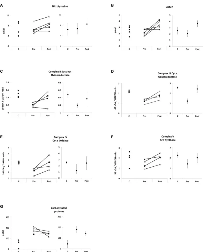
treatment) was contaminated with Tissue-Tek© optimum cutting temperature (OCT) com-pound. Repeated analyses of control muscle with and without OCT contamination revealed that OCT did not interfere with the cGMP and nitrotyrosine ELISA but with carbonylated pro-tein ELISA (not shown). Therefore, carbonyl ELISA analysis contained 4 sample pairs only. Western blot extracts of one biopsy (MA3 after treatment) did not include a sufficient amount of protein therefore OXPHOS expression could only be analyzed in four out of five sample pairs. Muscle biopsy results (mean values, standard error of the mean, changes, andp-values) are shown inTable 2, individual changes inFig 2.
While at baseline, mean cGMP and nitrotyrosine concentrations did not differ significantly between DMD and control muscle (Table 2,Fig 2A and 2B), distinct impaired mitochondrial protein expression in DMD muscle was evident (Table 2,Fig 2C–2F). Mean OXPHOS protein / GAPDH ratios of complexes II, III, IV, and V were all between -37 and -58% lower in DMD (p<0.05) compared to control muscles. Carbonylated proteins were four times higher
com-pared to control samples (p= 0.016) demonstrating high levels of oxidative stress in DMD muscle (Table 2,Fig 2G).
After 16 weeks of treatment with L-arginine and metformin, a significant increase of mean nitrotyrosine concentrations by 26% (p= 0.043) and a consistent, but not significant cGMP increase of 58% (p= 0.068) were observed in DMD patients. In line with a role for cGMP to control mitochondrial content, a relevant increase of all OXPHOS complexes between 41 to 99% was detected after treatment, albeit the increased expression observed for complexes II (85%,p= 0.144), III (47%,p= 0.068), IV (99%,p= 0.068) and V (41%,p= 0.068) was not sig-nificant (Table 2,Fig 2D and 2F). This change of mitochondrial content was associated with a trend towards reduced levels of carbonylated proteins in DMD patients (19% reduction after treatment (p= 0.144)) (Table 2,Fig 2G). Individual analysis of nitrotyrosine, cGMP, OXPHOS, and ROS concentrations showed consistent responses for the non-significant changes, too.
As expected from the molecular changes observed above, cGMP levels correlated positively with the expression of mitochondrial protein complexes III (R = 0.66) and IV (R = 0.57) in DMD patients. Furthermore, cGMP concentrations after treatment showed a positive correla-tion with nitrotyrosine concentracorrela-tion (R = 0.99), and positively correlated with changes of
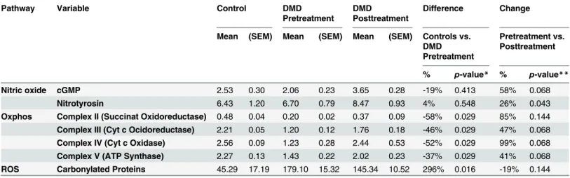
Table 2. Muscle biopsy analysis.
Pathway Variable Control DMD
Pretreatment
DMD
Posttreatment
Difference Change
Mean (SEM) Mean (SEM) Mean (SEM) Controls vs. DMD Pretreatment
Pretreatment vs. Posttreatment
% p-value* % p-value**
Nitric oxide cGMP 2.53 0.30 2.06 0.23 3.65 0.28 -19% 0.413 58% 0.068
Nitrotyrosin 6.43 1.20 6.70 0.79 8.47 0.93 4% 0.548 26% 0.043
Oxphos Complex II (Succinat Oxidoreductase) 0.48 0.04 0.20 0.02 0.37 0.09 -58% 0.029 85% 0.144
Complex III (Cyt c Ocidoreductase) 2.21 0.05 1.20 0.12 1.76 0.18 -46% 0.029 47% 0.068
Complex IV (Cyt c Oxidase) 2.56 0.09 1.23 0.28 2.44 0.53 -52% 0.029 99% 0.068
Complex V (ATP Synthase) 2.27 0.13 1.43 0.22 2.02 0.23 -37% 0.029 41% 0.068
ROS Carbonylated Proteins 45.29 17.19 179.10 15.32 145.34 10.52 296% 0.016 -19% 0.144
DMD = Duchenne Muscular Dystrophy; cGMP = cyclic guanosinmonophosphat; OXPHOS = oxidative phosphorylation; ROS = reactive oxygen species *Pvalues were calculated using the Mann-Whitney U test
**Pvalues were calculated using the Wilcoxon signed-rank test
Fig 2. Muscle biopsy.Muscle biopsy findings, individual control (C) concentrations and individual DMD changes before (PRE) and after (POST) treatment as well as point estimates with 95% confidence intervals. A = nitrotyrosine ELISA, concentration is provided in nmol / 7.9μg protein, B = cGMP ELISA, concentration is provided in pmol / 6.4μg protein, C = western blot succinat oxidoreductase (complex II) / GAPDH ratio, D = western blot cytochrome c oxidoreductase (complex III) / GAPDH ratio, E = western blot cytochrome c oxidase (Complex IV) / GAPDH ratio, F = western blot ATP synthase (Complex V) / GAPDH ratio, G = carbonylated protein ELISA (nmol / 1μg protein).
complex III (R = 0.86) and complex IV (R = 0.92) concentrations. In DMD muscles, cGMP (R = -0.33), complex III (R = -0.46), and complex IV (R = -0.39) concentrations showed nega-tive correlation with carbonylated proteins, indicating that cGMP-dependent increase in mito-chondrial content is not associated with oxidative stress in DMD.
Indirect calorimetry, DEXA, and quantitative muscle MRI
To assess the metabolic consequences of stimulated mitochondrial protein expression indirect
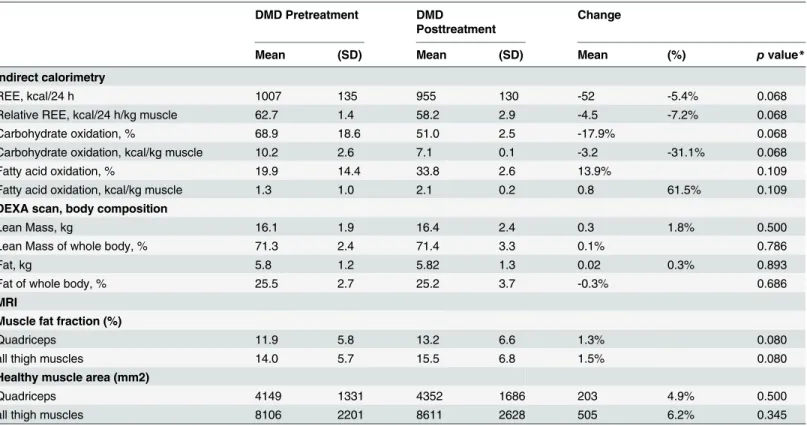
in vivocalorimetry was performed in DMD patients before and after treatment. Only data from four patients were analyzed before and after treatment, since one patient failed to finish the first examination (before treatment). A considerable, but statistically not significant decrease of the relative resting energy expenditure (REE) / kg muscle (mean change 7.2%,
p= 0.068), associated with a clear change of energy substrate use (Table 3,Fig 3A) was detected after treatment. Treatment decreased the mean relative carbohydrate oxidation rate (Fig 3B) while increasing the mean relative fatty acid oxidation (Fig 3C).
It is important to point out that in line with increased consumption of fatty acids, we also did not observe a significant worsening of the muscle-to-fat ratio in treated patients. DEXA scans showed that average whole body lean content and fat content did not change signifi-cantly. Quantitative muscle MRI did not show relevant changes to the muscle fat fraction (MFF) measured by the 2PD method after 16 weeks of treatment. Though not significant, there was an unexpected trend towards increased healthy muscle area (HMA) of all thigh muscles by 6.2% (Table 3).
Table 3. Summary of indirect calorimetry, DEXA, and muscle MRI data.
DMD Pretreatment DMD
Posttreatment
Change
Mean (SD) Mean (SD) Mean (%) pvalue*
Indirect calorimetry
REE, kcal/24 h 1007 135 955 130 -52 -5.4% 0.068
Relative REE, kcal/24 h/kg muscle 62.7 1.4 58.2 2.9 -4.5 -7.2% 0.068
Carbohydrate oxidation, % 68.9 18.6 51.0 2.5 -17.9% 0.068
Carbohydrate oxidation, kcal/kg muscle 10.2 2.6 7.1 0.1 -3.2 -31.1% 0.068
Fatty acid oxidation, % 19.9 14.4 33.8 2.6 13.9% 0.109
Fatty acid oxidation, kcal/kg muscle 1.3 1.0 2.1 0.2 0.8 61.5% 0.109
DEXA scan, body composition
Lean Mass, kg 16.1 1.9 16.4 2.4 0.3 1.8% 0.500
Lean Mass of whole body, % 71.3 2.4 71.4 3.3 0.1% 0.786
Fat, kg 5.8 1.2 5.82 1.3 0.02 0.3% 0.893
Fat of whole body, % 25.5 2.7 25.2 3.7 -0.3% 0.686
MRI
Muscle fat fraction (%)
Quadriceps 11.9 5.8 13.2 6.6 1.3% 0.080
all thigh muscles 14.0 5.7 15.5 6.8 1.5% 0.080
Healthy muscle area (mm2)
Quadriceps 4149 1331 4352 1686 203 4.9% 0.500
all thigh muscles 8106 2201 8611 2628 505 6.2% 0.345
REE = Resting Energy Expenditure; DEXA = Dual-Energy X-Ray absorptiometry; 2PD = two point Dixon method; MFF = muscle fat fraction *Pvalues were calculated using the Wilcoxon signed-rank test
Fig 3. Indirect calorimetry.Indirect calorimetry results demonstrating consistently decreased rates of resting energy expenditure per kg muscle in 24 hours (A), reduced usage of carbohydrates as oxidative fuel (B), and increased usages of fatty acids (C) in oxidative energy metabolism.
Clinical scores
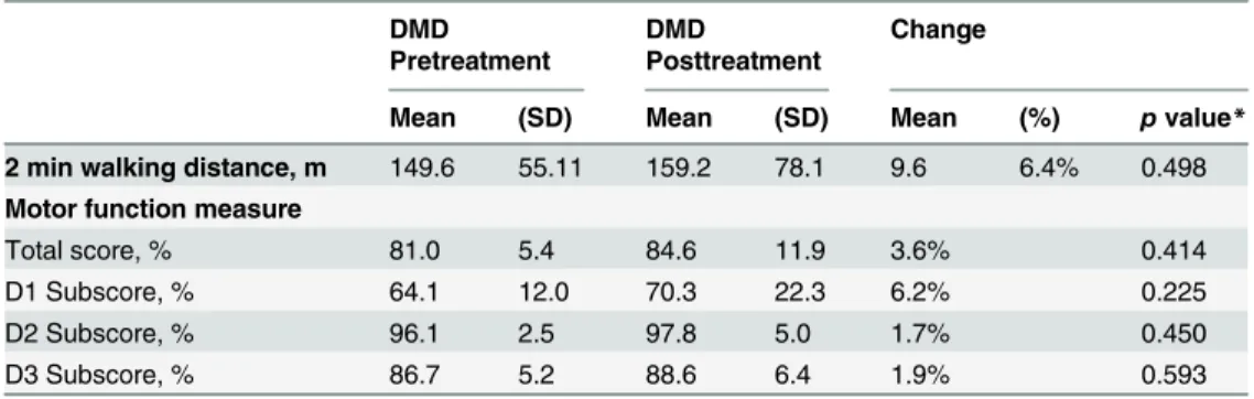
In four of the five treated patients motor function and walking distances improved, while the oldest and most severely affected patient worsened after the treatment period. The mean total MFM and the MFM D1 subscore (standing and transfers) improved in the treated DMD patients over the period of 16 weeks by 3.6% (total MFM) and +6.2% (MFM D1). Also, the mean two minute walking distance improved (+9.6 meter) (Table 4). Individual changes are presented inFig 4.
Discussion
While patients with primary mitochondrial dysfunction disorders frequently display impaired muscle function [3], mitochondrial dysfunction is not generally considered to play a major role in the pathophysiology of DMD. However, our baseline data demonstrate a significantly reduced expression of mitochondrial proteins in DMD muscle samples, confirming the pres-ence of severe mitochondrial abnormalities in DMD. Children with DMD have elevated syn-thesis of ADMA, an endogenous nNOS inhibitor, diminished arginine synsyn-thesis, and reduced NO bioavailability compared to healthy children [17]. Aim of this proof-of-concept investiga-tor driven pilot-trial was to examine if increasing intramuscular NO concentrations would ameliorate DMD via stimulation of mitochondrial biogenesis. Therefore, we treated five ambu-latory DMD patients for 16 weeks with L-arginine [34] a NO precursor, and metformin, a pharmacological AMPK activator and by this, indirect stimulator of nNOS [22,35,36]. Our treatment significantly increased nitrotyrosine concentrations and led to consistent cGMP increases in skeletal muscle biopsies of DMD patients indicating an increased NO formation in DMD muscle. In addition, a consistent increase of mitochondrial protein expression was observed. cGMP concentrations were positively correlated with mitochondrial protein concen-trations in DMD muscle. Even more important, we also observed a positive correlation between the increase in NO concentration markers and mitochondrial protein expression, indicating a direct relation between intramuscular NO and subsequent mitochondrial protein expression in DMD muscle. It is often thought that NO, in general, might be harmful to skele-tal muscle. NO generated by inducible nitric oxide synthase (iNOS) induces muscle atrophy via regulation of several transcription factors [37]. NO can react with superoxide anions (O2-) to form the toxic molecule peroxynitrite (ONOO-), which is believed to increase oxidative stress and muscle damage [38]. In contrast, neuronal and endothelial nitric oxide synthase
Table 4. Summary of clinical data. DMD Pretreatment
DMD
Posttreatment
Change
Mean (SD) Mean (SD) Mean (%) pvalue*
2 min walking distance, m 149.6 55.11 159.2 78.1 9.6 6.4% 0.498
Motor function measure
Total score, % 81.0 5.4 84.6 11.9 3.6% 0.414
D1 Subscore, % 64.1 12.0 70.3 22.3 6.2% 0.225
D2 Subscore, % 96.1 2.5 97.8 5.0 1.7% 0.450
D3 Subscore, % 86.7 5.2 88.6 6.4 1.9% 0.593
D1 Subscore = standing and transfers, D2 Subscore = axial and proximal motor capacity, D3 Subscore = distal motor capacity
*Pvalues were calculated using the Wilcoxon signed-rank test
Fig 4. Clinical changes.Clinical changes over treatment period of 16 weeks are shown. Four of the five patients showed improvements in 2 min walking distance (A), the MFM total score (B) and the MFM D1 subscore (C).
(nNOS and eNOS, respectively) are expressed in the skeletal muscle and released NO in physi-ological amounts is known to protect cells from damage due to increased oxidative stress. There is no evidence that exclusively nNOS is stimulated in our study but the increase of NO markers but the parallel decrease of ROS markers in DMD muscle after treatment as well as the negative correlation of cGMP to carbonylated proteins indicates that L-arginine and met-formin treatment decreases oxidative stress in muscle. If otherwise iNOS derived NO predomi-nated to the observed CGMP and nitrotyrosine we would have expected an increase of
of annual muscle fat fraction (MFF) increase in thigh muscles (10.1% in quadriceps) in age-matched, untreated, ambulatory DMD patients[45]. While age-matched ambulant DMD patients typically show a reduction of -17.8% per year (quadriceps -19.5%), in our treated patients we surprisingly observed a trend towards an increase in healthy muscle area of 5.9% (quadriceps +4.7%); this should be further examined in a larger scale clinical trial.
In addition to the approach utilized in this trial, other possible NO-based strategies to treat DMD might exist. Since a major function of nNOS-derived NO is to stimulate cGMP produc-tion, it is conceivable that impaired nNOS function might be partially restored using phospho-diesterase 5 (PDE5) inhibitors such as sildenafil and tadalafil. In line with this model, PDE5 inhibition has been shown to increase cytosolic cGMP [46]. However, PDE5 inhibition using sildenafil could not show improved muscle function in Becker muscular dystrophy [47]. Another cGMP-independent, NO-dependent pathway involves peroxynitrite (ONOO-) forma-tion, which is important for muscle protein synthesis and muscle hypertrophy [48]. Another potential strategy to stimulate NO-formation in DMD is the use of NO donors. In the mdx mouse model, a combination of ibuprofen and ISDN (isosorbide dinitrate, a NO donor) reduced muscle necrosis and inflammation and improved voluntary movements and resistance to exercise [49]. In contrast however, the combined approach of an NO-donating NSAID in human DMD patients resulted in no significant clinical improvement [50]. As our approach interferes more upstream in this pathway, it can also positively influence cGMP-independent NO-pathways. Finally, the advantage of L-arginine or L-citrulline is to show fewer side effects compared to PDE5 inhibitors that are typically associated with side effects such as headaches. Our study has clear limitations due to the small number of patients, the short observation period, and the lack of placebo-treated controls. Unfortunately direct measurements of NO are not possible. As a consequence indirect NO markers have to be used. As nitrite / nitrate are only reliably measurable by mass spectroscopy we analysed cGMP (as second messenger of NO) and nitrotyrosine that increases when NO concentration increase. This approach has lim-itations as cGMP can also be influenced by other substances or phosphodiesterase inhibition. The measurement of nitrotyrosine has limitations, too. Nitrotyrosine levels can also be elevated during inflammation and increased oxidative stress when inducible NOS (iNOS) is activated leading to elevated NO concentrations. As L-arginine and metformin treatment decreased car-bonylated proteins (marker of oxidative stress) the increase of cGMP and nitrotyrosine is unlikely to be linked to iNOS and inflammation, but this has not been proven. A further limita-tion of our study is that, despite the increase of cGMP, nitrotyrosine, and mitochondrial pro-teins after treatment we can’t exclude the possibility that the effects seen in our study may have also partially resulted from L-arginine effects beyond NO synthesis. L-arginine is involved in many physiological processes as recently investigated by Kayacelebi et al [51]. Thus other path-ways could have been involved and could have contributed to the observed effects on muscle function. For example L-Arg is the substrate of AGAT which produces guanidine acetate, the substrate of GAMT and precursor of creatine, which can improve muscle energetics via increased phosphocreatine concentrations. One patient was treated with steroids. The treat-ment with metformin and L-arginine showed the same clinical and paraclinical results in this boy as in the other“responders”. However, a synergistic or additive effect to steroids is possible but cannot be substantiated or excluded at this time.
in DMD patients [52] to validate our therapeutic approach. Additionally we perform a pilot trial to extend this approach to ambulant patients with Becker muscular dystrophy [53].
Supporting Information
S1 Appendix. CONSORT checklist. (PDF)
S2 Appendix. Study protocol in German. (PDF)
S3 Appendix. Study protocol in English. (PDF)
Author Contributions
Conceived and designed the experiments: DF PH UB AF NG M. Sinnreich OB SD. Performed the experiments: PH UB M. Schmid ER CN UP DR. Analyzed the data: PH UB DF BE TP SF MG OB AF M. Sinnreich. Wrote the paper: PH DF.
References
1. Yoshida M, Ozawa E. Glycoprotein complex anchoring dystrophin to sarcolemma. Journal of biochem-istry. 1990; 108(5):748–52. PMID:2081733.
2. Campbell KP, Kahl SD. Association of dystrophin and an integral membrane glycoprotein. Nature. 1989; 338(6212):259–62. doi:10.1038/338259a0PMID:2493582.
3. Smeitink J, van den Heuvel L, DiMauro S. The genetics and pathology of oxidative phosphorylation. Nature reviews Genetics. 2001; 2(5):342–52. doi:10.1038/35072063PMID:11331900.
4. Scholte HR, Luyt-Houwen IE, Busch HF, Jennekens FG. Muscle mitochondria from patients with Duchenne muscular dystrophy have a normal beta oxidation, but an impaired oxidative phosphoryla-tion. Neurology. 1985; 35(9):1396–7. PMID:4022395.
5. Kemp GJ, Taylor DJ, Dunn JF, Frostick SP, Radda GK. Cellular energetics of dystrophic muscle. Jour-nal of the neurological sciences. 1993; 116(2):201–6. PMID:8393092.
6. Sperl W, Skladal D, Gnaiger E, Wyss M, Mayr U, Hager J, et al. High resolution respirometry of permea-bilized skeletal muscle fibers in the diagnosis of neuromuscular disorders. Molecular and cellular bio-chemistry. 1997; 174(1–2):71–8. PMID:9309668.
7. Kuznetsov AV, Winkler K, Wiedemann FR, von Bossanyi P, Dietzmann K, Kunz WS. Impaired mito-chondrial oxidative phosphorylation in skeletal muscle of the dystrophin-deficient mdx mouse. Molecu-lar and celluMolecu-lar biochemistry. 1998; 183(1–2):87–96. PMID:9655182.
8. Braun U, Paju K, Eimre M, Seppet E, Orlova E, Kadaja L, et al. Lack of dystrophin is associated with altered integration of the mitochondria and ATPases in slow-twitch muscle cells of MDX mice. Biochi-mica et biophysica acta. 2001; 1505(2–3):258–70. PMID:11334790.
9. Millay DP, Sargent MA, Osinska H, Baines CP, Barton ER, Vuagniaux G, et al. Genetic and pharmaco-logic inhibition of mitochondrial-dependent necrosis attenuates muscular dystrophy. Nature medicine. 2008; 14(4):442–7. doi:10.1038/nm1736PMID:18345011; PubMed Central PMCID: PMC2655270.
10. Hankard R, Gottrand F, Turck D, Carpentier A, Romon M, Farriaux JP. Resting energy expenditure and energy substrate utilization in children with Duchenne muscular dystrophy. Pediatric research. 1996; 40(1):29–33. Epub 1996/07/01. doi:10.1203/00006450-199607000-00006PMID:8798242.
11. Gaeta M, Messina S, Mileto A, Vita GL, Ascenti G, Vinci S, et al. Muscle fat-fraction and mapping in Duchenne muscular dystrophy: evaluation of disease distribution and correlation with clinical assess-ments. Preliminary experience. Skeletal radiology. 2012; 41(8):955–61. Epub 2011/11/10. doi:10. 1007/s00256-011-1301-5PMID:22069033.
12. Hollingsworth KG, Garrood P, Eagle M, Bushby K, Straub V. Magnetic resonance imaging in Duchenne muscular dystrophy: longitudinal assessment of natural history over 18 months. Muscle & nerve. 2013; 48(4):586–8. doi:10.1002/mus.23879PMID:23620230.
14. MacAllister RJ, Whitley GS, Vallance P. Effects of guanidino and uremic compounds on nitric oxide pathways. Kidney international. 1994; 45(3):737–42. PMID:7515129.
15. Nisoli E, Carruba MO. Nitric oxide and mitochondrial biogenesis. Journal of cell science. 2006; 119(Pt 14):2855–62. doi:10.1242/jcs.03062PMID:16825426.
16. McGee SL, Hargreaves M. AMPK-mediated regulation of transcription in skeletal muscle. Clin Sci (Lond). 2010; 118(8):507–18. doi:10.1042/CS20090533PMID:20088830.
17. Horster I, Weigt-Usinger K, Carmann C, Chobanyan-Jurgens K, Kohler C, Schara U, et al. The L-argi-nine/NO pathway and homoarginine are altered in Duchenne muscular dystrophy and improved by glu-cocorticoids. Amino acids. 2015; 47(9):1853–63. doi:10.1007/s00726-015-2018-xPMID:26066683.
18. Garbincius JF, Michele DE. Dystrophin-glycoprotein complex regulates muscle nitric oxide production through mechanoregulation of AMPK signaling. Proceedings of the National Academy of Sciences of the United States of America. 2015; 112(44):13663–8. doi:10.1073/pnas.1512991112PMID: 26483453; PubMed Central PMCID: PMC4640723.
19. Jahnke VE, Van Der Meulen JH, Johnston HK, Ghimbovschi S, Partridge T, Hoffman EP, et al. Meta-bolic remodeling agents show beneficial effects in the dystrophin-deficient mdx mouse model. Skeletal muscle. 2012; 2(1):16. doi:10.1186/2044-5040-2-16PMID:22908954; PubMed Central PMCID: PMC3482394.
20. Ljubicic V, Miura P, Burt M, Boudreault L, Khogali S, Lunde JA, et al. Chronic AMPK activation evokes the slow, oxidative myogenic program and triggers beneficial adaptations in mdx mouse skeletal mus-cle. Human molecular genetics. 2011; 20(17):3478–93. doi:10.1093/hmg/ddr265PMID:21659335.
21. Pauly M, Daussin F, Burelle Y, Li T, Godin R, Fauconnier J, et al. AMPK activation stimulates autop-hagy and ameliorates muscular dystrophy in the mdx mouse diaphragm. The American journal of pathology. 2012; 181(2):583–92. doi:10.1016/j.ajpath.2012.04.004PMID:22683340.
22. Musi N, Hirshman MF, Nygren J, Svanfeldt M, Bavenholm P, Rooyackers O, et al. Metformin increases AMP-activated protein kinase activity in skeletal muscle of subjects with type 2 diabetes. Diabetes. 2002; 51(7):2074–81. PMID:12086935.
23. Ljubicic V, Jasmin BJ. Metformin increases peroxisome proliferator-activated receptor gamma Co-acti-vator-1alpha and utrophin a expression in dystrophic skeletal muscle. Muscle & nerve. 2015; 52 (1):139–42. doi:10.1002/mus.24692PMID:25908446.
24. Langone F, Cannata S, Fuoco C, Lettieri Barbato D, Testa S, Nardozza AP, et al. Metformin protects skeletal muscle from cardiotoxin induced degeneration. PloS one. 2014; 9(12):e114018. doi:10.1371/ journal.pone.0114018PMID:25461598; PubMed Central PMCID: PMC4252070.
25. Koga Y, Akita Y, Nishioka J, Yatsuga S, Povalko N, Tanabe Y, et al. L-arginine improves the symptoms of strokelike episodes in MELAS. Neurology. 2005; 64(4):710–2. doi:10.1212/01.WNL.0000151976. 60624.01PMID:15728297.
26. Bennett-Richards KJ, Kattenhorn M, Donald AE, Oakley GR, Varghese Z, Bruckdorfer KR, et al. Oral L-arginine does not improve endothelial dysfunction in children with chronic renal failure. Kidney interna-tional. 2002; 62(4):1372–8. Epub 2002/09/18. doi:10.1111/j.1523-1755.2002.kid555.xPMID: 12234308.
27. Berard C, Payan C, Hodgkinson I, Fermanian J. A motor function measure for neuromuscular diseases. Construction and validation study. Neuromuscular disorders: NMD. 2005; 15(7):463–70. Epub 2005/ 08/18. PMID:16106528.
28. http://www.ema.europa.eu/docs/en_GB/document_library/Scientific_guideline/2013/03/ WC500139508.pdf.
29. Mazzone E, Vasco G, Sormani MP, Torrente Y, Berardinelli A, Messina S, et al. Functional changes in Duchenne muscular dystrophy: a 12-month longitudinal cohort study. Neurology. 2011; 77(3):250–6. Epub 2011/07/08. doi:10.1212/WNL.0b013e318225ab2ePMID:21734183.
30. Elliott SA, Davidson ZE, Davies PS, Truby H. Predicting resting energy expenditure in boys with Duchenne muscular dystrophy. European journal of paediatric neurology: EJPN: official journal of the European Paediatric Neurology Society. 2012; 16(6):631–5. doi:10.1016/j.ejpn.2012.02.011PMID: 22497714.
31. Skalsky AJ, Han JJ, Abresch RT, Shin CS, McDonald CM. Assessment of regional body composition with dual-energy X-ray absorptiometry in Duchenne muscular dystrophy: correlation of regional lean mass and quantitative strength. Muscle & nerve. 2009; 39(5):647–51. doi:10.1002/mus.21212PMID: 19347922.
33. Fischmann A, Hafner P, Gloor M, Schmid M, Klein A, Pohlman U, et al. Quantitative MRI and loss of free ambulation in Duchenne muscular dystrophy. Journal of neurology. 2013; 260(4):969–74. Epub 2012/11/10. doi:10.1007/s00415-012-6733-xPMID:23138982.
34. de Castro Barbosa T, Jiang LQ, Zierath JR, Nunes MT. L-Arginine enhances glucose and lipid metabo-lism in rat L6 myotubes via the NO/ c-GMP pathway. Metabometabo-lism: clinical and experimental. 2013; 62 (1):79–89. doi:10.1016/j.metabol.2012.06.011PMID:22889511.
35. Ouyang J, Parakhia RA, Ochs RS. Metformin activates AMP kinase through inhibition of AMP deami-nase. The Journal of biological chemistry. 2011; 286(1):1–11. doi:10.1074/jbc.M110.121806PMID: 21059655; PubMed Central PMCID: PMC3012963.
36. Lee-Young RS, Griffee SR, Lynes SE, Bracy DP, Ayala JE, McGuinness OP, et al. Skeletal muscle AMP-activated protein kinase is essential for the metabolic response to exercise in vivo. The Journal of biological chemistry. 2009; 284(36):23925–34. doi:10.1074/jbc.M109.021048PMID:19525228; PubMed Central PMCID: PMC2781986.
37. Dudley RW, Danialou G, Govindaraju K, Lands L, Eidelman DE, Petrof BJ. Sarcolemmal damage in dystrophin deficiency is modulated by synergistic interactions between mechanical and oxidative/nitro-sative stresses. The American journal of pathology. 2006; 168(4):1276–87; quiz 404–5. doi:10.2353/ ajpath.2006.050683PMID:16565501; PubMed Central PMCID: PMC1606574.
38. Hall DT, Ma JF, Marco SD, Gallouzi IE. Inducible nitric oxide synthase (iNOS) in muscle wasting syn-drome, sarcopenia, and cachexia. Aging. 2011; 3(8):702–15. PMID:21832306; PubMed Central PMCID: PMC3184974.
39. Kruszelnicka O, Chyrchel B, Golay A, Surdacki A. Differential associations of circulating asymmetric dimethylarginine and cell adhesion molecules with metformin use in patients with type 2 diabetes melli-tus and stable coronary artery disease. Amino acids. 2015; 47(9):1951–9. doi: 10.1007/s00726-015-1976-3PMID:25859650.
40. Bestermann WH Jr. The ADMA-Metformin Hypothesis: Linking the Cardiovascular Consequences of the Metabolic Syndrome and Type 2 Diabetes. Cardiorenal medicine. 2011; 1(4):211–9. doi:10.1159/ 000332382PMID:22135630; PubMed Central PMCID: PMC3222110.
41. Radley-Crabb HG, Marini JC, Sosa HA, Castillo LI, Grounds MD, Fiorotto ML. Dystropathology increases energy expenditure and protein turnover in the mdx mouse model of duchenne muscular dys-trophy. PloS one. 2014; 9(2):e89277. doi:10.1371/journal.pone.0089277PMID:24586653; PubMed Central PMCID: PMC3929705.
42. Suhr F, Gehlert S, Grau M, Bloch W. Skeletal muscle function during exercise-fine-tuning of diverse subsystems by nitric oxide. International journal of molecular sciences. 2013; 14(4):7109–39. doi:10. 3390/ijms14047109PMID:23538841; PubMed Central PMCID: PMC3645679.
43. Vuillerot C, Girardot F, Payan C, Fermanian J, Iwaz J, De Lattre C, et al. Monitoring changes and pre-dicting loss of ambulation in Duchenne muscular dystrophy with the Motor Function Measure. Develop-mental medicine and child neurology. 2010; 52(1):60–5. Epub 2009/05/21. doi:10.1111/j.1469-8749. 2009.03316.xPMID:19453691.
44. Silva EC, Machado DL, Resende MB, Silva RF, Zanoteli E, Reed UC. Motor function measure scale, steroid therapy and patients with Duchenne muscular dystrophy. Arquivos de neuro-psiquiatria. 2012; 70(3):191–5. Epub 2012/03/07. PMID:22392111.
45. Bonati U, Hafner P, Schadelin S, Schmid M, Naduvilekoot Devasia A, Schroeder J, et al. Quantitative muscle MRI: A powerful surrogate outcome measure in Duchenne muscular dystrophy. Neuromuscular disorders: NMD. 2015; 25(9):679–85. doi:10.1016/j.nmd.2015.05.006PMID:26096788.
46. Percival JM, Whitehead NP, Adams ME, Adamo CM, Beavo JA, Froehner SC. Sildenafil reduces respi-ratory muscle weakness and fibrosis in the mdx mouse model of Duchenne muscular dystrophy. The Journal of pathology. 2012; 228(1):77–87. doi:10.1002/path.4054PMID:22653783; PubMed Central PMCID: PMC4067455.
47. Witting N, Kruuse C, Nyhuus B, Prahm KP, Citirak G, Lundgaard SJ, et al. Effect of sildenafil on skeletal and cardiac muscle in Becker muscular dystrophy. Annals of neurology. 2014; 76(4):550–7. doi:10. 1002/ana.24216PMID:25042931.
48. Ito N, Ruegg UT, Kudo A, Miyagoe-Suzuki Y, Takeda S. Activation of calcium signaling through Trpv1 by nNOS and peroxynitrite as a key trigger of skeletal muscle hypertrophy. Nature medicine. 2013; 19 (1):101–6. doi:10.1038/nm.3019PMID:23202294.
49. Sciorati C, Staszewsky L, Zambelli V, Russo I, Salio M, Novelli D, et al. Ibuprofen plus isosorbide dini-trate treatment in the mdx mice ameliorates dystrophic heart structure. Pharmacological research: the official journal of the Italian Pharmacological Society. 2013; 73:35–43. doi:10.1016/j.phrs.2013.04.009 PMID:23644256.
a safety study with pilot efficacy measures in adult dystrophic patients. Pharmacological research: the official journal of the Italian Pharmacological Society. 2012; 65(4):472–9. doi:10.1016/j.phrs.2012.01. 006PMID:22306844.
51. Kayacelebi AA, Langen J, Weigt-Usinger K, Chobanyan-Jurgens K, Mariotti F, Schneider JY, et al. Bio-synthesis of homoarginine (hArg) and asymmetric dimethylarginine (ADMA) from acutely and chroni-cally administered free L-arginine in humans. Amino acids. 2015; 47(9):1893–908. doi:10.1007/ s00726-015-2012-3PMID:26031828.
52. ClinicalTrials.govNCT01995032.