Session editor: Alfredo José Mansur
Associate editors: Desiderio Favarato Vera Demarchi Aiello
Mailing address: Alfredo José Mansur – InCor – Av. Dr. Enéas C. Aguiar, 44 – 05403-000 – São Paulo, SP - Brazil
Case 2 / 0 0 – A 1 5 -year-old male w ith progressive muscular dystrophy of the Becker type and severe heart failure - Instituto do Coração of the Hospital das Clínicas - FM USP
Clinicopathologic Session
The patient is a 15-year-old male admitted to the hospi-tal due to abdominal pain, vomiting, dark urine, and edema.
The patient had a history of good health until the age of 10 years when progressive muscular weakness, mainly in the lower limbs, began. He sought medical assistance and was diagnosed of progressive muscular dystrophy of the Becker type. His mother, a 34-year-old woman, is considered an asymptomatic carrier of the gene for progressive muscu-lar dystrophy, and his 4-year-old brother has signs of pro-gressive muscular dystrophy of the Duchenne type.
The patient also had bronchial asthma.
He reported fatigue on exertion six months earlier and daily crises of tachycardic palpitations 4 months earlier with cold sweating and perioral paleness of approximately 15 mi-nutes of duration. He denied syncope. Two months earlier, dyspnea became worse, being triggered on minimum exerti-on, and edema appeared. To the patient was prescribed 40mg of furosemide and 0.25mg of digoxin daily. One month earlier, the patient required hospitalization to control heart failure, and he was discharged with symptom improvement and the same medicamentous prescription. Chest X-ray at that time showed cardiomegaly (+++/4+), and the patient was then referred to InCor for medical treatment.
On physical examination (1/24/97), the patient was in a wheelchair, thin, eupneic, with a regular pulse of 92bpm, and blood pressure of 100/70mmHg. The precordium was mildly bulgy and the lung examination was normal. The ictus cordis was palpable on the 7th intercostal space, no thrill was audible,
the first cardiac sound was split on the tricuspid region, the 4th
cardiac sound was audible, and no murmur could be heard. The liver was palpable 1cm from the right costal margin and no ede-ma existed. All pulses were palpable and symmetric.
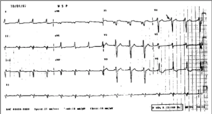
Electrocardiographic assessment (1/10/97) showed si-nus rhythm, heart rate of 100bpm a PR interval of variable duration 0.16s or 0.12s. Internimtent delta wave was pre-sent, QRS duration was 0.12s and the QRS axis was shifted upwards and to the left. The delta wave was positive in I, aVL, and from V2 to V6, and negative in II, III, aVF and V1 (fig. 1). The diagnosis (was preexcitation of Wolf-Parkin-son-White syndrome).
A chest X-ray showed cardiomegaly (+++/4+) and a mild increase in the pulmonary trunk.
The medications being used were maintained and 12.5mg daily of captopril was added. Assessment of cardiac function and arrhythmias was started.
Three months later (4/11/97), the patient sought medi-cal assistance due to edema, which had started in the lower limbs and had proceeded to the face in the 2 previous mon-ths, with cough and hemoptysis, reduction in appetite, and weight loss. Two weeks earlier, in addition to edema, pain in the right hypochondrium, vomiting, and dark urine appea-red. The patient denied dyspnea and fecal acholia.
On physical examination (4/11/97), the patient was in regular condition, eupneic, with a regular pulse of 100bpm and blood pressure of 100/70mmHg. Lung examination sho-wed reduction in the respiratory sounds in both bases and no rales. Heart examination showed no abnormal cardiac so-unds and no murmurs. The liver was palpated 5cm from the right costal margin.
The electrocardiogram (4/12/97) showed sinus rhy-thm, heart rate of 125bpm, QRS axis of +140° parallel, left and right atrial hypertrophy and left and right ventricular hyper-trophy.
The laboratory tests at admission are shown in table I. The echocardiogram (4/13/97) showed left ventricular diastolic and systolic diameters of 58mm and 52mm, respec-tively, and a circumferential shortening fraction of 10%. The left ventricle was diffusely hypokinetic and signs of a throm-bus of 23mm were detected in the left ventricular apex.
Ventilation and perfusion pulmonary scintigraphies (4/ 14/97) showed hypoperfusion in the right pulmonary middle 1/3 and base, and in smaller areas of the lateral segment of the middle lobe, and part of the anterior basal and posterior basal segments of the lower lobe of the same lung. In the ventilation scintigraphy as compared with the perfusion study, a concordant pattern was observed in the lateral segment of the middle lobe and a discordant pattern in the remaining segments. Those images suggested right pulmonary thromboembolism with a probable infarct in the middle lobe. The patient evolved with clinical improvement, requi-ring a continuous use of dobutamine and dopamine, in addi-tion to 80 mg of furosemide daily. On April 4th, 24,000 units of
heparin through intravenous via were started.
On the night of April 20th, the patient had general
tonic-clonic convulsions followed by bradycardia and cardiac arrest that did not respond to resuscitation maneuvers and died.
the biceps brachii muscle and serial frozen sections were stained by hematoxylin and eosin and the modified Gomo-ri’s trichrome. The sections also underwent histochemical reactions, such as NADH, SDH, acid phosphatase, and 4.3 and 9.4 ATPase. The biopsy showed a variation in the dia-meter of the muscle fibers, presence of round hyaline fibers, degenerating fibers, predominance of type I fibers, and pro-liferation of the endo- and perimysial connective tissue.
Dystrophin analysis by immunostaining and the quantitative Western blot analysis showed a reduced amo-unt and abnormal molecular weight of that protein.
(Dr. Sueli Kazue Nagahashi Marie)
Discussion
Clinical features – The patient is a 15-year-old male
diagnosed with Becker muscular dystrophy and progressi-ve heart failure of recent onset.
On physical examination, the cardiac silhouette was
enlarged and the 4th cardiac sound was audible, indicating
cardiac dilation and myocardial dysfunction. These findin-gs were confirmed on echocardiographic analysis, which showed a low circumferential shortening fraction and diffu-se hypokinesia.
Appearance of edema in the lower limbs and face in association with pain in the right hypochondrium, vomi-ting, and enlargement of the liver on palpation, indicate ag-gravation of the heart failure and visceral congestion. Be-cause the serum levels of albumin, total proteins, and creati-nine were normal, the possibility of the edema being due to hypoproteinemia or renal failure was eliminated.
Another cause of vomiting could be the patient’s low cardiac output.
The facial edema could also be caused by an obstruc-tion of the superior vena cava; however, as enlargement of the liver occurred as well as edema of the lower limbs, the most probable cause is an increase in right ventricular filling pressures.
Hemoptysis could be a consequence of pulmonary
Fig. 1 - Electrocardiogram – preexcitation syndrome, intermittent delta waves.
Table I – Admission laboratory tests
Laboratory tests 4/11/97 4/14/97 4/20/97
Red blood cells/mm3 6.500.000 - 4.700.000
Hemoglobin g/dL 15.5 - 11.5
Hematocrit % 49 - 35
MCV ((m3) 75 - 74
MCHC (g/dL) 32 - 33
Leukocytes/mm3 11.100 - 12.200
Rod (%) 8 - 6
Segmented (%) 70 - 61
Eosinophils (%) 0 - 0
Basophils (%) 1 - 0
Lymphocytes (%) 20 - 28
Monocytes (%) 1 - 5
Platelets/mm3 273.000 - 448.000
Prothrombin time (s) 15.3 (N=12,2) 16.9 (12.2) 15.7 (12.2)
INR 1,62 1.99 1.29
APTT(s) 29.4 (N=28) 59.4 (28) 35.1 (28)
Fibrin dimer (N <500ng/mL) - positivo
-Fibrinogen (mg/dL) - 326(200-400)
-Activity of the factors II, VII and X (%) - 50 (100)
-Activity of factor V (%) - 76 (68-150)
-Platelet aggregation - normal
-Presence of lupus anticoagulant - negativa
-Urea(mg/dL) 49 - 40
Creatinine (mg/dL) 0.5 - 0.5
Total protein (g/dL) 6.9 -
-Albumin (g/dL) 3.7 -
-Total bilirubin (mg/dL) 1.4 -
Direct bilirubin (mg/dL) 0.62 -
-GOT/AST (U/L) 23 -
-GPT/ALT (U/L) 15 -
-Lactic dehydrogenase (U/L) - 341 (240)
-Sodium (mEq/L) 134 - 130
dystrophy, dystrophin is present but has an abnormal mole-cular weight.
Clinically, it is possible to distinguish the 2 forms of muscular dystrophy. Duchenne muscular dystrophy has the onset of its manifestations at about the age of 2 years, and it is rapidly progressive with loss of strength, mainly in the muscles of the pelvic and shoulder girdles (proximal muscles of the limbs), involving rather the lower limbs than the upper limbs. Waddling gait, frequent falls, pseudohy-pertrophy of the calves, lumbar lordosis, kyphoscoliosis, and shortening of the Achilles tendon are commonly found. Between the 8th and the 10th year of life, walking requires the
use of crutches and by the age of 12 most patients are confi-ned to wheelchairs.
Becker muscular dystrophy is slowly progressive and usually starts around the age of 5 years (from 5 to 15 years), but it may start in the 3rd or 4th decades of life.
Patients manage to walk until after the age of 15, which constitutes a clinical difference between the two forms of
muscular dystrophy 3.
Cardiac involvement occurs in both forms of muscular dystrophy. In Duchenne muscular dystrophy, heart failure is rapidly progressive, but it may stabilize after some time, and the only evidence of cardiac involvement may be
elec-trocardiographic alterations 4. Thoracic deformities may
render the clinical examination of the heart difficult. Pulmo-nary and systemic thromboembolism have been reported in the final stages of the disease.
Cardiac involvement is segmentary, with dystrophy of the posterobasal segment and lateral extension of the left ventricular wall. This may cause dysfunction of the postero-lateral papillary muscle and mitral regurgitation 5.
Electrocardiographic alterations are present from chil-dhood onward and consist of the following: appearance of wide R waves in the right leads, increase in the R/S relation, and Q waves in I, aVL, V5 and V6.
Inappropriate sinus tachycardia is the most common arrhythmia and, in the final phase of the disease, atrial flutter is the most common supraventricular arrhythmia. Atrial extrasystoles and ectopic atrial rhythm have also been reported. Fifty per cent of the cases have a short PR interval without delta waves, which may represent fascicular bands or accelerated conduction of the atrio-ventricular node 6.
In Becker muscular dystrophy, heart disease is severe and rapidly progressive with a frequent impairment of the heart after adolescence. Dilation of the 4 cardiac chambers occurs and cardiac involvement may be more intense than muscle incapacitation. Cardiac involvement in Becker mus-cular dystrophy is characterized by a precocious involve-ment of the right ventricle in association or not with left
ventricular dysfunction 1. Yet, abnormality in infranodal
conduction may occur, which may be expressed through fascicular and atrioventricular blocks.
(Dr. José Leão de Sousa Jr)
congestion only but pulmonary thromboembolism should always be eliminated because the patient had congestive heart failure and remained in bed, which are known factors associated with that complication. Confirming this fact, in the present case, in addition to hemoptysis, aggravation of dyspnea and electrocardiographic alterations suggesting hypertrophy of the right chambers of sudden onset occur-red. These findings together with those of the ventilation and perfusion pulmonary scintigraphies and the presence of fibrin dimers strongly suggest the diagnosis of pulmo-nary thromboembolism.
The patient had crises of tachycardic palpitations in association with sweating and paleness. Electrocardiogra-phic analysis showed a PR interval of variable duration; when it was short, a negative delta wave appeared in V1, II, III, and aVF, as well as a sudden transition of the QRS complex from V1 to V2, with an alteration in the QS pattern to RS. These findings suggest the presence of a preexcitation syndrome with an anomalous via of right posteroseptal location.
The convulsions preceding the cardiopulmonary ar-rest could have resulted from embolization of the left ventri-cle thrombus; the patient, however, had been under anticoa-gulation therapy with heparin for six days.
The patient’s cause of death was a natural evolution of the cardiomyopathy of progressive Becker muscular dystrophy leading to severe and terminal heart failure. The final event might have been ventricular tachyarrhythmia or supraventricular arrhythmia with rapid conduction through the anomalous via, both causing ventricular fibrillation and death.
Considering the syndromic point of view and the pathophysiology of the cardiovascular events, those are the significant findings in this case. Now I begin to discuss the etiology and pathophysiology of the patient’s underly-ing disease, Becker muscular dystrophy.
Muscular dystrophy belongs to a group of hereditary and progressive diseases. Cardiac involviment is an
inhe-rent part of the management1. The X-linked muscular
trophy has 2 variants as follows: Duchenne muscular dys-trophy and Becker muscular dysdys-trophy.
The disease is recessive and sex-linked, being transmit-ted from the mother to half of her male offsprings as a manifest disease and to half of her female offsprings as gene carriers. Therefore, genetic counseling is important because of the high probability that a mother carrying the gene will give birth to a female carrier (50%) or a sick male (50%).
The gene for Duchenne muscular dystrophy was iden-tified in the short arm of the X chromosome in the Xp21 locus and it has a high mutation rate. Cardiac involvement in Becker muscular dystrophy occurs preferentially when deletion of the gene includes a specific segment of intron located between the 48 and 49 exon 1.
That gene produces the dystrophin protein that is lo-cated in the sarcolemma of the muscle fibers and accounts
for their stability 2. In Duchenne muscular dystrophy, that
Diagnostic hypotheses – Cardiomyopathy of Becker
muscular dystrophy, preexcitation syndrome, and pulmo-nary thromboembolism.
Neurologist comments – Anatomicopathological
fin-dings of muscular dystrophy in skeletal muscles are charac-terized by the presence of excessively stained necrotic or degenerating hyaline fibers, with a marked variation in the diameter of the muscle fibers and replacement of these fi-bers by adipose tissue and connective tissue, which prolife-rates in the endomysium and perimysium.
Dystrophin, which is usually distributed in the subsarco-lemmal zone of skeletal muscle fibers, is absent in Duchenne muscular dystrophy (DMD) and present in variable amounts with a discontinuous marking in Becker muscular dystrophy (BMD). Usually, the amount of dystrophin present in BMD is inversely proportional to the severity of clinical findings 7, 8.
Dystrophin production depends on the maintenance of the mutation ability of protein transcription and synthe-sis. Usually, out-of-frame deletions, which rupture the rea-ding frame of the mRNA of dystrophin, lead to the formation of a very abnormal protein, which is rapidly destroyed by the cell. This happens in DMD. If the deletion preserves the reading frame of the mRNA (in-frame mutation), a qualitati-vely or quantitatiqualitati-vely altered protein is formed, which is par-tially functional. This occurs in more moderate and varied
cases, such as BMD 9.
Diagnosis of dystrophy linked to the X chromosome is made by the following methods: 1) dosage of CK – elevated levels up to 300 times the normal value, even in preclinical stages; 2) detection of deletion in the gene of dystrophin in DNA of peripheral lymphocytes; 3) muscle biopsy, which is considered the gold standard for diagnosis in cases where deletion is not detected. Immunostaining and quantification of dystrophin by Western blot are performed in the biopsy 10, 11.
Prenatal diagnosis may be made in pregnant women carriers or those at risk through the demonstration of deleti-on deleti-on the Xp21 in DNA extracted from cells of the chorideleti-onic
villi obtained through biopsy performed by the 10th
gesta-tional week 12.
Detection of a female carrier is the most important way of preventing new cases of DMD/BMD. CK level helps in this detection because it is increased in about 80% of female carriers. Among the most recent techniques used in detec-ting female carriers, we can cite the following: restriction fragment length polymorphism study (RFLP), the use of probe linkage for determining polymorphic DNA sequen-ces; identification of the mutation through analysis of DNA dosage; detection of the junction of fragments through in situ hybridization; DNA sequencing and mRNA amplifica-tion in lymphocytes. If the mutaamplifica-tion of the family is known, the carrier condition may be determined with 100% of accu-racy 13, 14. Immunohistochemical analysis of dystrophin in
female carriers shows mosaic staining with positive and ne-gative fibers 15. Usually, asymptomatic female carriers have
the stain of normal dystrophin.
(Dr. Sueli Kazue Nagahashi Marie)
Autopsy
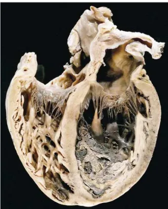
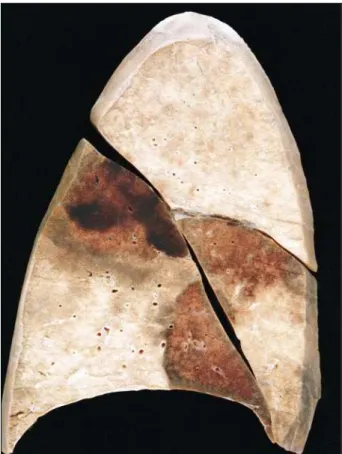
The heart showed dilation of the 4 chambers and bi-ventricular cavitary thrombosis (fig. 2). The left bi-ventricular wall was not thickened (7mm). Microscopically, narrowing of the myocardial fibers occurred as well as scarce foci of fibrosis (fig. 3) and a mononuclear inflammatory cell infiltra-tion, with no aggression to the cardiomyocytes. Despite fo-cal disarrangement of myocardial fibers, the amount was not enough to characterize hypertrophic cardiomyopathy. Thromboembolism with recent hemorrhagic infarct in the lower lobe of the left lung and in the middle and lower lobes of the right lung (fig. 4) was observed. Death resulted from
Fig. 2 – Section showing dilation of the 4 cardiac chambers and biventricular thrombosis.
1. Melacini P, Fanin M, Danieli GA, et al. Cardiac involvement in Becker muscular dystrophy. J Am Coll Cardiol 1993; 22: 1927-34.
2. Stevenson S, Rothery S, Cullen MJ, Severs NJ. Spatial relationship of the C-termi-nal domains of dystrophin and beta-dystroglycan rect molecular interaction at the plasma membrane interface. Circ Res 1998; 82: 82-93.
3. Aicardi J. Primary Muscle Disease. In: Aicardi J. Diseases of the Nervous in Chil-dhood.: Mac Keith Press, 1992; 1172-237.
4. De Vissier M, de Voogt WG, la Riviere GVl. The heart in Becker muscular dystro-phy, fascioscapulohumeral dystrophy and Bethlem myopathy. Muscle Nerve 1992; 15: 591-6.
5. Comi LI, Nigro G, Politano L, Petretta VR. The cardiomyopathy of Duchenne/ Becker consultands. Int J Cardiol 1992; 34: 297-305.
6. Hassanz, Fastabend CP, Mohanty PK, Isaacs ER. Atrioventricular block and supraventricular linked muscular dystrophy. Circulation 1979; 60: 1365-9.
7. Baumbach LL, Chamberlain JS, Ward PA, Farwell NJ, Caskey CT. Molecular and clinical correlations of deletions leading to Duchenne and Becker muscular dys-trophies. Neurology 1989; 465-474.
8. Hoffman EP, Fishbeck KH, Brown RH. Characterization of dystrophin in mus-cle-biopsy specimens from patients with Duchenne’s or Becker’s muscular dys-trophy. N Engl J Med 1988; 318: 1363-8.
9. Bushby KMD, Gardner-Medwin D. The clinical, characteristics of Becker mus-cular dystrophy. 1. Natural History. J Neurol 1993;240:98-140.
References
10. Bushby KMD, Thambyayah M, Gardner-Medwin D. Prevalence and incidence of Becker muscular dystrophy. Lancet 1991; 337: 1022-4.
11 Hoffman EP. Genotype/phenotype correlations in Duchenne/Becker dystrophy. In: Patridge T, ed. Molecular and Cell Biology of Muscular Dystrophy. London: Chapman Hall, 1993: 12-36.
12. Laing NG. Molecular genetics and genetic counselling for Duchenne/Becker muscular dystrophy. In: Patridge T, ed. Molecular and Cell Biology of Muscular Dystrophy. London: Chapman Hall, 1993: 37-84.
13. Yoshida K, Ikeda SIO, Nakamura A. Molecular anlysis of the Duchenne muscu-lar dystrophy gene in patients with Becker muscumuscu-lar dystrophy presenting with dilated cardiomyopathy. Muscle Nerve 1993; 16: 1161-6.
14. Wilton SD, Johnsen RD, Pedretti JR, Laing NG. Two distinct mutations in a sin-gle dystrophin gene: identification of an altered splice-site as the primary Becker muscular dystrophy mutation. J Med Genet 1993; 46: 563-9.
15. Vainzof M, Passos-Bueno MR, Zatz M. Dystrophin and DNA findings in Du-chenne and Becker carriers. In: Lane RJM. Handbook of Muscle Disease, ed. Marcel Dekker Inc, 1996; 265-74.
16. Oldfors A, Eriksson BO, Kyllerman M, Martinsson T, Wahlstrom J. Dilated car-diomyopathy and the dystrophin gene: an illustrated review. Br Heart J 1994; 72: 344-8.
17. Maeda M, Nakao S, Miyazato H, et al. Cardiac dystrophin abnormalities in Be-cker muscular dystrophy assessed by endomyocardial biopsy. Am Heart J 1995; 129: 702-7.
the aggravation of heart failure due to the pulmonary lesions. The rectus abdominis muscle showed mild atro-phy. Muscle changes might have been more exuberant if a muscle of the lower limbs had been examined.
(Dr. Paulo Sampaio Gutierrez)
Fig. 4 - Hemorrhagic infarcts in the middle and lower lobes of the right lung.
Anatomicopathological diagnoses –
Cardiomyopa-thy of Becker muscular dystrophy; biventricular thrombo-sis; bilateral pulmonary thromboembolism.
Comments
Becker muscular dystrophy, like the Duchenne va-riant, is related to a defect in the short arm of the X
chromosome in the Xp21 locus 1. Both may show cardiac
involvement, which is characterized by arrhythmia or congestive heart failure with dilated cardiomyopathy, or both. Absence of cardiac symptoms in some patients is believed to result from a limitation in physical activities directly caused by the skeletal muscle disease. In some series of patients with Becker muscular dystrophy,
car-diac changes are found in 60% to 70% of cases 15. No
stu-dy exists of the incidence of muscle disease among pa-tients with dilated cardiomyopathy or with arrhythmias, who probably constitute a small fraction of patients. Therefore, the neurologist managing a patient with muscle disease should proceed to the appropriate and careful cardiac evaluation. In the same way, the cardio-logist examining a patient with dilated cardiomyopathy or arrhythmias with no demonstrable cause should in-vestigate, including genetic counseling, whether a di– sease of that type exists or not. For this purpose, dosage of creatine kinase should be performed, or as a last reso-urce, the immunohistochemical analysis of the myocar-dial biopsy, once the material is frozen and not fixed in for-malin 16, 17.