INTRODUCTION
Address to: Dra Marcelle Aquino Rabelo. Deptº de Medicina Tropical/UFPE. Av. Prof. Moraes Rego 1235, Cidade Universitária, 50670-901 Recife, PE, Brasil. Phone: 55 81 2126-8526
e-mail: [email protected] Received 28 April 2014
Accepted 30 July 2014
The occurrence and dissemination of methicillin and
vancomycin-resistant
Staphylococcus
in samples
from patients and health professionals of a university
hospital in Recife, State of Pernambuco, Brazil
Marcelle Aquino Rabelo
[1], Armando Monteiro Bezerra Neto
[1], Stéfany Ojaimi Loibman
[1],
Jailton Lobo da Costa Lima
[2], Ewerton Lucena Ferreira
[3], Nilma Cintra Leal
[4]and Maria Amélia Vieira Maciel
[1][1]. Departamento de Medicina Tropical, Universidade Federal de Pernambuco, Recife, PE. [2]. Instituto de Ciências Biológicas, Universidade de Pernambuco, Recife, PE. [3]. Departamento de Ciências Biológicas (Microbiologia), Universidade de São Paulo, São Paulo, SP. [4]. Centro de Pesquisas Aggeu Magalhães, Fundação Oswaldo Cruz, Recife, PE.
ABSTRACT
Introduction: Methicillin-resistant Staphylococcus aureus (MRSA) strains have been responsible for many nosocomial outbreaks. Within hospitals, colonized employees often act as reservoirs for the spread of this organism. This study collected clinical samples of 91 patients admitted to the intensive care unit (ICU), hemodialysis/nephrology service and surgical clinic, and biological samples from the nasal cavities of 120 professionals working in those environments, of a University Hospital in Recife, in the State of Pernambuco, Brazil. The main objective of this study was to determine the occurrence and dissemination of methicillin- and vancomycin-resistant Staphylococcus spp. Methods: The isolates obtained were tested for susceptibility to oxacillin and vancomycin and detection of the mecA gene. In addition, the isolates were evaluated for the presence of clones by ribotyping-polymerase chain reaction (PCR). Results: MRSA occurrence, as detected by the presence of the mecA gene, was more prevalent among nursing technicians; 48.1% (13/27) and 40.7% (11/27) of the isolates were from health professionals of the surgical clinic. In patients, the most frequent occurrence of mecA-positive isolates was among the samples from catheter tips (33.3%; 3/9), obtained mostly from the hemodialysis/nephrology service. Eight vancomycin-resistant strains were found among
the MRSA isolates through vancomycin screening. Based on the amplifi cation patterns, 17 ribotypes were identifi ed, with some
distributed between patients and professionals. Conclusions: Despite the great diversity of clones, which makes it diffi cult to
trace the source of the infection, knowledge of the molecular and phenotypic profi les of Staphylococcus samples can contribute towards guiding therapeutic approaches in the treatment and control of nosocomial infections.
Keywords: MRSA. Vancomycin. Patients. Health professionals. Ribotyping-PCR.
Staphylococcus remains one of the most common pathogens in systemic infections in communities and hospitals. With the advent of resistance to methicillin in the 1960s, Staphylococcus
began to receive special attention, especially with regard to controlling the spread of this microorganism. Since then, the therapeutic options have become increasingly restricted1,2.
In most cases, this methicillin resistance is determined by the presence of the mecA gene, located in the chromosome and responsible for the synthesis of PBP2a or PBP2', a
penicillin-binding protein (PBP), which regulates bacterial cell wall synthesis in the presence of beta-lactam antibiotics2,3. This gene is widely distributed among Staphylococcus aureus and between species of coagulase-negative Staphylococcus, and its detection by molecular methods is considered the gold standard for a qualitative assessment of resistance to methicillin2,4,5.
METHODS
The identification of MRSA carriers is a step towards establishing a control policy, thereby helping to identify the measures needed to reduce the colonization pressure9. The knowledge of the molecular epidemiology of the diseases
caused by these bacteria may assist in developing more effi cient
strategies for reducing the infection, as genetic relationships
among the different clones can be inferred, the gene fl ow route
can be detected and the spread of infection can be traced from
the molecular profi les 9,13.
Appropriate systems for typing are needed to determine the genetic variability of the isolates, thus providing an effective epidemiological control. The ribotyping-polymerase chain reaction (PCR) is often referenced and used because of its high taxonomic and epidemiological value. This technique is a valuable tool for identifying and differentiating isolates of the
Staphylococcus genus13-15.
Despite the relevance of these microorganisms as important pathogens associated with health care-related infections, studies that provide data regarding the colonization of health care workers, especially multi-professional assessments in Pernambuco, Brazil, associating them with the spread of such infections, are still scarce. Hence, this study aims to describe the occurrence and spread of methicillin- and vancomycin-resistant
Staphylococcus spp. in samples from the nasal cavities of health professionals and clinical samples from patients admitted to the intensive care unit (ICU), hemodialysis/nephrology service and surgical clinic of a university hospital in the City of Recife by analyzing epidemiological/bacterial MRSA marker data to assist in promoting control actions at the hospital.
Collection of biological material
The study included all patients and all health professionals from the sectors of the general ICU, surgical clinic and hemodialysis/nephrology service of the Hospital das Clínicas, Federal University of Pernambuco (UFPE), Brazil during the period from April to August 2011 based on the list of patients and staff in each sector.
Samples from the nasal cavities of health professionals were collected using sterile swabs after each individual signed a Statement of Free and Informed Consent (SFIC) and completed a questionnaire on information about their professional activities. A previous study showed that this site was most likely to be colonized8. These samples were introduced into brain heart infusion (BHI) and transported to the Bacteriology Laboratory, Department of Tropical Medicine/UFPE.
The patient samples were collected according to the standard procedures used in the hospital for catheter tips, drains, blood, abscesses, surgical wounds, tracheal secretions and so forth, and the isolates were obtained after culture in the Bacteriology Laboratory of the Hospital das Clínicas of the Federal University of Pernambuco. Patients who did not have any areas with characteristics of bacterial infection were submitted to the collection of nasal swabs after signing an SFIC.
Isolation and identifi cation of Staphylococcus The isolates of Staphylococcus spp., identified by the Bacteriology Laboratory of the hospital, from the various biological samples collected from patients and the nasal swab samples from health professionals were placed in BHI broth and then inoculated into 5% sheep blood agar and incubated for 24-48 h at 37°C. Colonies with macroscopic characteristics of the genus Staphylococcus were Gram stained, and when confi rmed
by morphology and staining, were submitted for identifi cation
using deoxyribonuclease (DNase), catalase and coagulase tests and mannitol fermentation. The remaining samples of secretions from patients were seeded according to the standard protocol in 5% sheep blood agar and MacConkey agar. However, only the colonies with characteristics of the genus Staphylococcus
were identifi ed16.
Cefoxitin and oxacillin susceptibility
After identifi cation, susceptibility testing of Staphylococcus
was performed using the disk-diffusion method on Mueller-Hinton agar17 with 1μg oxacillin and 30μg cefoxitin. The Clinical and Laboratory Standards Institute (CLSI) 2013 interpretive breakpoints were considered.
Screening for oxacillin resistance
The isolates selected were those that showed resistance
or had intermediate profi les to oxacillin and/or cefoxitin in a
disk-diffusion test. Colonies from 5% blood agar plates were resuspended in BHI to obtain a turbidity equivalent to 0.5 on the McFarland scale. A 1µL platinum wire loop was dipped in the suspension, and the bacteria were seeded in a 10-15mm area on plates with Mueller-Hinton agar medium containing NaCl
(4% v/v; 0.68mol/L) and 6μg/mL oxacillin. These plates were
incubated at 35°C for 24h and then read, considering that >1 colony = resistance17. For quality control, the standard MRSA strain ATCC 33591 was used for the positive control (MRSA), and the standard Methicillin-sensitive S. aureus (MSSA) strain ATCC 29213 was used for the negative control.
Screening for vancomycin resistance
The isolates that possessed the mecA gene, assayed by PCR, were subjected to screening for vancomycin resistance. Colonies from 5% blood agar plates were resuspended in BHI to obtain a turbidity equivalent to 2.0 on the McFarland scale. A 1µL platinum wire loop was dipped in the suspension, and the bacteria were seeded in an area with a 10-15mm diameter on
plates with BHI agar medium containing 6μg/mL vancomycin
(Oxoid). The plates were incubated at 35°C for 24h and 48h and then read, considering that >1 colony = resistance18-20. For quality control, the standard strains Enterococcus faecalis
ATCC 29212 – sensitive and Enterococcus faecalis ATCC 51299 – resistant were used.
DNA preparation
RESULTS
Identifi cation by PCR of the mecA gene
PCR was performed utilizing the primers described by Petinaki et al.22. The amplifi cation reaction mixture was prepared in a total volume of 25µL containing 50mM KCl, 10mM Tris-HCl, 1.5mM MgCl2, 200mM dNTP (Promega), 20pmol of each primer, 20ng of genomic DNA, and 1U Taq DNA polymerase (Promega). The reactions were performed in a thermocycler (Biometra), programmed initially for 30 thermal cycles, with denaturation of 1 min at 94°C, annealing of 1 min at 50°C and
extension of 1 min at 72°C, followed by a fi nal step of 10min
at 72°C. The negative control contained all of the components of the reaction mixture except DNA. ATCC 33591 S. aureus
(MRSA) was used for the positive control. The amplifi cation product was submitted to 1% agarose gel electrophoresis with ethidium bromide staining and was visualized with an ultraviolet (UV) transilluminator and then digitized (Kodak Digital Science).
Ribotyping-PCR
The isolates that were positive for the mecA gene by PCR were subjected to ribotyping-PCR to assess the genetic relationship of the isolates following the protocol described by Cuny et al.23. For the 16S-23S ribosomal ribonucleic acid (rRNA) spacer region amplifications, the primers rRNA1 (5'- TTG TAC ACA CCG CCC GTC A-3') and rRNA2 (5'- GGT ACC TTA GAT GTT TCA GTT C-3') were used.
The amplifi cation reaction mixture was prepared in a total
volume of 25µL containing 50mM KCl, 10mM Tris-HCl, 1.5mM MgCl2, 200mM dNTP (Promega), 20pmol of each primer, 20ng of genomic DNA, and 1U Taq DNA polymerase (Promega). The reactions were performed in a thermocycler (Biometra), programmed initially for 30 thermal cycles, with denaturation of 1min at 94°C, annealing of 1 min at 55°C and
extension of 1min at 72°C, followed by a fi nal step of 7min at
72°C. The amplifi cation product was submitted to 2% agarose gel electrophoresis with ethidium bromide staining (2µg/mL) and visualized with a UV transilluminator and then digitized (Kodak Digital Science). A 100bp Ladder (Invitrogen) was used as a molecular weight standard to estimate the sizes of
the amplifi ed fragments.
Statistical analysis
The clinical and microbiological data were statistically analyzed using Epi Info (version 6.04.) according to the frequency distribution. The dendrogram was constructed using Darwin 5.0.158 software (Cirad - Department: Systèmes Biologiques (BIOS), Research Unit: Genetic improvement of vegetatively propagated crops, Team: BioMathematics, Avenue Agropolis - TA A75/02, 34398 Montpellier Cedex 5 – France).
Ethical considerations
This study was approved by the Ethics Committee on Research of the Federal University of Pernambuco (CEP/CCS/ UFPE - Comitê de Ética em Pesquisa/Centro de Ciências da Saúde/Universidade Federal de Pernambuco), CAAE number 0490.0.172.000-11.
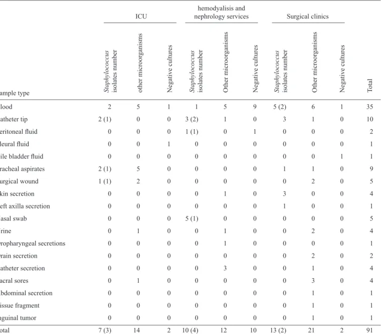
Samples were collected from 91 patients (Table 1) and 120 health professionals, including physicians, nurses, nursing technicians, physiotherapists, nutritionists and psychologists (Table 2) from the sectors of the ICU, surgical clinic and the hemodialysis/nephrology service of the Hospital das Clínicas of the Federal University of Pernambuco in the period from April to August 2011.
A sample was obtained from each patient and, after
identifi cation tests, 30 bacteria of the genus Staphylococcus were isolated. The most frequent sample type from patients was blood culture (37.4%), followed by catheter tip (11%). In all, 14 negative cultures were obtained (Table 1).
Among the isolates from patients that were classifi ed as the
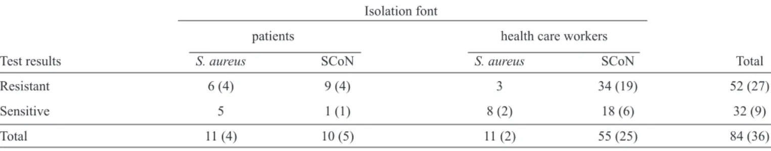
genus Staphylococcus, 11 were identifi ed as coagulase-negative Staphylococcus and 19 as S. aureus. Using the oxacillin and/ or cefoxitin disk-diffusion tests, 21 Staphylococcus spp. with
resistance profi les were selected and submitted for detection of
the mecA gene by PCR. Nine positive isolates were detected after this step. The Staphylococcus spp. isolates from the health professionals were also subjected to this test, with 63 resistant isolates being selected (Table 3); of these, 27 isolates encoded the mecA gene, with a total of 36 MRSA isolates (Table 4).
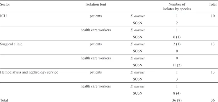
Of the isolates subjected to oxacillin screening, 61.9% were resistant; of these, 75% of the isolates had the mecA gene (Table 3). The greatest occurrence of mecA- positive isolates in the samples from patients was among the isolates from catheter tips (33.3%) (Table 1). Despite similar percentages, the sector that was the most frequent source for positive isolates in this group was the hemodialysis/nephrology service (44.4%) (Table 4). On conducting the vancomycin screening, eight isolates were determined to be resistant (Table 4).
In this study, there was no statistically signifi cant difference
observed among health professionals when the prevalence of MRSA in females was compared to males. Considering age groups, individuals between 20 and 28 years old were the most colonized by MRSA, these microorganisms being most prevalent among nursing technicians (48.1% among the positive isolates). Considering hospital sectors, the surgical clinic accounted for the highest incidence of positive isolates (40.7% among health professionals).
The prevalence of MRSA was high (77.8%) among professionals who simultaneously used a medical coat, gloves
and a mask only in specifi c situations of contact with fl uids or
secretions of patients. The group that reported that they most often used a medical coat, gloves and a mask together from the personal protective equipment (PPE) available was also the one most colonized by MRSA (29.6%). The occurrence of MRSA was also highest among the professionals who performed their activities during the day (24/100) compared with those who performed their duties during the night (3/20).
Blood 2 5 1 1 5 9 5 (2) 6 1 35
Catheter tip 2 (1) 0 0 3 (2) 1 0 3 1 0 10
Peritoneal fl uid 0 0 0 1 (1) 0 1 0 0 0 2
Pleural fl uid 0 0 1 0 0 0 0 0 0 1
Bile bladder fl uid 0 0 0 0 0 0 0 0 1 1
Tracheal aspirates 2 (1) 5 0 0 0 0 1 1 0 9
Surgical wound 1 (1) 2 0 0 0 0 0 2 0 5
Skin secretion 0 0 0 0 1 0 3 0 0 4
Left axilla secretion 0 0 0 0 0 0 1 0 0 1
Nasal swab 0 0 0 5 (1) 0 0 0 0 0 5
Urine 0 1 0 0 1 0 0 2 0 4
Oropharyngeal secretions 0 0 0 0 1 0 0 0 0 1
Drain secretion 0 0 0 0 0 0 0 2 0 2
Catheter secretion 0 0 0 0 3 0 0 1 0 4
Sacral sores 0 1 0 0 0 0 0 3 0 4
Abdominal secretion 0 0 0 0 0 0 0 1 0 1
Tissue fragment 0 0 0 0 0 0 0 1 0 1
Inguinal tumor 0 0 0 0 0 0 0 1 0 1
Total 7 (3) 14 2 10 (4) 12 10 13 (2) 21 2 91
Staphylococcus isolates number other microor
ganisms
Negative cultures Staphylococcus isolates number Other microor
ganisms
Negative cultures Staphylococcus isolates number Other microor
ganisms
Negative cultures Total hemodyalisis and
ICU nephrology services Surgical clinics
Sample type
ICU: intensive care unit. Note: the parenthesis data are the number of mecA postive isolates.
TABLE 1 - Distribution of Staphylococcus spp. isolates from the patients by sample related the hospital sectors.
frequencies of resistant strains were similar among those who worked only in the hospital under study and those who worked in another hospital (Table 2).
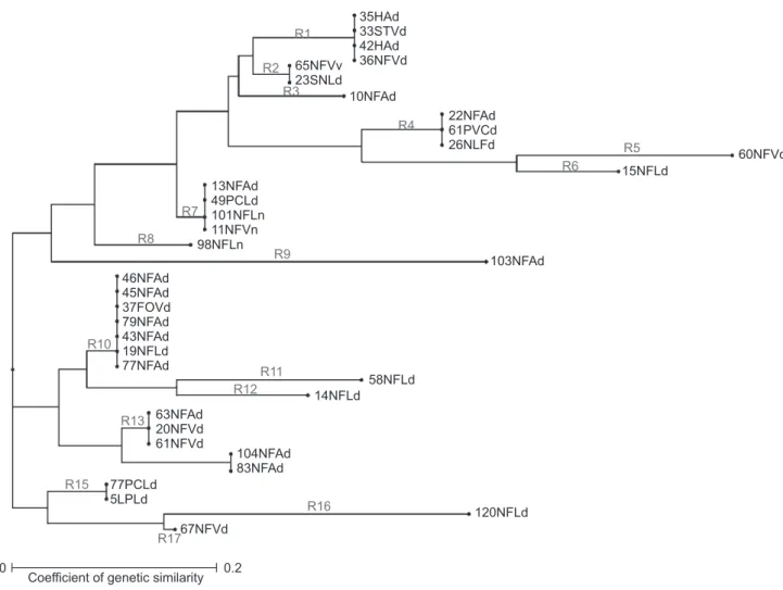
In the ribotyping-PCR reactions, two to fi ve fragments of
approximately 500-900bp in size were observed. Based on the
amplifi cation patterns, the 36 isolates were classifi ed into 17
ribotypes, designated in this study as R1 to R17 (Figure 1). Six isolates of S. aureus (four patients and two professionals) were distributed in three ribotypes (R1, R7, R15). The
coagulase-negative Staphylococcus (CoNS) isolates from fi ve patients
were distributed in four different ribotypes (R2, R4, R10, R15), and 25 isolates from health professionals were distributed in 15 professional ribotypes, with a prevalence of ribotypes R10 (six isolates), R7 and R13 (three isolates each) and R4 and R14 (two isolates each). The ribotypes R3, R5, R6, R8, R9, R11, R12, R16 and R17 each occurred in only one isolate (Figure 1).
The profi les R1, R2, R4, R7 and R10 were observed in isolates
TABLE 2 - Distribution of variables related to the presence of MRSA in health professionals from the Hospital of UFPE, 2011.
Total MRSA Non MRSA
Variables n % n % n %
Gender
male 20 16.7 5 18.5 15 16.1
female 100 83.3 22 81.5 78 83.9
Age (years)
20-28 43 35.8 9 33.3 34 36.6
29-33 26 21.7 4 14.8 22 23.7
34-44 27 22.5 7 25.9 20 21.5
45-60 24 20.0 7 25.9 17 18.3
Professional activity
nurse 43 35.8 7 25.9 36 38.7
nurse technician 45 37.5 13 48.1 32 34.4
physician 26 21.7 6 22.2 20 21.5
physiotherapist 3 2.5 1 3.7 2 2.1
nutritionist 2 1.7 0 0 2 2.1
psychologist 1 0.8 0 0 1 1.1
Sector
ICU 23 19.2 7 25.9 16 17.2
surgical clinic 50 41.7 11 40.7 39 41.9
hemodialysis and nephrology service 47 39.2 9 33.3 38 40.9
Use of PPE
always 27 22.5 5 18.5 22 23.7
sometimes 87 72.5 21 77.8 66 71.0
never 6 5.0 1 3.7 5 5.4
PPE used most often
medical coat+gloves+mask 32 26.7 8 29.6 24 25.8
medical coat 8 6.7 1 3.7 7 7.5
gloves 5 4.2 2 7.4 3 3.2
gloves+mask 9 7.5 2 7.4 7 7.5
gloves+medical coat 21 17.5 7 25.9 14 15.0
medical coat+gloves+mask +cap+glasses 3 2.5 0 0 3 3.2
medical coat+gloves+mask+cap 29 24.2 3 11.1 26 28.0
medical coat+gloves+mask+glasses 3 2.5 1 3.7 2 2.1
gloves+mask+cap 3 2.5 2 7.4 1 1.1
medical coat+cap 3 2.5 1 3.7 2 2.1
gloves+medical coat+cap 2 1.7 0 0 2 2.1
mask+medical coat 1 0.8 0 0 1 1.1
mask+medical coat+cap 1 0.8 0 0 1 1.1
TABLE 2 - Continuation.
Total MRSA Non MRSA
Variables n % n % n %
Shift
Diurnal 100 83.3 24 88.9 76 81.7
Nocturnal 20 16.7 3 11.1 17 18.3
Length of time in the profession (years)
<1 25 20.8 4 14.8 21 22.6
1-5 30 25.0 9 33.3 21 22.6
5-10 22 18.3 4 14.8 18 19.4
10-15 13 10.8 2 7.4 11 11.8
15-20 9 7.5 3 11.1 6 6.4
>20 21 17.5 5 18.5 16 17.2
Length of time in the sector (years)
<1 54 45.0 11 40.7 43 46.2
1-3 25 20.8 7 25.9 18 19.3
3-7 12 10.0 3 11.1 9 9.7
7-11 12 10.0 2 7.4 10 10.7
11-15 5 4.2 0 0 5 5.4
15-20 8 6.7 4 14.8 4 4.3
>20 4 3.3 0 0 4 4.3
Number of hospitals worked in
1 57 47.5 11 40.7 46 49.5
2 49 40.8 11 40.7 38 40.9
3 10 8.3 3 11.1 7 7.5
>3 4 3.3 2 7.4 2 2.1
MRSA: Methicillin-resistant Staphylococcus aureus; UFPE: Universidade Federal de Pernambuco; ICU: intensive care unit; PPE: personal
protective equipment. Note: The variable "Use of Personal Protective Equipment (PPE)" for this study was defi ned as the frequency of use of
all personal protective equipment (Medical coat+gloves+mask) during activities. Categorized as: always (in all situations of patient contact); sometimes (only in situations that manipulate biological fl uids, such as blood and secretions, and/or when the patient has infectious disease symptoms); or never (do not use all PPEs in any situation).
TABLE 3 - Results of oxacillin screening distributed by isolation font, showing the number of mecA positive isolates.
Isolation font
patients health care workers
Test results S. aureus SCoN S. aureus SCoN Total
Resistant 6 (4) 9 (4) 3 34 (19) 52 (27)
Sensitive 5 1 (1) 8 (2) 18 (6) 32 (9)
Total 11 (4) 10 (5) 11 (2) 55 (25) 84 (36)
DISCUSSION
The prevalences of MRSA were 10% (9/91) for patients and 22.5% (27/120) for health professionals; both groups carried primarily coagulase-negative Staphylococcus, with prevalences of 5.5% and 20.8%, respectively. These percentages may be considered low, as the prevalence of isolation of MRSA strains ranges from 40 to 80%2,9,24 in Brazilian hospitals, and the data from the Antimicrobial Surveillance Program (SENTRY) show that MRSA corresponds to 31% of the causes of nosocomial and community infections and is considered the most common among the most prevalent pathogens25. However, the fact that professionals have a higher percentage of MRSA points to contamination in the hospital itself. The SENTRY survey conducted in Brazilian hospitals showed that the resistance of CoNS in blood cultures is 80%25;and in our study, this was the most frequent type of sample.
Among ICU patients from another university hospital in Recife, a prevalence of S. aureus colonization of 37.7% was reported, of which MRSA accounted for 13%26, similar to the prevalence of 10% (3/30) for MRS in the ICU that was measured in this study. As to the health-care team, in a study conducted in a university hospital in Londrina, colonization by S. aureus
among the medical staff was close to that normally detected in the community: 17.7%, of whom 1.2% were MRSA carriers27, which is lower than the MRSA colonization percentage of 23.1% (6/26) detected in this study. In a study at another university hospital in Recife, the colonization of health workers reached 25.7%, but the percentage of MRSA was considered below
TABLE 4 - Distribution of mecA positive isolates by isolation sector and isolates by isolation font.
Sector Isolation font Number of Total
isolates by species
ICU patients S. aureus 1 10
SCoN 2
health care workers S. aureus 1
SCoN 6 (1)
Surgical clinic patients S. aureus 2 (1) 13
SCoN 0
health care workers S. aureus 0
SCoN 11 (2)
Hemodialysis and nephrology service patients S. aureus 1 13
SCoN 3
health care workers S. aureus 1
SCoN 8 (4)
Total 36 (8) 36
ICU: intensive care unit; S.: Staphylococcus; SCoN: coagulase-negative Staphylococcus. Note: the data in parentheses are the numbers of
resistant strains in the screening of vancomycin distributed by isolation font.
the limits described in the literature (only three nurses among the 202 professionals from whom samples were collected had positive samples10) and was below the percentage that we detected with respect to MRSA. In another study conducted in the same hospital as our study, the colonization of nursing staff accounted for 25.8% of positive samples and; once again, the percentage of MRSA was considered below the limits described in the literature (3.3% of the samples8).
A study in a public hospital in the interior of the State of São Paulo suggested that nurses and nursing technicians are the professional class that is most colonized by MRSA, citing prevalences of 7.1% among nurses28 and 10.8% among nursing technicians.
The age group of MRSA occurrence among health professionals was the same as that described in other studies conducted in the same hospital in previous years8,10,29. The frequency of MRSA infection was highest in the 20- to 28-year-old age group. These studies suggest that this incidence may be due to the need for improvements in professional practice, such as washing hands before and after procedures and avoiding contacting nostrils with hands. This same deficiency in professional practice could also explain the higher prevalence in the group who had been health-care professionals for between 1 and 5 years.
In this study, a higher incidence of methicillin-resistant samples was observed only by phenotypic methods, such as oxacillin screening, compared to genetic screening, which suggests the presence of other resistance mechanisms independent of the mecA gene22,30,31.
10 35HAd
33STVd 42HAd 36NFVd
R1
R2
R3
R4
R5 R6
R7
R8
R9
R10
R11 R12
R13
R15
R16
R17
98NFLn
58NFLd 14NFLd
120NFLd 67NFVd
Coefficient of genetic similarity 0 0.2
104NFAd 83NFAd 77PCLd
5LPLd 63NFAd 20NFVd 61NFVd 46NFAd 45NFAd 37FOVd 79NFAd 43NFAd 19NFLd 77NFAd
13NFAd 49PCLd 101NFLn 11NFVn
15NFLd
103NFAd
60NFVd 10NFAd
22NFAd 61PVCd 26NLFd 65NFVv
23SNLd
FIGURE 1 - Z estimated by ribotyping-PCR for 36 methicillin-resistant Staphylococcus spp. isolates from patients and health care workers from a university hospital in Recife, State of Pernambuco, Brazil. Note: The letter R indicates ribotype. In the description of the isolates, the number indicates the isolate number, the fi rst two letters indicate the type of sample, the third letter
indicates the hospital sector and the last letter indicates the collection period. Sample types: LP: peritoneal fl uid; PC: catheter tip;
SN: nasal swab from patient; ST: tracheal aspirates, H: blood; FO: surgical wound; NF: nasal swab from health professional. Sectors:
A: Surgical clinic; I: Hemodialysis/Nephrology Service. V: ICU. Collection period: d: day (7h to 19h) and n: night (19h to 7h).
PCR: polymerase chain reaction; ICU: intensive care unit.
culture (isolate 35HAd, Figure 1). The remaining samples were
from professionals, two of which were classifi ed as the same
ribotype (isolates 101NFLn and 110NFVn, Figure 1), suggesting that they are the same strain. Studies indicate that resistance to vancomycin should be determined by more sensitive techniques, such as plate screening, E-test, microdilution and genotype detection17-20. In Brazil, intermediate resistance to vancomycin in patients has been described, but few studies have reported colonization of health professionals by these strains32. The fi rst case of transferable vancomycin resistance in a community-associated MRSA strain was reported in a Brazilian hospital, indicating that the presence of MRSA containing the vanA gene could be a future public problem33.
Some studies that use ribotyping-PCR to assess genetic similarity also present ample polymorphisms, as in our study,
considering the number of ribotypes observed, thus indicating dispersion in the hospital sectors34,35, but these studies do not make comparisons of dispersion between classes of patients and professionals.
In the ribotyping-PCR reactions, few ribotypes (R1, R2, R4, R7 and R10) were distributed among samples from patients and health professionals, suggesting a low spread between these
classes. Pulsed-fi eld gel electrophoresis (PFGE) was used to
that health professionals are the source of transmission, as they are carriers of several ribotypes not found in patients.
This approach is useful to once again raise the question of the role of health professionals in spreading nosocomial infections. Despite the fact that the contribution of health professionals in
the spread of resistant strains has not yet been confi rmed36,37, various studies based on molecular techniques do suggest they are a vehicle of dissemination7,38,39. The diversity of ribotypes
identifi ed, despite the stability of the internal transcribed spacer
(ITS) 16S-23S region, suggests the presence of several clones circulating in the hospital during the study period; thus, it is possible that there may be multiple sources of contamination.
In view of these fi ndings, routine screenings of health care
professionals for MRSA colonization is not necessary; likewise, their decolonization, mainly due to the associated cost36, should only be conducted in situations in which the epidemiological data suggest that they are serving as the transmission source6,7 – or, as a last resort, to contain transmission when other measures have already been taken in an outbreak40. In these situations, the
identifi cation of MRSA carriers is a step towards establishing
a control policy and helps to identify the measures needed to reduce the colonization pressure9. Despite the fact that the low spread of methicillin-resistant isolates between classes has been demonstrated, other factors may also contribute to the spread of the microorganism, such as its capacity to colonize, to multiply itself and to invade the mucosal epithelia cells of the host, along with the capacity to withstand the selective pressure of hospital environments. This situation highlights the importance of monitoring the distribution and routes of the dissemination of MRSA clones in hospitals41, the emphasis being on identifying isolates resistant to vancomycin in samples of colonization. Thus, measures to contain the spread of infections associated with health care should be further developed and applied.
ACKNOWLEDGMENTS
The authors declare that there is no confl ict of interest.
CONFLICT OF INTEREST
FINANCIAL SUPPORT
REFERENCES
Thank you to the bacteriology laboratory of the Hospital das Clínicas for providing some samples and to the Centro de Pesquisas Aggeu Magalhães (CPqAM) for providing space for performing the molecular experiments.
This work was supported by FACEPE (Fundação de Amparo Ciência do Estado de Pernambuco) grant no APQ 0579-2.12/07 and PROPESQ (Pró-Reitoria para Assuntos de Pesquisa e Pós-Graduação-Universidade Federal de Pernambuco) for the Edital
PQ-Multiusuário-2010/Propesq no 23076.024115/2010-91.
1. Tverdek FP, Crank CW, Segreti J. Antibiotic Therapy of Methicillin-Resistant Staphylococcus aureus in Critical Care. Crit Care Clin2008; 24:249-260.
2. Moellering Jr RC. MRSA: the fi rst half century. J Antimicrob Chemother
2012; 67:4-11.
3. Schito GC. The importance of development of antibiotic resistance in
Staphylococcus aureus. Clin Microbiol Infect 2006; 12:3-8.
4. Swenson JM, Lonsway D, MCallister S, Thompson A, Jevitt L, Zhu W, et al. Detection of mecA-mediated resistance using reference and commercial testing methods in a collection of Staphylococcus aureus
expressing boderline oxacillin MICs. Diag Microbiol Infect Dis 2007; 38:1346-1350.
5. Shariati L, Validi M, Tabatabaiefar MA, Karimi A, Nafi si MR.
Comparison of Real-Time PCR with Disk Diffusion, Agar Screen and E-test Methods for Detection of Methicillin-Resistant Staphylococcus aureus. Curr Microbiol2010; 61:520-524.
6. Siegel JD, Rhinehart E, Jackoson M, Chiarello L. Healthcare Infection Control Practices Advisory Committee. Management of multidrug-resistant organisms in health care settings, 2006. Am J Infect Control 2007; 35:165-193.
7. Ben-David D, Mermel LA, Parenteau S. Methicillin-resistant
Staphylococcus aureus transmission: the possible importance of unrecognized health care worker carriage. Am J Infect Control 2008; 36:93-97.
8. Silva ECBF, Samico TM, Cardoso RR, Rabelo MA, Bezerra Neto AM, de Melo FL, et al. Colonization by Staphylococcusaureus among the nursing staff of a teaching hospital in Pernambuco. Rev Esc Enferm-USP 2012; 46:132-137.
9. Santos HB, Machado DP, Camey AS, Kuchenbecker RS, Barth AL, Wagner MB. Prevalence and acquisition of MRSA amongst patients admitted to a tertiary-care hospital in Brazil. BMC Infect Dis 2010; 10:328-334.
10. Silva ECBF, Maciel MAV, Melo FL, Lopes ACS, Aca IS. Epidemiological surveillance and susceptibility of Staphylococcus aureus among healthcare workers at a reference hospital: preliminary assessment. Rev Inst Adolfo Lutz 2010; 69:126-130.
11. Ellis MW, Hospenthal DR, Dooley DP, Gray PJ, Murray CK. Natural history of community-acquired methicillin-resistant Staphylococcus aureus colonization and infection in soldiers. Clin Infect Dis 2004; 39:971-979.
12. Rafee Y, Abdel-Haq N, Asmar B, Salimnia T, Pharm CV, Rybak Pharm MJ, et al. Increased prevalence of methicillin-resistant Staphylococcus aureus nasal colonization in household contacts of children with community acquired disease. BMC Infect Dis 2012; 12:45.
13. Oliveira AM, Ramos MC. PCR-based ribotyping of Staphylococcus aureus. Braz J Med Biol Res 2002; 35:175-180.
14. Kostman JR, Alden MB, Mair M, Edlind TD, LiPuma JJ, Stull TL, et al. A universal approach to bacterial molecular epidemiology by polymerase chain reaction ribotyping. J Infect Dis1995; 171:204-208.
15. Forsman P, TiIsala-Timisjarvi A, Alatossava T. Identifi cation of
staphylococcal and streptococcal causes of bovine mastitis using 16S-23S rRNA spacer regions. Microbiol 1997; 143:3491-3500.
16. Winn WC, Allen SD, Janda WM, Koneman EW, Procop GW, Schreckenberger PC, et al. Koneman’s color atlas and textbook of diagnostic microbiology. 6th edition. Guanabara Koogan. Rio de Janeiro. 2008; p. 617-665.
17. Clinical and Laboratory Standards Institute (CLSI). Performance standards for antimicrobial susceptibility testing, twenty-three informational supplement, documentM100-S23.Wayne, PA, USA: CLSI; 2013.
18. Nunes APF, Schuenck RP, Bastos CCR, Magnanini MM, Long JB, Iorio NL, et al. Heterogeneous resistance to vancomycin and teicoplanin among
19. Burnham CAD, Weber CJ, Dunne Jr WM. Novel screening agar for detection of vancomycin-nonsusceptible Staphylococcus aureus.
J Clin Microbiol2010; 48:949-951.
20. Howden BP, Davies JK, Johnson PDR, Stinear TP, Grayson ML. Reduced vancomycin susceptibility in Staphylococcus aureus, including intermediate and heterogeneous vancomycin-intermediate strains: resistance mechanisms, laboratory detection, and clinical implications. Clin Microbiol Rev 2010; 23:99-139.
21. Freitas MFL, Luz IS, Silveira-Filho VM, Junior JWP, Stamford TLM, Mota RA, et al. Staphylococcal toxin genes in strains isolated from cows with subclinical mastitis. Pesq Vet Bras2008; 28:617-621.
22. Petinaki E, Arvaniti A, Dimitracopoulos G, Spiliopoulou I. Detection of mecA, mecR1 and mecI genes among clinical isolates of methicillin-resistant staphylococci by combined polymerase chain reactions. J Antimicrob Chemother 2001; 47:297-304.
23. Cuny C, Claus H, Witte W. Discrimination of S. aureus by PCR for r-RNA gene spacer size polymorphism and comparison to SmaI macrorestriction patterns. Zent Bakt 1996; 83:466-476.
24. Trindade PDA, Pacheco RL, Costa SF, Rossi F, Baroni AA, Mamizuka EM, et al. Prevalence of SCCmec type IV in nosocomial bloodstream isolate of methicillin-resistant Staphylococcus aureus clone. J Clin Microbiol2005; 43: 3435-3437.
25. Gales AC, Sader HS, Ribeiro J, Zoccoli C, Barth A, Pignatari AC. Antimicrobial susceptibility of gram-positive bacteria isolated in Brazilian hospitals participating in the SENTRY program (2005-2008). Braz J Infect Dis 2009; 13:90-98.
26. Cavalcanti SMM, França ER, Cabral C, Vilela MA, Montenegro F, Menezes D, et al. Prevalence of Staphylococcus aureus introduced into intensive care units of a University Hospital. Braz J Infect Dis 2005; 9:56-63.
27. Heshiki Z, Quesada RMB, Heshiki RE, Joaquim DM, Brandão LG.
Nasal bacteriological fl ora: a study among medical residents of Londrina
University Hospital-Parana State-Brazil. Semina: Ciências biológicas e da saúde 2002; 23:3-10.
28. Moura JP, Pimenta FC, Hayashida M, Cruz ED, Canini SR, Gir E. Colonization of nursing professionals by Staphylococcus aureus.
Rev Lat Am Enf 2011; 19:325-331.
29. Silva ECBF, Antas MGC, Bezerra Neto AM, Melo FL, Maciel MAV. Prevalence and risk factors for Staphylococcus aureus in health care workers at a University Hospital of Recife-PE. Braz J Infect Dis 2008; 12:504-508.
30. Yoshida R, Kuwahara-Arai K, Baba T, Cui L, Richardson JF, Hiramatsu K. Physiological and molecular analysis of a mecA-negative
Staphylococcus aureus clinical strain that expresses heterogeneous methicillin resistance. J Antimicrob Chemother 2003; 51:247-255.
31. Cuny C, Layer F, Strommenger B, Witte W. Rare occurrence of methicillin-resistant Staphylococcus aureus CC130 with a novel mecA homologue in humans in Germany. PLoS ONE 2011; 6:e24360.
32. Palazzo IC, Araujo ML, Darini AL. First report of vancomycin-resistant staphylococci isolated from healthy carriers in Brazil. J Clin Microbiol 2005;43:179-185.
33. Rossi F, Diaz L, Wollam A, Panesso D, Zhou Y, Rincon S, et al. Transferable Vancomycin Resistance in a Community-Associated MRSA Lineage. N Engl J Med 2014; 370:1524-1531.
34. Pereira MSV, Leal NC, Leal TCA, Sobreira M, de Almeida AM, Siqueira-Júnior JP, et al. Typing of human and bovine Staphylococcus aureus by RAPD and ribotyping-PCR. Lett Appl Microbiol 2002; 35:32-36.
35. McAleese F, Murphy E, Babinchak T, Singh G, Said-Salim B, Kreiswirth B, et al. Use of ribotyping to retrospectively identify methicillin-resistant Staphylococcus aureus isolates from phase 3 clinical trials for tigecycline that are genotypically related to community-associated isolates. Antimicrob Agent Chemother 2005; 49:4521-4529.
36. Albrich WC, Harbarth S. Health-care workers: source, vector, or victim of MRSA? Lancet Infect Dis2008; 8:289-301.
37. Gordon R, Lowy FD. Pathogenesis of methicillin-resistant
Staphylococcus aureus infection. Clin Infect Dis 2008; 46:350-359. 38. Eveillard M, Martin Y, Hidri N, Boussougant Y, Joly-Guillou ML.
Carriage of methicillin-resistant Staphylococcus aureus among hospital employees: prevalence, duration, and transmission to households. Infect Control Hosp Epidemiol 2004; 25:114-120.
39. Eveillard M, De Lassence A, Lancien E, Barnaud G, Ricard JD, Joly-Guillou ML. Evaluation of a strategy of screening multiple anatomical sites for methicillin-resistant Staphylococcus aureus at admision to a teaching hospital. Infect Control Hosp Epidemiol 2006; 27:181-184.
40. Wenzel RP, Reagan DR, Bertino JS, Baron JE, Arias K. Methicillin-resistant Staphylococcus aureus outbreak: a consensus panel´s defi nition
and management guidelines. Am J Infect Dis 1998; 26:102-110.