InTrODUcTIOn
Address to: Dr. Eduardo Sergio da Silva. Campus Centro-Oeste Dona Lindu/ UFSJ. R. Sebastião Gonçalves Coelho 400, 35501-296 Divinópolis, MG, Brasil. Phone: 55 37 3221-1584
e-mail: [email protected] Received 21 October 2013 Accepted 29 January 2014
Laboratory diagnosis of amebiasis in a sample of
students from southeastern Brazil and a comparison
of microscopy with enzyme-linked immunosorbent assay
for screening of infections with
Entamoeba
sp.
Valeriana Valadares Pereira
[1], Abiqueila da Silva Conceição
[1],
Leandro Henrique Silva Maximiano
[1], Leonardo de Queiroz Gomes Belligoli
[1]and Eduardo Sergio da Silva
[1][1]. Campus Centro-Oeste Dona Lindu, Universidade Federal de São João del-Rei, Divinópolis, MG.
ABStRACt
introduction: Epidemiological studies on amebiasis have been reassessed since Entamoeba histolytica and E. dispar were i rst
recognized as distinct species. Because the morphological similarity of these species renders microscopic diagnosis unreliable, additional tools are required to discriminate between Entamoeba species. The objectives of our study were to compare microscopy
with ELISA kit (IVD®) results, to diagnose E. histolytica infection, and to determine the prevalence of amebiasis in a sample of
students from southeastern Brazil. Methods: In this study, diagnosis was based on microscopy due to its capacity for revealing
potential cysts/trophozoites and on two commercial kits for antigen detection in stool samples. Results: For 1,403 samples
collected from students aged 6 to 14 years who were living in Divinópolis, Minas Gerais, Brazil, microscopy underestimated the number of individuals infected with E. histolytica/E. dispar (5.7% prevalence) compared with the ELISA kit (IVD®)-based
diagnoses (15.7% for E. histolytica/E. dispar). A comparison of the ELISA (IVD®) and light microscopy results returned a 20%
sensitivity, 97% specii city, low positive predictive value, and high negative predictive value for microscopy. An ELISA kit (TechLab®) that was specii c for E. histolytica detected a 3.1% (43/1403) prevalence for E. histolytica infection. Conclusions:
The ELISA kit (IVD®) can be used as an alternative screening tool. The high prevalence of E. histolytica infection detected in
this study warrants the implementation of actions directed toward health promotion and preventive measures.
Keywords:Entamoeba histolytica. Entamoeba dispar. Microscopy. ELISA.
Amebiasis is a human infection caused by Entamoeba histolytica, a protozoan of cosmopolitan distribution, with
or without clinical manifestations1. Infection by the species
Entamoeba dispar is approximately 10 times more common than
infection by E. histolytica2. Given the morphological similarity
of these species, diagnosis based on light microscopy can yield
either under- or overestimation of infection rates, leading to unnecessary treatment3. The sensitivity of microscopy ranges
from 5% to 60%, and its specii city ranges from 10% to 50%4.
Due to the invasive behavior of E. histolytica and the noninvasive nature of E. dispar, coupled with the inability of
microscopy to distinguish between the species, the World Health Organization (WHO) recommends that diagnoses attained by microscopy be recorded as “E. histolytica/E. dispar”1.
In 1997, the WHO also advocated procedures capable of ensuring differentiation between these species so that treatment
is restricted to confirmed cases of E. histolytica infection.
Biochemical, immunological, and molecular biology methods are now capable of differentiating between Entamoeba species5. Among these methods, tests for antigen detection in stool samples
are advantageous in terms of speed, accuracy, and reliability3,5.
The objectives of our study were to compare the parasitological examination of stools with ELISA kit (IVD®) results as a screening test for the diagnosis of infections by
Entamoeba sp., to diagnose E. histolytica using an enzyme
immunoassay for the detection of a specii c antigen, and to determine the prevalence of amebiasis in a sample of students
from southeastern Brazil.
MeTHODs
This cross-sectional epidemiological study with a stratii
ed-sampling design included a total of 1,403 male and female
students aged 6 to 14 years who attended 15 public schools in Divinópolis county, State of Minas Gerais, Brazil. The subjects lived in urban neighborhoods and rural communities, thus
Pereira VV et al - Laboratory diagnosis of amebiasis
resULTs
TABLE 1 - Comparison of samples by light microscopy and ELISA (E. histolytica/E. dispar ).
ELISA
E. histolytica/E. dispar
Presence Absence Total
Microscopy n % n % n %
Positive 45 3.2 35 2.5 80 5.7
Negative 175 12.5 1,148 81.8 1,323 94.3
Total 220 15.7 1,183 84.3 1,403 100.0
ELISA: Enzyme-linked immunosorbent assay; E: entamoeba.
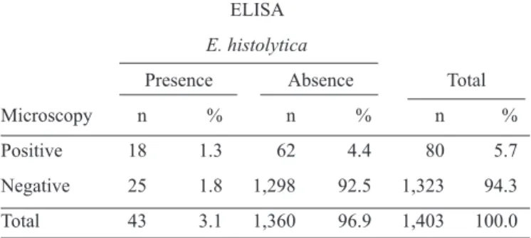
TABLE 2 - Light microscopy and ELISA (E. histolytica II )results. ELISA
E. histolytica
Presence Absence Total
Microscopy n % n % n %
Positive 18 1.3 62 4.4 80 5.7
Negative 25 1.8 1,298 92.5 1,323 94.3
Total 43 3.1 1,360 96.9 1,403 100.0
ELISA: Enzyme-linked immunosorbent assay; E: entamoeba.
study period, Divinópolis had 10,656 students aged 6 to 14 years who were enrolled in 36 municipal schools. The city is
approximately 100km from Belo Horizonte, the state capital.
Of its 213,016 residents, 207,516 live in urban neighborhoods,
and 5,500 live in rural areas6.
Students whose parents or guardians agreed to i ll in a questionnaire and to sign a consent form were given a collection cup with no preservatives. The samples (one per student) were
transported on ice to the Universidade Federal de São João
del-Rei (UFSJ) Laboratory of Immunology and Parasitology,
prepared on the day of collection, and processed using the Hoffmann-Pons-Janer (HPJ, or Lutz) method7. To increase the
likelihood of parasite detection, four qualii ed professional examined each sample (100% of i elds read). An aliquot of each
sample was stored at -20°C for later coproantigen testing using an
E. histolytica/E. dispar ELISA (Enzyme-linked immunosorbent
assay) kit (IVD® Research, Carlsbad, CA, USA) for the in vitro
detection (but not discrimination) of E. histolytica and E. dispar. According to the manufacturer’s instructions, the immunoassay
is based on the interaction of monoclonal antibodies conjugate with peroxidase that bind to antigens of E. dispar and E. histolytica, and the reaction is revealed by the addition of a
substrate containing tetramethylbenzidine and peroxide. The kit has a sensitivity of 88% and a specii city of 100%8. In comparison, the E. histolytica II kit (TechLab®, Blacksburg, VA,
USA) is an immunoassay based on the interaction of monoclonal antibodies with the single antigenic determinant adhesin present at the galactose afi nity E. histolytica. The kit has a sensitivity
of 96.9% and a specii city of 100%9. All tests were run and interpreted according to the manufacturer’s instructions.
The data were encoded and processed using Statistical Package for the Social Sciences (SPSS) software, version
19.0, American University in Cairo - Department of University Academic Computing Technologies (UACT). The
Chi-squared test was used to compare proportions, and the adopted signii cance level was 5% (p-value < 0.05). To compare
the microscopy test and the ELISA kit (IVD®), the sensitivity
and specii city, positive predictive value (PPV), and negative predictive value (NPV) were computed, assuming that the
ELISA kit (IVD®) can adequately serve as the gold standard, using a dichotomous approach10.
Ethical considerations
The investigation was approved by the research ethics committee (opinion 56/2009) and was performed from February 2010 to October 2011.
The microscopy results revealed a 5.7% (80/1,403) prevalence
of infection for the E. histolytica/E. dispar complex. ELISA
(IVD®) testing returned a 15.7% (220/1,403) infection rate for
E. histolytica/E. dispar. A total of 45 (3.2%) samples were positive
by both tests, whereas 35 (2.5%) were positive only by direct microscopy, and 175 (12.5%) were positive only by ELISA (IVD®). Both tests were negative for 1148 (81.8%) samples (table 1).
In comparison with ELISA (IVD®), light microscopy showed 20% sensitivity and 97% specii city, with 56% PPV, 87% NPV, 44% false positives (1 – PPV), and 13% false
negatives.
The E. histolytica II kit (TechLab®, Blacksburg, VA, USA),
specii c for E. histolytica,returned a 3.1% (43/1403) infection rate for E. histolytica. The results of the ELISA (TechLab®)
and microscopy were positive in 18 (1.3%) samples (table 2).
Of the 1,403 samples, 52% (728/1403) were from females, and 48% (675/1403) were from males. The ages and genders of the subjects were evenly distributed. A signii cant association (p-value = 0.01) was observed for E. histolytica with females
but with not males.
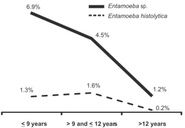
E. histolytica infection was detected in all age groups, with
the highest number of cases in individuals aged > 9 and ≤ 12
years (Figure 1). When the study population was segmented
by age range, no signii cant association was observed for the
age groups.
Divinópolis county is composed of 11 so-called planning regions, 2 of which are rural and 9 of which are urban.
E. histolytica cases were detected in 7 regions (the Southeast,
DIscUssIOn
1.3% 1.6%
0.2% 6.9%
4.5%
1.2%
< 9 years > 9 and<12 years >12 years
Entamoeba sp.
Entamoeba histolytica
FIGURE 1 - Prevalence of E. histolytica and Entamoeba sp. in 6- to 14-year-old students, by age. Divinópolis, Minas Gerais, Brazil, from February 2010 to October 2011 (n = 1,403).
Data on the prevalence of E. histolytica have emerged
from studies on helminths and protozoans that were based
on microscopic investigation of cysts and/or trophozoites in clinical specimens. Until relatively recently, E. histolytica and
E. dispar were not differentiated, and infection with either of
the two species was referred to as amebiasis, resulting in an
overestimation of the true prevalence11.
However, the procedure for distinction has been reevaluated
since Clark and Diamond12 demonstrated that E. histolytica and E. dispar are distinct species that, despite morphological similarities, differ in pathogenicity. The genus Entamoeba
contains many morphologically similar species, several of
which, including E. histolytica, E. dispar, E. moshkovskii,
E. polecki, and E. hartmanni, can be found in human stools13,14.
Therefore, the epidemiology of amebiasis is confusing, mainly because of the recently appreciated distinctions between
E. histolytica, E. dispar,and E. moshkovskii11. Because light
microscopy does not efi ciently identify E. histolytica, other
tools have been developed to differentiate between Entamoeba
species, including polymerase chain reaction (PCR), isoenzyme
analysis, and ELISA, which are well suited for estimations of
true prevalence and the treatment of patients5. Malatyali et al.15 advocated the use of sensitive and effective tests, such as ELISA, for antigen detection in stools.
Several epidemiological studies have been conducted to estimate the incidence and prevalence of amebiasis by testing commercially available antigens from various manufacturers11.
Our study distinguished E. histolytica from other amebae
and strengthened existing epidemiological data. Moreover, the study evaluated the ELISA kit (IVD®) as a screening test.
In the present investigation, microscopy underestimated the
number of subjects infected with E. histolytica/E. dispar (5.7%),
in contrast to the E. histolytica/E. dispar ELISA kit (IVD®)
(15.7%). Whereas the ELISA (IVD®) was positive for 220 of the patients who had E. histolytica/E. dispar cysts in their
stools, only 45 samples were positive in both tests. Additionally, 175 samples with negative results by direct microscopy were positive in the ELISA (IVD®) antigen detection test. This difference may be attributed to the quantity of the pathogen in the samples. Stools with a low number of cysts may be negative by direct microscopic examination but may yield
positive results using ELISA3.
A comparison of the samples by light microscopy and ELISA (IVD®) revealed a low sensitivity (20%) and a high specii city (97%) for light microscopy. The high NPV of 87% reduced the likelihood of false-negative results, yet the low PPV of 56% rendered the test unreliable. However, positivity on microscopy does not rule out the possibility that 44% of the samples are
negative. Delialioglu et al.3 reported that microscopy provided
53.8% sensitivity and 94% specii city, with 78% PPV and 17% NPV, relative to an ELISA kit (Ridascreen Entamoeba, R-Biopharm AG, Darmstadt, Germany). In another study,
compared with an ELISA triage kit (ProSpecT EIA, Alexon Inc., Sunnyvale, CA, USA), microscopy was more specii c (92.1%) but less sensitive (68.4%)16. The ELISA kit (Alexon-Trend,
Inc., Sunnyvale, CA, USA) had a sensitivity of 54.5% and a specii city of 94%. If matched with culture and microscopy, the sensitivity of direct microscopic examination was 66%, and the specii city was 83.7%. However, the results should be coni rmed with a larger number of fecal samples17.
Considering these data, the low sensitivity of microscopy may have been inl uenced by the collection of a single stool
sample per student. According to Ravdin18, theexamination
of three separate stool specimens is required to attain 90% sensitivity, and a single examination identii es only 40% to 60% of infections. If feces were collected more than once and were i xed in preservatives, a higher prevalence of E. histolytica/E. dispar would be expected11. These data suggest that the ELISA
(IVD®) can be used as a screen for the immediate testing of
stools. The performance of antigen detection assays suggests that
they may be considered as reference standards for the detection
of E. histolytica and E.dispar5,19.
However, microscopy should still be considered as a
screening method for the detection of Entamoeba found in human stools, despite the fact that this technique cannot
differentiate between E. histolytica, E. moshkovskii, and
E. dispar, although E. polecki, E. coli, and E. hartmanni can be
differentiated morphologically from E.histolytica20.
One of the problems with screening kits is that they cannot differentiate between the amebae. However, the ELISA kit (TechLab®) is commercially available for the specii c, direct
detection of an E.histolytica antigen in stool specimens5,19. In the
1,403 samples subjected to ELISA (TechLab®), the prevalence
of E. histolytica was 3.1%. Haque et al.21, based on isoenzyme analysis of 202 samples from symptomatic individuals seen at the International Center for Diarrheal Disease Research in
Dhaka, Bangladesh, obtained 52 culture-positive results using
ensured faster diagnosis than when using isoenzyme analysis
and achieved a higher sensitivity and specificity than did microscopy. The E. histolytica II ELISA (TechLab®) correctly
identii ed 21 of 22 cases of E. histolytica infection and 28 of 30 for E. dispar cases. Haque et al.19, examining 2000 samples
using two TechLab® kits (the E. histolytica II ELISA kit and an
Entamoeba ELISA kit), reported prevalence rates of 4.2% for E. histolytica and 6.5% for E. dispar in children aged 1 to 14
years who were living in the vicinity of Dhaka and presenting with diarrhea. In contrast, in asymptomatic children, the percentages were 1% for E. histolytica and 7% for E. dispar.
Following the same strategy, Nesbitt et al.22 examined 842
samples from Kilimanjaro, Tanzania, and detected prevalence values of 1% for E. histolytica and 7.3% for E. dispar. The high prevalence of E. histolytica infection detected in the present
study warrants the implementation of actions directed toward
health promotion and preventive measures.
The present results also corroborate previous i ndings23 that indicated that females are more prone than males to
E. histolytica infection. In Brazil, amebiasis rates are highest
in the northern region of the country, where both intestinal and extraintestinal forms of the disease exist, with serious public health implications24. In Belém, the capital city of the
northern state of Pará, a prevalence rate of 29.35% has been
reported using the E. histolytica II ELISA (TechLab®)25. The highest reported E. histolytica prevalence rates in Brazil that
were detected using the E. histolytica II kit (TechLab®) were
36.6% (30/82) for stool samples from the state of Rondônia in Ariquemes and 19.4% (26/134) for stools taken from residents
of Monte Negro26.
In Pernambuco state, in northeastern Brazil, Dourado et al.27 detected only E. dispar, whereas in Macaparana county, within
the same state, all samples investigated by Pinheiro et al.28 tested negative in an E. histolytica-specii c ELISA (TechLab®) and
positive for E. dispar using a molecular biology method.
In southeastern Brazil, using light microscopy, Santos et al.29 detected a 21% prevalence of the E. histolytica/E. dispar
complex in urban and rural areas of Rio de Janeiro State, yet only two samples tested positive for E. histolytica by PCR and E. histolytica II ELISA (TechLab®). However, in São Leopoldo,
within the southern State of Rio Grande do Sul, Tomé and
Tavares30 found no cases of E. histolytica infection using the
E. histolytica II ELISA (TechLab®).
Light microscopy has several limitations when applied to the diagnosis of amebiasis, given factors such as examiner experience and similarities between Entamoeba cysts3, which can increase the likelihood of false-positive results4. Despite the
low cost of light microscopy compared with culture, isoenzyme
analysis, antigen detection, and PCR, the method’s dependence
on subjective diagnosis limits its reliability3. Therefore, microscopy is not appropriate for either rapid disease diagnosis or prevalence studies.
According Ngui et al.31, molecular techniques are indeed promising tools for epidemiological studies, particularly in discriminating the pathogenic from the non-pathogenic species of Entamoeba. E. moshkovskii, another morphologically
indistinguishable human parasitic Entamoeba, has not
been mentioned, nor has it been considered a contributor to prevalence i gures in endemic areas11. Molecular techniques that can differentiate all studied species of Entamoeba, including E. moshkovskii, in human specimens have already
been reported in Italy, Bangladesh, India, Australia, Turkey,
Iran, and Malaysia31-36.
It is necessary to use new techniques to differentiate
Entamoeba diagnoses37 and to establish a readily available and
cost-effective test for the specii c diagnosis of amebiasis caused by E. histolytica in public laboratories26.
Diagnostic methods that are more sensitive and specii c than light microscopy are required to establish the true distributions
of E. histolytica and to reduce the rates of unnecessary treatment,
thereby discouraging the development of drug resistance,
precluding the risks of side effects, and reducing the costs
of hospitalization. The present i ndings demonstrate that the ELISA kit (IVD®) can be used as an alternative screening tool. In addition, this assay could be utilized by personnel who do
not have extensive training in manual parasitological methods. The determination of the true prevalence of E. histolytica
infection among students from southeastern Brazil is very
crucial, as this information will lead to a better understanding of the public health problem and will help outline measures for controlling amebiasis.
AcKnOWLeDGMenTs
The authors wish to thank the Program for Health Education
through Work of the Brazilian Ministry of Health, the University Extension Program (ProExt) of the Higher Education Division of the Brazilian Ministry of Education, and the Universidade Federal de São João del- Rei in Minas Gerais.
references
The authors declare that there is no conl ict of interest.
cOnfLIcT Of InTeresT
1. World Health Organization (WHO). Amoebiasis. Weekly Epidemiological Record 1997; 72:97-100.
2. Huston CD, Petri WA. Amebiasis: clinical Implications of the recognition of Entamoeba dispar. Curr Infect Dis 1999; 1:441-447.
3. Delialioglu N, Aslan G, Sozen M, Babur C, Kanik A, Emekdas G. Detection of Entamoeba histolytica/Entamoeba dispar in Stool Specimens by Using Enzyme-linked Immunosorbent Assay. Mem Inst Oswaldo Cruz 2004; 99:769-772. 4. Petri WA, Haque R, Lyerly D, Vines RR. Estimating the impact of
amebiasis on health. Parasitol Today 2000; 16:320-321.
5. Haque R, Petri WA. Diagnosis of amebiasis in Bangladesh. Arch Med Res 2006; 37:273-276.
6. Instituto Brasileiro de Geograi a e Estatística (IBGE). Cidades [Internet]. [Cited 2011 July 10] Available at: http://www.ibge.gov.br/cidadesat/topwindow.htm?1. 7. Hoffmann WA, Pons JA, Janer JL. The sedimentation concentration
method in Schistosomiasis mansoni. Puert Rico J Publ Health 1934; 2:283-298.
8. ELISA kits Entamoeba histolytica/Entamoeba dispar -IVD®® Research, Carlsbad, CA, USA - kit label; 2011.
9. ELISA kits E. histolytica II – Techlab®, Blacksburg, VA, USA - kit label; 2011.
10. Fletcher RH, Fletcher SW. Epidemiologia Clínica Elementos Essenciais, Porto Alegre: Artmed; 2006.
11. Tengku SA, Norhayati M. Public health and clinical importance of amoebiasis in Malaysia: A review.Trop Biomed 2011; 28:194-222. 12. Clark CG, Diamond LS. A redescription of Entamoeba histolytica
Schaudinn, 1903 (Emended Walker, 1911) separating it from Entamoeba dispar Brumpt, 1925. J Eukaryot Microbiol 1993; 40:340-344.
13. Clark CG, Diamond LS. Ribosomal RNA genes of ‘pathogenic’ and ‘nonpathogenic’ Entamoeba histolytica are distinct. Mol Biochem Parasitol 1991; 49:297–302.
14. Silva EF, Gomes MA. Parasitologia Humana - Amebíase: Entamoeba histolytica/Entamoeba díspar. 12rd ed. São Paulo: Atheneu; 2011. 15. Malatyalı E, Ozçelik S, Celiksöz A. The investigation of Entamoeba
histolytica prevalence in some villages of Sivas by ELISA method. Türkiye Parazitol Derg 2011; 35:6-9.
16. Pillai DR, Keystone JS, Sheppard DC, MacLean JD, MacPherson DW, Kain KC. Entamoeba histolytica & Entamoeba dispar: epidemiology and comparison of diagnostic methods in a setting of non endemicity. Clin Infect Dis 1999; 29:1315-1318.
17. Gatti S, Swierczynski G, Robinson F, Anselmi M, Corrales J, Moreira J, et al. Amebic infections due to the Entamoebahistolytica-Entamoeba dispar complex: a study of the incidence in a remote rural area of Ecuador. Am J Trop Med Hyg 2002; 67:123-127.
18. Ravdin JI. Diagnosis of invasive amoebiasis - time to end the morphology era. Gut 1994; 35:1018-1021.
19. Haque R, Faruque ASG, Hahn P, Lyerly DM, Petri WA. Entamoeba histolytica and Entamoeba dispar infection in children in Bangladesh. J Infect Dis1997; 175:734-736.
20. Santos HLC, Bandyopadhyay K, Bandea R, Peralta RHS, Peralta JM, Silva AJ. LUMINEXW: a new technology for the simultaneous identii cation of i ve Entamoeba spp. commonly found in human stools. Parasites & Vectors 2013; 6:1-9.
21. Haque R, Neville LM, Hahn P, Petri WA. Rapid diagnosis of Entamoeba infection by using Entamoeba and E. histolytica stool antigen detection kits. J Clin Microbiol1995; 33:2558-2561.
22. Nesbitt RA, Mosha FW, Katki HA, Ashraf M, Assenga C, Lee CM, et al. Amebiasis and comparison of microscopy to Elisa technique in detection of Entamoeba histolytica and Entamoeba dispar. J Nat Med Assoc 2004; 96:671-677.
23. Blessmann J, Van LP, Nu PAT, Thi HD, Muller-Myhsok B, Buss H, et al. Epidemiology of amebiasis in a region of high incidence of amebic liver abscess in central Vietnam.Am J Trop Med Hyg2002;66:578-583. 24. Cunha AS, Silva EF, Raso P, Melo SM. Patogenia da amebíase I. Aspectos
clínicos da amebíase no Brasil com especial referência aos estudos
realizados em 3 grupos populacionais de regiões geográi cas distintas. Rev Inst Med Trop São Paulo 1977; 19:289-300.
25. Silva MCM, Monteiro CSP, Araújo BAV, Silva JV, Póvoa MM. Determinação da infecção por Entamoeba histolytica em residentes da área metropolitana de Belém, Pará, Brasil, utilizando ensaio imunoenzimático (ELISA) para detecção de antígenos. Cad Saúde Pública 2005; 21:969-973.
26. Santos RV, Nunes JS, Camargo JASA, Rocha EMM, Fontes G, Camargo LMA. High occurrence of Entamoeba histolytica in the municipalities of Ariquemes and Monte Negro, State of Rondônia, Western Amazonia-Brazil. Rev Inst Med Trop Sao Paulo 2013;55:193-196.
27. Dourado A, Maciel A, Aça IS. Occurrence of Entamoeba histolytica/ Entamoeba dispar in ambulatory patients of Recife, PE.Rev Soc Bras Med Tropl 2006; 39:388-389.
28. Pinheiro SMB, Carneiro RM, ACA IS, Irmão JI, Morais JRMA, Coimbra MRM, et al. Determination of the prevalence of Entamoeba histolytica and E. dispar in the Pernambuco state of Northeastern Brazil by a polymerase chain reaction.Am J Trop Med Hyg 2004; 70:221-224.
29. Santos HL, Peralta RH, Macedo HW, Barreto MG, Peralta JM. Comparison of multiplex-PCR and antigen detection for differential diagnosis of Entamoeba histolytica. Braz J Infect Dis 2007; 11:365-370. 30. Tomé JBS, Tavares RG. Differentiation between Entamoeba histolytica
and Entamoeba dispar by means of immunoenzymatic assay for antigen detection in children fecal samples. Rev Inst Adolfo Lutz 2007; 66:305-307. 31. Ngui R, Angal L, Fakhrurrazi SA, Ai Lian YL, Ling LY, Ibrahim J, et al.
Differentiating Entamoeba histolytica, Entamoeba dispar and Entamoeba moshkovskii using nested polymerase chain reaction (PCR) in rural communities in Malaysia. Parasit Vectors 2012; 5:187-193.
32. Ali IK, Hossain MB, Roy S, Ayeh-Kumi PF, Petri Jr WA, Haque R, et al. Entamoeba moshkovskii infections in children, Bangladesh. Emerg Infect Dis 2003; 9:580-584.
33. Fotedar R, Stark D, Beebe N, Marriott D, Ellis J, Harkness J. Laboratory diagnostic techniques for Entamoeba species. Clin Microbiol Rev 2007; 20:511-532.
34. Fotedar R, Stark D, Marriott D, Ellis J, Harkness J. Entamoeba moshkovskii infections in Sydney, Australia. Eur J Clin Microbiol Infect Dis 2008; 27:133-137.
35. Solaymani-Mohammadi S, Rezaian M, Babaei Z, Rajabpour A, Meamar AR, Pourbabai AA, et al. Comparison of a stool antigen detection kit and PCR for diagnosis of Entamoeba histolytica and Entamoeba dispar infections in asymptomatic cyst passers in Iran. J Clin Microbiol 2006; 44: 2258-2261.
36. Tanyuksel M, Ulukanligil M, Guclu Z, Araz E, Koru O, Petri WA Jr. Two cases of rarely recognized infection with Entamoeba moshkovskii. Am J Trop Med Hyg 2007; 76:723-724.