InTrODUcTIOn
Address to: Dra Maria do Desterro Soares Brandão Nascimento. Depto de Patologia/NIBA/UFMA. Av. dos Portugueses 1966, Prédio do CCBS, Bloco 3,
Sala 3A, Cidade Universitária do Bacanga, 65080-040 São Luis, MA, Brasil.
Phone: 55 98 3272-8535; Fax: 55 98 3272-8535
e-mail:[email protected] Received 10 November 2013 Accepted 30 January 2014
Diversity and dynamics of airborne fungi in São Luis,
State of Maranhão, Brazil
Geusa Felipa de Barros Bezerra
[1],
Silvio Monteiro Gomes
[2],
Marcos Antonio Custódio Neto
da Silva
[3],
Ramon Moura dos Santos
[1],
Walbert Edson Muniz Filho
[1],
Graça Maria de Castro Viana
[1]and Maria do Desterro Soares Brandão Nascimento
[1],[4][1]. Departamento de Patologia, Núcleo de Imunologia Básica e Aplicada, Universidade Federal do Maranhão, São Luis, MA. [2]. Departamento de Biologia,
Universidade Federal do Maranhão, São Luis, MA. [3]. Curso de Medicina, Universidade Federal do Maranhão, São Luis, MA. [4]. Centro de Estudos Superiores de Caxias, Universidade Estadual do Maranhão, Caxias, MA.
ABStRACt
introduction: This study aimed to identify airborne fungi in São Luis, Maranhão, Brazil, to determine the prevalent genera
and to correlate these genera with the area and season. Methods: In total, 1,510 colony-forming units (CFUs) of airborne fungi
were isolated from the north, south, east and west sides and from the center of the city from January to December 2007. The samples were collected on Petri dishes that were exposed to the fungi by the gravitational method. Results: Twenty genera of
fungi were isolated; the most common were Aspergillus (33.5%), Penicillium (18.8%), Cladosporium (14.2%), Curvularia
(10.6%) and Fusarium (7.6%). The CFUs of the fungi were statistically signii cant (p < 0.0001). Fungal biological diversity was
present all year, without any large seasonal variations but with slight increases in May, August and September. Conclusions:
The fungal genera identii ed in this study were correlated with natural systems and could be useful when evaluating the impact
of environmental changes on the region.
Keywords: Fungi. Environment.Biodiversity.
Fungi, especially i lamentous fungi, which are common aeroallergens, form a major part of bioaerosols1. Fungi are
ubiquitous in outdoor air, and their concentrations, aerodynamic
diameters and taxonomic compositions have potentially important implications for human health2.
The diversity and abundance of anemophilous microorganisms can be inl uenced by and can interfere with environmental conditions. These microorganisms are inl uenced by factors such
as season, temperature, the relative humidity of the air and other
parameters that exhibit seasonal variation3,4.
The relationships among allergic exposure, the fungal presence in indoor and outdoor environments and consequent allergic diseases are not fully understood5,6. Therefore, it is important
to know both the frequency of certain airborne fungi and their distributions according to the season and the main environment
(i.e., indoor or outdoor) in order to evaluate their correlations
with respiratory symptoms related to allergic processes7-9.
These fungi can be used to assess effects on the environment and could contribute to determining the principal changes. The spores of fungi can be present in air particles and can
potentially influence the hydrological cycle and climatic
changes. In addition, humans are exposed daily to bioaerosols in their personal and professional lives, and these airborne particles represent a potential biological occupational hazard10.
Biological particles in the air are approximately 40% organic carbon by mass and can be an important source of bioaerosols in the atmosphere above continents4. Despite the importance
of airborne fungi, very little is known about their diversity11, especially of the fungi in São Luis, Maranhão.
For this reason, it was important to perform further observations to determine and characterize the frequencies of the main airborne fungi in outdoor environments and to identify possible correlations with seasonality and the possibility of
monitoring allergic respiratory diseases.
MeTHODs
Airborne fungi were collected between January and December 2007 in i ve outdoor areas of the City of São Luis (northern, southern, eastern, western and central), located in the
State of Maranhão, Brazil. São Luis is located at a latitude of
2º31’47” S and a longitude of 44º18’10’’W, and it is 24m above
sea level. This city covers an area of approximately 830km2 and
resULTs An analysis of the mycobiota was performed using Petri
dishes (10cm by 2cm) containing 10mL of Sabouraud agar medium. The dishes were exposed to open air, in selected districts and in a predetermined region, for 15min every month, while placed at a height of 1.5m from the ground to collect fungal spores by the action of gravity5,10. Three dishes were exposed each month in each area, resulting in a total of 15 samples per month and 180 samples per year. It is important to emphasize that the areas where the research was conducted were randomly selected. The samples used for microculture and taxonomic analysis were obtained from the colony-forming units (CFUs)13,14.
Statistical analysis
The results were analyzed using SPSS software, Chicago, USA, SPSS Inc® version 16.0 for Windows (2007). The study was cross-sectional and observational, and descriptive statistical techniques were used to assess all of the variables, with the aid of graphs and tables of frequencies. Two-tailed analysis of variance was performed to determine the relationships of the frequencies of the species (from among the i ve most frequent species) and the months of the year with the number of CFUs.
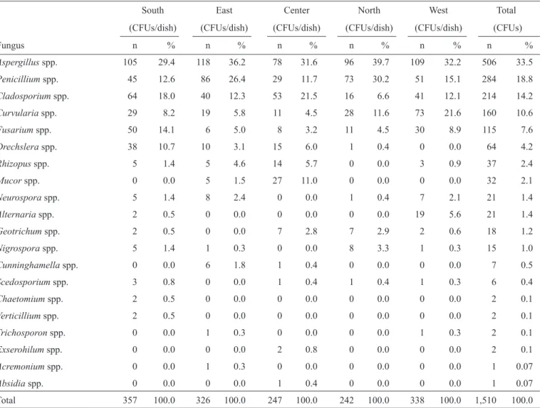
Twenty genera of fungi were isolated in this study. Depending on the region, it was possible to isolate between 10 and 14 genera, as shown in table 1 (χ2 = 535.95, p < 0.0001).
The main genera found in all of the regions were Aspergillus (33.5%), Penicillium (18.8%), Cladosporium (14.2%),
Curvularia (10.6%) and Fusarium (7.6%), and the detailed
distributions are shown in table 1.
Figure 1 shows that the median number of CFUs/dish during
the rainy season (January to June) was 20 (maximum = 279 and minimum = 0), and the corresponding number during the dry season was 14 (maximum = 227 and minimum = 0), using the Mann-Whitney test (p = 0.96).
A comparison of the average number of CFUs for the i ve most frequent fungi (table 2), using Tukey’s test, showed that the number for Aspergillus differed signii cantly (p < 0.05) from the numbers for the genera Cladosporium, Curvulariaand Fusarium.
TABLE 1 - Frequency distribution of colony-forming units of fungi by area in São Luis, State of Maranhão, between January and December 2007.
South East Center North West Total
(CFUs/dish) (CFUs/dish) (CFUs/dish) (CFUs/dish) (CFUs/dish) (CFUs)
Fungus n % n % n % n % n % n %
Aspergillus spp. 105 29.4 118 36.2 78 31.6 96 39.7 109 32.2 506 33.5
Penicillium spp. 45 12.6 86 26.4 29 11.7 73 30.2 51 15.1 284 18.8
Cladosporium spp. 64 18.0 40 12.3 53 21.5 16 6.6 41 12.1 214 14.2
Curvularia spp. 29 8.2 19 5.8 11 4.5 28 11.6 73 21.6 160 10.6
Fusarium spp. 50 14.1 6 5.0 8 3.2 11 4.5 30 8.9 115 7.6
Drechslera spp. 38 10.7 10 3.1 15 6.0 1 0.4 0 0.0 64 4.2
Rhizopus spp. 5 1.4 5 4.6 14 5.7 0 0.0 3 0.9 37 2.4
Mucor spp. 0 0.0 5 1.5 27 11.0 0 0.0 0 0.0 32 2.1
Neurospora spp. 5 1.4 8 2.4 0 0.0 1 0.4 7 2.1 21 1.4
Alternaria spp. 2 0.5 0 0.0 0 0.0 0 0.0 19 5.6 21 1.4
Geotrichum spp. 2 0.5 0 0.0 7 2.8 7 2.9 2 0.6 18 1.2
Nigrospora spp. 5 1.4 1 0.3 0 0.0 8 3.3 1 0.3 15 1.0
Cunninghamella spp. 0 0.0 6 1.8 1 0.4 0 0.0 0 0.0 7 0.5
Scedosporium spp. 3 0.8 0 0.0 1 0.4 1 0.4 1 0.3 6 0.4
Chaetomium spp. 2 0.5 0 0.0 0 0.0 0 0.0 0 0.0 2 0.1
Verticillium spp. 2 0.5 0 0.0 0 0.0 0 0.0 0 0.0 2 0.1
Trichosporon spp. 0 0.0 1 0.3 0 0.0 0 0.0 1 0.3 2 0.1
Exserohilum spp. 0 0.0 0 0.0 2 0.8 0 0.0 0 0.0 2 0.1
Acremonium spp. 0 0.0 1 0.3 0 0.0 0 0.0 0 0.0 1 0.07
Absidia spp. 0 0.0 0 0.0 1 0.4 0 0.0 0 0.0 1 0.07
Total 357 100.0 326 100.0 247 100.0 242 100.0 338 100.0 1,510 100.0
January to June July to December
3 2
1 0
CFU/dish
250
200
150
100
50
0
TABLE 2 - Comparison of the average number of CFUs for the i ve
most frequent fungi and the frequency distribution of the recovered colony-forming units of fungi by area in São Luis, State of Maranhão, between January and December 2007.
Fungus Average ± SD
Aspergillus 42.17 ± 19.6
Penicillium 23.67 ± 12.5
Cladosporium 17.83 ± 22.1
Curvularia 13.33 ± 10.0
Fusarium 9.58 ± 10.8
Region Total CFUs
South 357 23.6
West 338 22.4
East 326 21.6
North 256 17.0
Center 233 15.4
Total 1,510 100.0
CFUs: colony-forming units; SD: standart deviation.
Regarding seasonality, we observed the occurrence of fungi throughout the year, with a slight increase in the percentages of fungal genera in the months of May, August and September
(Figures 2 and 3).
When the percentages of CFUs collected in each of the
i ve (northern, southern, central, eastern and western) areas of São Luis were analyzed, a statistically signii cant difference was observed (Table 2; χ2 = 38.99, p < 0.0001). The prevalence in
the central and northern areas was lower than in the other areas.
FIGURE 1 - Frequency distribution of the different genera of fungi isolated in
the rainy season (January to June) and the dry season (July to December) in São Luis, State of Maranhão, between January and December 2007. Legend
for Figure 1: Box plot. Medians and quartiles. CFUs per period/season.
Mann-Whitney test, p = 0.96. CFUs: colony-forming units.
FIGURE 2 - Fungal growth and the climate variables humidity, temperature and rainfall, recorded monthly in São Luis, State of Maranhão, Brazil, in 2007. CFUs: colony-forming units.
0 20 40 60 80 100
Max
Mean
Min
H
u
m
idity(%
)
0 5 10 15 20
Rainfall (mm/24h)
Rainfall(
m
m
/2
4
h
)
0 2 4 6 8 10 12
Fungi - CFU/dish (%)
F
u
n
g
i-C
F
U
/d
is
h
(%
)
0 5 10 15 20 25 30 35 40
Jan Feb Mar Apr May Jun Jul Aug Sep Oct Nov Dec
Max
Mean
Min
T
e
m
p
e
ra
tu
re
(
˚C
0 10 20 30 40 50 60 70 80
J
a
n
F
e
b
Ma
r
Apr Ma
y
J
u
n
J
u
l
Aug Se
p
Oct No
v
D
e
c
CFUs
Month
Aspergillusspp.
Penicilliumspp.
Cladosporium spp.
Curvulariaspp.
Fusariumspp.
FIGURE 3 - Seasonal distribution of the i ve most prevalent genera isolated in
São Luis, State of Maranhão, in 2007. CFUs: colony-forming units.
DIscUssIOn
Aspergillus was the most commonly isolated genus in the
current study, and Penicillium was the second most commonly isolated genus. In Mexico City, Penicillium is also the second most common genus15. However, in studies performed in
other countries, such as France, the USA, Chile and Cuba,
Cladosporium has stood out as the most prevalent genus16-18.
Aspergillus, Penicillium, Cladosporium, Curvularia and
Fusarium are the most frequent outdoor species, according
to previous research19,20. The genera of fungi identii ed in the
present study were correlated with natural systems and could be useful in assessing the impact of environmental changes on
the region studied.
In Brazil, the occurrence of airborne fungi in indoor and outdoor areas has been investigated in different regions3,21,22. In the Northeast, Fortaleza, Natal and Recife are climatically
similar cities. In Fortaleza, Ceará reported that the genera
Aspergillus and Penicillium prevailed10. Additionally,
Aspergillus and Penicillium were more frequent genera in
Recife and Natal, respectively23,24. Curvularia appeared with
the highest frequency only in Fortaleza8. In Belém, Pará,
Aspergillus, Penicillium and Cladosporium were reported to be
the most prevalent genera isolated25.
Despite differences in climate, in Porto Alegre, Rio Grande do Sul, Aspergillus was the second most frequent genus26. In Botucatu, São Paulo, Cladosporium was the most frequent
genus27. In the metropolitan area of São Paulo, Penicilliumspp and Aspergillus spp. were the dominant species both indoors
and outdoors28.
High relative humidity is essential for the development of
fungi, and sunny weather favors the release of spores29. High temperature and humidity can result in increased concentrations of fungi19. A high concentration of spores in the air is important
because this situation can result in increases in allergic diseases
of the respiratory system10.
In the South and the West, the greatest numbers of airborne fungal genera were isolated (table 1). These regions have greater areas of vegetation covering them. The North is near the sea and
presents a low level of air pollution. In Centro (CE), a small number of airborne fungi were isolated, possibly due to higher
levels of pollution.
The quantitative analysis of colony counts in the northern,
southern, central, eastern and western areas was statistically signii cant (p = 0.0002) when assessing the i ve most frequent genera relative to the months of the year. Seasonal l uctuations were also reported in Santiago, Chile30.
The literature reveals that Cladosporium has been repeatedly
found indoors, particularly in house dust13. However, in the current paper, Cladosporium was found outdoors considerably more
often, given that it was the second most isolated genus. The occurrence of a great number of airborne fungi emphasizes the importance of studying airborne fungi in São Luis, Maranhão. The climate of tropical areas supports the growth of airborne fungi, resulting in high levels of fungal spores in the air, which
can increase the incidence of allergic respiratory diseases related to these fungi.
AcKnOWLeDGMenTs
The authors would like to express their gratitude to Maranhão Federal University (UFMA) for allowing the use of
the Applied and Basic Immunology Center (NIBA) facilities.
The authors declare that there is no conl ict of interest.
cOnfLIcT Of InTeresT
fInAncIAL sUPPOrT
This research was supported by Fundação de Amparo à Pesquisa e ao Desenvolvimento Cientíi co e Tecnológico do
references
1. Lanier C, André V, Séguin V, Heutte N, El Kaddoumi A, Bouchart V,
et al. Recurrence of Stachybotrys chartarum during mycological and toxicological study of bioaerosols collected in a dairy cattle shed. Ann
Agric Environ Med 2012; 19:61-67.
2. Yamamoto N, Bibby K, Qian J, Hospodsky D, Rismani-Yazdi H,
Nazaroff WW, et al. Particle-size distributions and seasonal diversity of
allergenic and pathogenic fungi in outdoor air. ISME J 2012; 6:1801-1811. 3. Schoenlein-Crusius IH, Trufem SFB, Grandi RAP, Milanez AI,
Pires-Zottarelli CLA. Airborne fungi in the region of Cubatão, São Paulo State, Brazil. Braz J Microbiol 2001; 32:61-65.
4. Bowers RM, Fierer N, Horanyi E, Hannigan M, Hallar AG, Mccubbin I,
et al. The contribution of biological particles to observed particulate organic carbon at a remote high altitude. Atmos Environ 2009; 43:4278-4282.
5. Bernardi E, Nascimento JS. Airborne fungi at Laranjal beach, Pelotas, Rio Grande do Sul, Brazil. Arq Inst Biol 2005; 72:93-97.
6. Pongracic JA, O’Connor JP, Muilenberg ML, Vaughn B, Gold DR, Kattan M, et al. Differential effects of outdoor versus indoor fungal spores on asthma
morbidity in inner-city children. J Allergy Clin Immunol 2010; 125:
593-599.
7. Chapman JA. How relevant are pollen and mold spore counts to clinical practice? Ann Allergy Asthma Immunol 2000; 84:467-468.
8. Bush RK, Portnoy JM, Saxon A, Terr AI, Wood RA. The medical effects of mold exposure. J Allergy Clin Immunol 2006; 117:326-333. 9. Kalyoncu F. Relationship between airborne fungal allergens and
meteorological factors in Manisa City, Turkey. Environ Monit Assess 2010; 165:553-558.
10. Menezes EA, Trindade ECR, Costa MM, Freire CCF, Cavalcante MS,
Cunha FA. Airborne fungi isolated from Fortaleza city, State of Ceará,
Brazil. Rev Inst Med Trop São Paulo 2004; 46:133-137.
11. Fröhlich-Nowoisky J, Pickersgill DA, Després VR, Pöschl U. Hight diversity of fungi in air particulate matter. Proc Natl Acad Sci U S A 2009; 106:12814-12819.
12. Instituto Brasileiro De Geograi a e Estatística (IBGE). Cidades. [Cited
2007 October 10]. Available at: http://www.ibge.g.,ov.br/cidadesat/ painel/ painel.php?codmun=210300#/.
13. Riddel RW. Permanent stained mycological preparations obtained by slide culture. Mycologia 1950; 42:265-270.
14. De Hoog GS, Guarro J, Gené J, Figueras MJ. Atlas of clinicalfungi. 2nd edition. Utrecht, The Netherlands: Centraal bureau voor Schimmelcultures; 2000.
15. Rosas I, Calderón C, Ulloa M, Lacey J. Abundance of airborne Penicillium
CFU in relation to urbanization in México City. Appl Environ Microbiol
1993; 59:2648-2652.
et al. Analysis of fungal l ora in indoor dust by ribosomal DNA sequence analysis, quantitative PCR, and culture. Appl Environ Microbiol 2008;
74:233-244.
17. Sautour M, Sixt N, Dalle F, L'ollivier C, Fourquenet V, Calinon C, et al.
Proi les and seasonal distribution of airborne fungi in indoor and
outdoor environments at a French hospital. Sci Total Environ 2009; 407: 3766-3771.
18. Almaguer M, Aira MJ, Rodriguez, Rajo FJ, Rojas TI. Study of airborne
fungus spores by viable and non-viable methods in Havana, Cuba. Taylor
and Francis Online 2013; 52:289-298.
19. Kasprzyk I. Aeromycology - Main research i elds of interest during the last 25 years. Ann Agric Environ Med 2008; 15:1-7.
20. Traid-Hoffman C, Jakob T, Behrendt H. Determinants of allergenicity. J Allergy Clin Immunol 2009; 123:558-566.
21. Faria A. Ecological aspects and clinical mycotic l ora of anemophilus. [Doctors Thesis] [Belo Horizonte]: Universidade Federal de Minas Gerais; 1997.
22. Gambale W, Purchio A, Paula CR. Action of abiotic factors on airborne
fungi in the city of Sao Paulo, Brasil. Braz J Microbiol 1983; 14:204-214.
23. Oliveira MTB, Braz RFS, Ribeiro MAG.. Airborne fungi isolated from
Natal, State of Rio Grande do Norte, Brazil. Rev Microbiol (S. Paulo)
1993; 24:198-202.
24. Machado GMR. Fungos anemói los de áreas do grande Recife. [Masters
Dissertation] [Recife]: Universidade Federal de Pernambuco; 1979.
25. Pereira BFP, Melo LE, Costa PF. Fungos Anemói los isolados na cidade
de Belém, Estado do Pará – Brazil. Rev Eletr Bio 2013; 6:82-93.
26. Mezzari A, Perin C, Santos Junior SA, Bernd LAG, Di Gesu G. Airborne fungi and sensitization in atopic individuals in Porto Alegre, RS. Rev Assoc Med Bras 2003; 49:270-273.
27. Crocel J, Silva EGM, Furtado EL, Queluz THAT. Estudo dos fungos
anemói los da cidade de Botucatu e sua correlação com sensibilização em pacientes com doenças alérgicas respiratórias. Rev Bras Alerg
Imunopatol 2003; 26:95-109.
28. Gonçalves FL, Bauer H, Cardoso MR, Pukinskas S, Matos D, Melhem M, et al. Indoor and outdoor atmospheric fungal spores in the São Paulo metropolitan area (Brazil): species and numeric concentrations. Int J Biometeorol 2010; 54:347-355.
29. Godinho R, Lanza M, Godinho A, Rodrigues A, Assiz TML. Frequency
of positive skin tests for airborne allergic agents. Braz J Otorhinolaryngol
2003; 69:824-828.
30. Ibañez V, Thompson Moya L, Mañalich Muxi J. Fungal seasonal
l uctuation of anemophilus fungi in Northern Santiago-Chile. Boletin