Immunohistochemical evaluation of estrogen and progesterone
Immunohistochemical evaluation of estrogen and progesterone
Immunohistochemical evaluation of estrogen and progesterone
Immunohistochemical evaluation of estrogen and progesterone
Immunohistochemical evaluation of estrogen and progesterone
receptors of pre and post-neoadjuvant chemotherapy for breast
receptors of pre and post-neoadjuvant chemotherapy for breast
receptors of pre and post-neoadjuvant chemotherapy for breast
receptors of pre and post-neoadjuvant chemotherapy for breast
receptors of pre and post-neoadjuvant chemotherapy for breast
cancer
cancer
cancer
cancer
cancer
Avaliação imunoistoquímica dos receptores de estrogênio e progesterona no
Avaliação imunoistoquímica dos receptores de estrogênio e progesterona no
Avaliação imunoistoquímica dos receptores de estrogênio e progesterona no
Avaliação imunoistoquímica dos receptores de estrogênio e progesterona no
Avaliação imunoistoquímica dos receptores de estrogênio e progesterona no
câncer de mama, pré e pós-quimioterapia neoadjuvante
câncer de mama, pré e pós-quimioterapia neoadjuvante
câncer de mama, pré e pós-quimioterapia neoadjuvante
câncer de mama, pré e pós-quimioterapia neoadjuvante
câncer de mama, pré e pós-quimioterapia neoadjuvante
JAN PAWEL ANDRADE PACHNICKI1; NICOLAU GREGORI CZECZKO,TCBC-PR2; FILIPE TUON2 ; TEREZA SANTOS CAVALCANTI1 ; ANDRESSA BRESSAN
MALAFAIA1; ANA MARIA TULESKI3
A B S T R A C T A B S T R A C T A B S T R A C T A B S T R A C T A B S T R A C T
Objective Objective Objective Objective
Objective: To evaluate the immunoexpression of estrogen and progesterone receptors in biopsies and surgical specimens of patients with breast cancer before and after neoadjuvant chemotherapy and to correlate their changes with the pattern of response to chemotherapy and diagnosis of menopause. MethodsMethodsMethodsMethodsMethods: We selected 47 patients with histological diagnosis of locally advanced primary breast carcinoma. For each patient there were two blocks: the biopsy specimen and surgical resection one. From these blocks hormone receptor expression was assessed by immunohistochemistry using the technique of streptoavidin-biotin-immunoperoxidase and anti-ER and anti-PR primary antibodies. The statistical analysis used the Student’s t test and the nonparametric Fisher’s exact test, with significance level of 5%. ResultsResultsResultsResults: Of the 47 patients, 30 showed positive immunostainingResults for hormone receptors. There was significant reduction in the levels of both estrogen and progesterone receptors and in their immunoreactivity. In 53.3% we observed changes in levels of estrogen receptor expression, 56.6% in the progesterone receptor, 26.6% in the immunoexpression of estrogen receptor immunoreactivity and 33.3% in the immunoreactivity of the progesterone receptor. There was no statistical correlation between the influence of the response to chemotherapy and the diagnosis of menopause in women with variation in the expression of hormone receptors. ConclusionConclusionConclusionConclusionConclusion: Neoadjuvant chemotherapy has significantly altered hormone receptor immunoreactivity in patients in the sample, reducing its positivity in tumor cells.
Key words: Key words: Key words: Key words:
Key words: Colon. Colitis. Image processing, computer-assisted. Fatty acids, volatile.
From the Post-graduation Program in Principles of Surgery, Evangelic Faculty of Paraná / Evangelic University Hospital of Curitiba and Curitiba Medical Research Institute, Curitiba, Paraná, Brazil.
1. Master’s Degree, Post-Graduation Program in Principles of Surgery, Medical Research Institute of the Evangelic School of Paraná / Evangelic University Hospital of Curitiba; 2. PhD, Permanent Professor, Post-Graduation Program in Principles of Surgery, Medical Research Institute of the Evangelic School of Paraná / Evangelic University Hospital of Curitiba; 3. Medical School Graduate, the Evangelic School of Paraná / Evangelic University Hospital ofCuritiba.
INTRODUCTION
INTRODUCTION
INTRODUCTION
INTRODUCTION
INTRODUCTION
I
ncreasing numbers of patients are being diagnosed worldwide with invasive breast cancer and much is being done in drug development and improvement of operations directed to the individual characteristics of each patient. The expansion of knowledge, together with the implementation of these new techniques, enhances medical research in pursue of new perspectives.One of the biggest challenges for the study and treatment of breast cancer is the resolution of tumor heterogeneity characteristic of these carcinomas. By the end of the last decade the patients who had this diagnosis were treated as having similar diseases, an approach based primarily on morphological classification, what is impossible to justify why the cases with the same
diagnosis and staging could have markedly different outcomes.
In recent years there has been great progress in the conventional treatment of this tumor. Multidisciplinary strategies were developed based on clinical and laboratory evidence, indicating the systemic nature of the disease at diagnosis.
Surgical strategies evolved to less mutilation, since the extent of removal of the tumor showed little influence on prognosis. Conservative operations associated with radiotherapy became effective in local disease control. Chemotherapy and hormone therapy were shown to be important complements to the manifestations, reducing the risk of recurrence and death.
recommended that the first consideration in selecting the type of treatment should be the endocrine responsiveness. The recognition of this fact increased the relevance of pathologic evaluation as to the biological information.
For this reason the estrogen receptor has been the most extensively studied prognostic indicator to date. Several authors have reported a positive relationship between estrogen receptor, increased disease-free interval and better survival of patients. The estrogen and progesterone receptors are currently the most widely used predictive factors for the choice of hormonal treatment1.
Neoadjuvant chemotherapy is also frequently used in the treatment of breast cancer for the purpose of reducing the tumor size and estimate the sensitivity to chemotherapy. In the meantime, the effect of it in the expression of estrogen receptor, progesterone receptor and HER-2 remains uncertain 2. Changes in expression of these biomarkers during neoadjuvant chemotherapy may influence the clinical decision of adjuvant molecular and hormonal therapy 3.
Breast cancer is characterized by cellular heterogenicity, which gives the possibility of different responses to chemotherapy in different cell clones within the same tumor. This heterogenicity can be considered one of the reasons for the negative response to endocrine therapy in estrogen receptor-positive patients 4.
Analyzing biopsies and surgical specimens of patients with breast cancer before and after neoadjuvant chemotherapy, this study aims to: a) to determine the expression of estrogen receptor; b) to determine the expression of the progesterone receptor; c) to correlate the change in tumor size, due to the response to chemotherapy, with the immunoreactivity of estrogen receptors and progesterone; d) to correlate the state of pre-menopausal and post-menopausal women with immunoexpression of estrogen receptors and progesterone.
METHODS
METHODS
METHODS
METHODS
METHODS
This study was conducted at the Post-graduation Program in Principles of Surgery, Evangelic Faculty of Paraná / Evangelic University Hospital of Curitiba / Medical Research Institute, Curitiba, Paraná State, Brazil. The research project was approved by the Ethics Committee of the Evangelic Benevolent Society of Curitiba.
Data were collected for evaluation based on analysis of records and paraffin blocks of 47 patients with histological diagnosis of primary breast carcinoma stage III, regardless of their histological type.
As inclusion criteria, all had undergone core-biopsy or surgical core-biopsy, neoadjuvant chemotherapy with subsequent surgical treatment (radical or conservative surgery). The chemotherapy regimen used for all patients consisted of three cycles of cyclophosphamide - 600 mg/
m2 adriamycin - 60 mg/m2 and 5-fluorouracil - 600 mg/m2, followed by three cycles of docetaxel - 100 mg/m 2, with an interval of 21 days.
Exclusion criteria were: diagnosis of metastases, changing of the clinical stage initially marked, treatment with neoadjuvant chemotherapy regimen different from the established protocol, absence of residual tumor for analysis of hormone receptor immunoreactivity as a result of chemotherapy (complete response).
For comparison between groups, the sample was divided according to the pattern of response to chemotherapy and to the diagnosis of menopause. We did not use the histologic type of tumor for comparison of hormone receptor immunoreactivity. We performed diagnostic confirmation and qualitative analysis of the blocks, excluding the ones that had fixed defects, fungal contamination, a high percentage of necrosis or inconclusive diagnosis.
In the standardization of response to chemotherapy, we used the system proposed by the International Union for Cancer Control, dividing the sample into two groups: a) presence of response (complete response with complete disappearance of the tumor or partial response with a decrease greater than 50% of the tumor); b) non-response (stable disease with decreased below 50%, increase of the tumor less than 25% or disease increase with tumor progression greater than 25%)5.
In the diagnosis of menopause, we used the one proposed by the World Health Organization, dividing the sample into two groups: a) pre-menopausal (menstrual cycles, regular or not, with intervals of less than 12 months), b) postmenopausal (absence of menstrual cycles by a period equal to or greater than 12 months).
The expression of estrogen and progesterone receptors was evaluated with the aid of immunohistochemistry of streptoavidin-biotin-immunoperoxidase and anti-ER primary antibody (Dako, M7047, 1/30) and anti-PR (Novocastra, NCL-, 1/40). All these steps were made with the use of positive controls, tissues from breast cancer with patterns already known positive for estrogen and progesterone.
When ready for evaluation and reading, the slides were sent for analysis by two pathologists, without knowledge of the diagnosis, to classify as for the endocrine responsiveness6.
hypothesis of different probabilities. The significance level was 5%.
RESULTS
RESULTS
RESULTS
RESULTS
RESULTS
The patients had an average age of 50, 24 (51%) being pre-menopausal and 23 (49%) post-menopausal.
With regard to response to neoadjuvant chemotherapy, 28 (59.5%) had partial response and 19 (40.5%) no response to the proposed therapy; 12 (25.5%) were classified as stable disease and seven (15%) with disease progression.
Seventeen patients (36%) had negative immunoexpression of estrogen receptor in the diagnostic biopsy. The same number had no immunoreactivity of progesterone receptor. In these patients there was no change in expression of hormone receptors.
Thirty patients had positive immunostaining of hormone receptors. Of these, 16 (53.3%) had a decrease in the expression of estrogen receptors and 17 (56.6%), in the progesterone receptor. There was a negative immunoexpression of estrogen receptor and progesterone in 26.6% (n = 8) and 33.3% (n = 10), respectively.
When paired and compared by Student’s t test, the expressions of hormonal receptors before and after neoadjuvant chemotherapy showed a significant reduction in the levels of estrogen receptor (p <0.0001) and progesterone (p <0.0001). There was also a reduction in its immunoreactivity, with p = 0.0035 for the changes in
estrogen receptor and p = 0.001 for the progesterone receptor.
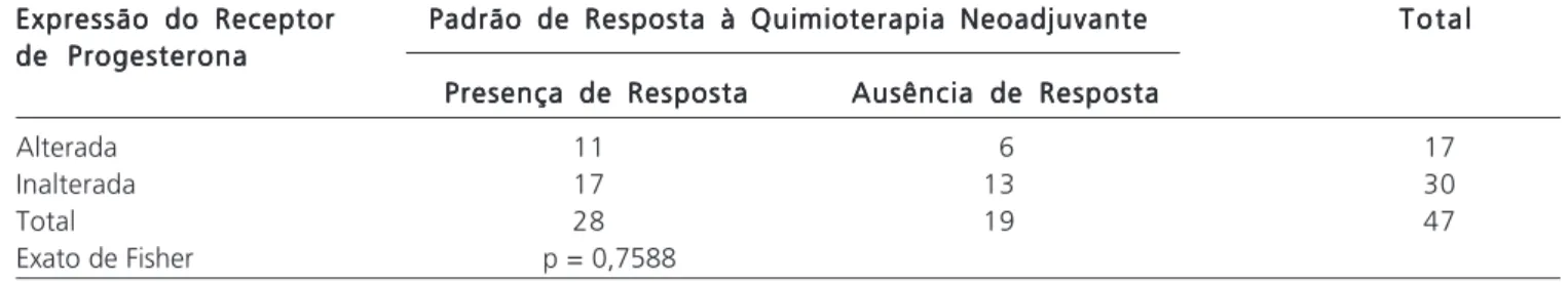
When analyzing the presence or absence of response to neoadjuvant chemotherapy, that is, the actual reduction of tumor size with the variation in the expression of the hormone receptor, there was no statistical significance (p = 0.7631) related to changes in the estrogen receptor immunoreactivity, nor (p = 0.7588) in relation to the progesterone receptor (Fisher’s exact test) (Tables 1 and 2).
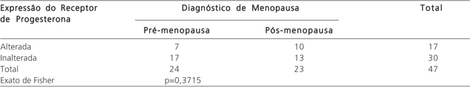
Regarding the diagnosis of pre and post-menopause, when compared with changes in the expression of hormone receptors, there was also no statistic significant differences (p = 0.2270) related to changes in immunoexpression of estrogen receptor, nor ( p = 0.3715) in relation to the progesterone receptor (Fisher’s exact test) (Tables 3 and 4).
DISCUSSION
DISCUSSION
DISCUSSION
DISCUSSION
DISCUSSION
With regard to clinical and pathological aspects of the study group, we did not considered differences in the histological classification of breast tumors in the sample: breast ductal and lobular adenocarcinoma. It would appear that breast carcinomas present in a wide range, not only on morphological aspect, but mainly clinical and in evolution. It expresses high heterogeneity with regard to clinical presentation and biological behavior to therapeutic response, irrespective of histological classification 7.
Tabela 1 Tabela 1Tabela 1 Tabela 1
Tabela 1 - - - Mudanças na imunoexpressão do receptor de estrogênio em relação ao padrão de resposta à quimioterapia neoadjuvante.
Expressão do Receptor Expressão do ReceptorExpressão do Receptor Expressão do Receptor
Expressão do Receptor Padrão de Resposta à Quimioterapia NeoadjuvantePadrão de Resposta à Quimioterapia NeoadjuvantePadrão de Resposta à Quimioterapia NeoadjuvantePadrão de Resposta à Quimioterapia NeoadjuvantePadrão de Resposta à Quimioterapia Neoadjuvante T o t a lT o t a lT o t a lT o t a lT o t a l de Estrogênio
de Estrogêniode Estrogênio de Estrogênio de Estrogênio
Presença de Resposta Presença de RespostaPresença de Resposta Presença de Resposta
Presença de Resposta Ausência de RespostaAusência de RespostaAusência de RespostaAusência de RespostaAusência de Resposta
Alterada 9 7 16
Inalterada 19 12 31
Total 28 19 47
Exato de Fisher P = 0,7631
Tabela 2 Tabela 2 Tabela 2 Tabela 2
Tabela 2 --- Mudanças na imunoexpressão do receptor de progesterona em relação ao padrão de resposta à quimioterapia neoadjuvante.
Expressão do Receptor Expressão do ReceptorExpressão do Receptor Expressão do Receptor
Expressão do Receptor Padrão de Resposta à Quimioterapia NeoadjuvantePadrão de Resposta à Quimioterapia NeoadjuvantePadrão de Resposta à Quimioterapia NeoadjuvantePadrão de Resposta à Quimioterapia NeoadjuvantePadrão de Resposta à Quimioterapia Neoadjuvante T o t a lT o t a lT o t a lT o t a lT o t a l de Progesterona
de Progesteronade Progesterona de Progesterona de Progesterona
Presença de Resposta Presença de RespostaPresença de Resposta Presença de Resposta
Presença de Resposta Ausência de RespostaAusência de RespostaAusência de RespostaAusência de RespostaAusência de Resposta
Alterada 11 6 17
Inalterada 17 13 30
Total 28 19 47
To determine the levels of hormone receptors in breast carcinomas we used immunohistochemistry, which allows the intranuclear identification of these receptors and quantitative determination. This technique is characterized by high sensitivity. However, it should be noted that the quantitative assessment of immunohistochemical results is dependent upon numerous intrinsic factors to the method itself (choice of specific antiserum, dilution to be used, effectiveness of the procedure for recovery of tissue antigenicity, choice of detection system, time and temperature of incubation).
The heterogeneity shown by patients diagnosed with locally advanced breast cancer leads to a vast system of treatments8. Several factors are therefore important in the pathology appraisal of mammary carcinomas, aiming to guide the physician regarding treatment options and prognosis. Hormone receptors, HER-2 expression and tumor histological grade are among these factors that are most commonly used in clinical practice. With the growing use of neoadjuvant chemotherapy has come the question of what effect it would have 9.
The influence of chemotherapy on the phenotype of breast carcinomas has been investigated by several authors for nearly three decades. Gompertz equation shows that, as the tumor grows, the doubling time of its volume increases, ie, it would display a slow exponential growth. As chemotherapeutic agents interact with mitotically active cells, there is less potential cell death in greater masses that, on their turn, have smaller fractions of growth. Therefore, there is an initial period on tumor growth during which healing is possible with chemotherapeutic agents. These will be ineffective in the later stages 4.
A pathologic complete response after neoadjuvant chemotherapy implies the absence of residual disease, invasive or in situ, and is correlated with prolonged disease-free intervals and overall survival. Review of several randomized trials of neoadjuvant chemotherapy in operable breast cancer reported response rates between 49% and 94%, with pathologic complete response of 4% to 34% 8. During formation of the sample for this research it was found that according to literature data, 28% of patients showed full pathological response, an exclusion criterion for the analysis.
Since the activity of chemotherapy is closely linked to the cell cycle, studies of cell proliferation markers could demonstrate changes under the action of chemotherapy 4. This understanding of tumor response to treatment may influence the use of more effective therapeutic strategies 10.
The present study demonstrated that 26.6% of the studied women whose biopsy showed to be estrogen receptor positive were estrogen receptor negative in the surgical specimen, and 33.3% that denoted a biopsy positive expression of progesterone receptor were negative for progesterone receptor in the surgical specimen. The decrease in immunoreactivity after neoadjuvant chemotherapy of both hormonal receptors had statistical significance. When the sample was divided into groups related to response to neoadjuvant chemotherapy, ie, reduction of tumor size and diagnosis of menopause, there were no statistical differences in the correlation between these groups and changes in hormone receptor immunoreactivity.
The literature provides conflicting articles r e g a r d i n g t h e p o t e n t i a l f o r c h a n g e s i n
Tabela 4 Tabela 4 Tabela 4 Tabela 4
Tabela 4 - Mudanças na imunoexpressão do receptor de progesterona em relação ao diagnóstico de menopausa.
Expressão do Receptor Expressão do Receptor Expressão do Receptor Expressão do Receptor
Expressão do Receptor Diagnóstico de MenopausaDiagnóstico de MenopausaDiagnóstico de MenopausaDiagnóstico de MenopausaDiagnóstico de Menopausa T o t a lT o t a lT o t a lT o t a lT o t a l de Progesterona
de Progesterona de Progesterona de Progesterona de Progesterona
P r é - m e n o p a u s a P r é - m e n o p a u s a P r é - m e n o p a u s a P r é - m e n o p a u s a
P r é - m e n o p a u s a P ó s - m e n o p a u s aP ó s - m e n o p a u s aP ó s - m e n o p a u s aP ó s - m e n o p a u s aP ó s - m e n o p a u s a
Alterada 7 10 17
Inalterada 17 13 30
Total 24 23 47
Exato de Fisher p=0,3715
Tabela 3 Tabela 3 Tabela 3 Tabela 3
Tabela 3 - - - Mudanças na imunoexpressão do receptor de estrogênio em relação ao diagnóstico de menopausa.
Expressão do Receptor Expressão do Receptor Expressão do Receptor Expressão do Receptor
Expressão do Receptor Diagnóstico de MenopausaDiagnóstico de MenopausaDiagnóstico de MenopausaDiagnóstico de MenopausaDiagnóstico de Menopausa T o t a lT o t a lT o t a lT o t a lT o t a l de Estrogênio
de Estrogênio de Estrogênio de Estrogênio de Estrogênio
P r é - m e n o p a u s a P r é - m e n o p a u s a P r é - m e n o p a u s a P r é - m e n o p a u s a
P r é - m e n o p a u s a P ó s - m e n o p a u s aP ó s - m e n o p a u s aP ó s - m e n o p a u s aP ó s - m e n o p a u s aP ó s - m e n o p a u s a
Alterada 6 10 16
Inalterada 18 13 31
Total 24 23 47
immunoexpression of estrogen and progesterone receptors after systemic treatment of primary or neoadjuvant chemotherapy.
Induction of menopause as a result of therapy is postulated as a possible mechanism for the decreased expression of estrogen receptors. However, it was also observed that the occurrence of it does not influence progesterone receptor immunoreactivity 11. In relation to the menstrual state, we found a significant reduction in the values of estrogen receptor in pre-menopausal patients. Breast carcinoma in young women show high proliferation rates (undifferentiated tumors). Premature ovarian failure in young patients and the consequent reduction of estrogen would be responsible for some of the effects of treatment, such as reduced levels of estrogen receptor. When assessing the behavior of the progesterone receptor, it was observed a significant increase after chemotherapy in post-menopausal patients. In these patients the levels of estrogen receptors after chemotherapy remained high, which justifies the proportional increase in the values of progesterone receptors 12. Katzenellenbogen and Norman, in 1990, had already shown that the progesterone receptor, often considered under extrinsic regulation, is also regulated by other hormones such as insulin and IGF-1, among others. In fact, it was demonstrated that the concentration of IGF-1 in cells of breast cancer correlates linearly with the levels of estrogen and progesterone receptors, thus suggesting an endocrine action, autocrine and paracrine, in over the stimulation of tumor growth and of the progesterone receptor rates themselves 4.
Choi and Lee 13 reported that tumor specimens for histological and tumor markers should be obtained before neoadjuvant chemotherapy, as the latter may influence the expression of prognostic markers in locally advanced breast tumors, thus influencing both the prognosis and decision-making regarding adjuvant systemic treatment.
Other series do not maintain the concept that the hormone receptor immunoreactivity may change after the administration of preoperative chemotherapy. In their article, Arens et al.14 compared a group of 25 patients who received neoadjuvant treatment with a control group of 30 patients who received no preoperative therapy. There were no significant differences between the biopsy specimens and the resection ones with respect to estrogen receptor expression. Rare patients in both groups showed an increase or decrease in expression of these receptors, but in general they did not reach statistical significance 14.
Nevertheless, there are many contradictory findings about changes in progesterone receptor immunoreactivity after neoadjuvant chemotherapy, ranging from 0% to 63.2% 2. Rody et al. 15 observed the greatest loss of this receptor gene expression after neoadjuvant chemotherapy in microarray analysis (= 63%) 15.
Everything suggests that neoadjuvant chemotherapy does not cause resistance to subsequent chemotherapy or hormone therapy, because the estrogen receptor remains the best predictor of response to endocrine therapy and the immunoexpression of this receptor displays no significant alteration2.
Several other authors reported the absence of significant changes in estrogen and progesterone receptor 5,16,17. As these changes can have a direct impact on the treatment, the immunohistochemistry assay is needed before and after the neoadjuvant chemotherapy in patients with breast cancer 18.
The immunoexpression of estrogen and progesterone receptors after neoadjuvant chemotherapy was significantly decreased. There is a need, however, of further studies with other immunohistochemical markers, creating an immunohistochemical panel and defining a pattern of changes after neoadjuvant chemotherapy.
As has often been cited in the literature, one must pay attention to the fact that breast cancer is heterogeneous. The patients have different developmental stages of the disease, and high variability inherent to the tumor, showing different rates of tumor growth, metastasis and other biological characteristics. Thus, the result of a given treatment may vary from one patient to another. Whether changes observed after the neoadjuvant chemotherapy are only changes of the cell phenotype or they reflect the new neoplastic clones, no one knows. However, if the cancer cells change during the primary chemotherapy, attention is needed to adapt our strategy of treatment to obtain better results.
R E S U M O R E S U M O R E S U M O R E S U M O R E S U M O
Objetivo: Objetivo: Objetivo: Objetivo:
Objetivo: Avaliar a imunoexpressão dos receptores de estrogênio e progesterona em biópsias e peças cirúrgicas de pacientes com câncer de mama pré e pós-quimioterapia neoadjuvante e correlacionar suas alterações com o padrão de resposta à quimioterapia e diagnóstico de menopausa. Métodos: Métodos: Métodos: Métodos: Métodos: Selecionaram-se 47 pacientes com diagnóstico histopatológico de carcinoma primário de mama localmente avançado. Para cada paciente existiam dois blocos: o espécime da biópsia e o da ressecção cirúrgica. A partir destes blocos foi avaliada a expressão dos receptores hormonais por imunoistoquímica com a técnica da streptoavidina-biotina-imunoperoxidase e anticorpos primários anti-RE e anti-RP. A análise estatística utilizou o teste paramétrico t de Student e o não-paramétrico exato de Fisher, com nível de significância de 5%. Resultados:Resultados:Resultados:Resultados:Resultados: Das 47 pacientes, 30 apresentavam imunoexpressão positiva dos receptores hormonais. Observou-se redução significativa tanto nos níveis de receptor de estrogênio e progesterona quanto em sua imunoexpressão. Em 53,3% observaram-se mudanças nos níveis expressos de receptor de estrogênio, 56,6% em receptor de progesterona, 26,6% na imunoexpressão do receptor de estrogênio e 33,3% na imunoexpressão do receptor de progesterona. Não foi encontrada significância estatística ao correlacionar-se a influência da resposta à quimioterapia e do diagnós-tico de menopausa nas pacientes com a variação na expressão dos receptores hormonais. Conclusão:Conclusão:Conclusão:Conclusão:Conclusão: A quimioterapia neoadjuvante alterou significativamente a imunoexpressão dos receptores hormonais nas pacientes da amostra, reduzindo sua positividade nas células tumorais.
Descritores: Descritores: Descritores: Descritores:
Descritores: Imunoistoquímica. Receptores estrogênicos. Receptores de progesterona. Neoplasias da mama. Terapia neoadjuvante.
REFERENCES
REFERENCES
REFERENCES
REFERENCES
REFERENCES
1. Orvieto E, Viale G. Receptores dos hormônios esteroides. In: Veronesi U. Mastologia Oncológica. Rio de Janeiro: Medsi; 2002. p. 267-71.
2. Kasami M, Uematsu T, Honda M, Yabuzaki T, Sanuki J, Uchida Y, et al. Comparison of estrogen receptor, progesterone receptor and Her-2 status in breast cancer pre- and post-neoadjuvant chemotherapy. Breast. 2008;17(5):523-7.
3. Shimizu C, Ando M, Kouno T, Katsumata N, Fujiwara Y. Current trends and controversies over pre-operative chemotherapy for women with operable breast cancer. Jpn J Clin Oncol. 2007;37(1):1-8.
4. Depes DB, Souza MA, Ribalta JCL, Alves MTS, Kemp C, Lima GR. Alterações na expressão do antígeno nuclear de proliferação celular e dos receptores de estrogênio e de progesterona provocadas pela quimioterapia primária no carcinoma de mama. Rev Bras Ginecol Obst. 2003;25(8):545-52.
5. Faneyte IF, Schrama JG, Peterse JL, Remijnse PL, Rodenhuis S, van de Vijver MJ. Breast cancer response to neoadjuvant chemotherapy: predictive markers and relation with outcome. Br J Cancer. 2003;88(3):406-12.
6. Goldhirsch A, Wood WC, Gelber RD, Coates AS, Thürlimann B, Senn HJ; 10th St. Gallen conference. Progress and promise: highlights of the international expert consensus on the primary therapy of early breast cancer 2007. Ann Oncol. 2007;18(7):1133-44. Erratum in: Ann Oncol. 2007;18(11):1917.
7. Coradini D, Daidone MG. Biomolecular prognostic factors in breast cancer. Curr Opin Obstet Gynecol. 2004;16(1):49-55.
8. Nolen BM, Marks JR, Ta’san S, Rand A, Luong TM, Wang Y, et al. Serum biomarker profiles and response to neoadjuvant chemotherapy for locally advanced breast cancer. Breast Cancer Res. 2008;10(3):R45.
9. Adams AL, Eltoum I, Krontiras H, Wang W, Chhieng DC. The effect of neoadjuvant chemotherapy on histologic grade, hormone receptor status, and HER2/neu status in breast carcinoma. Breast J. 2008;14(2):141-6.
10. Ellis MJ, Tao Y, Luo J, A’Hern R, Evans DB, Bhatnagar AS, et al. Outcome prediction for estrogen receptor-positive breast cancer based on postneoadjuvant endocrine therapy tumor characteristics. J Natl Cancer Inst. 2008;100(19):1380-8.
11. Taucher S, Rudas M, Gnant M, Thomanek K, Dubsky P, Roka S, et al. Sequential steroid hormone receptor measurements in primary breast cancer with and without intervening primary chemotherapy. Endocr Relat Cancer. 2003;10(1):91-8.
12. Katzenellenbogen BS, Norman MJ. Multihormonal regulation of the progesterone receptor in MCF-7 human breast cancer cells:
interrelationships among insulin/insulin-like growth factor-I, serum, and estrogen. Endocrinology. 1990;126(2):891-8.
13. Choi UJ, Lee KM. The changes of the histologic and biologic markers induced by neoadjuvant chemotherapy in locally advanced breast cancer. J Breast Cancer. 2009;12(1):41-6.
14. Hensel M, Schneeweiss A, Sinn HP, Egerer G, Kornacker M, Solomayer E, et al. Stem cell dose and tumorbiologic parameters as prognostic markers for patients with metastatic breast cancer undergoing high-dose chemotherapy with autologous blood stem cell support. Stem Cells. 2002;20(1):32-40.
15. Rody A, Karn T, Gätje R, Kourtis K, Minckwitz G, Loibl S, et al. Gene expression profiles of breast cancer obtained from core cut biopsies before neoadjuvant docetaxel, adriamycin, and cyclophoshamide chemotherapy correlate with routine prognostic markers and could be used to identify predictive signatures. Zentralbl Gynakol. 2006;128(2):76-81.
16. Burcombe RJ, Makris A, Richman PI, Daley FM, Noble S, Pittam M, et al. Evaluation of ER, PgR, HER-2 and Ki-67 as predictors of response to neoadjuvant anthracycline chemotherapy for operable breast cancer. Br J Cancer. 2005;92(1):147-55.
17. Bao H, Yu D, Wang J, Qiu T, Yang J, Wang L. Predictive value of serum anti-p53 antibodies, carcino-embryonic antigen, carbohydrate antigen 15-3, estrogen receptor, progesterone receptor and human epidermal growth factor receptor-2 in taxane-based and anthracycline-based neoadjuvant chemotherapy in locally advanced breast cancer patients. Anticancer Drugs. 2008;19(3):317-23.
18. Ha GW, Youn HJ, Jung SH. The effect of neoadjuvant chemotherapy on the biological prognostic markers in breast cancer patients. J Korean Surg Soc. 2008;74(6):412-7.
Received on 21/06/2011
Accepted for publication 29/08/2011 Conflict of interest: none
Source of funding: none
How to cite this article: How to cite this article: How to cite this article: How to cite this article: How to cite this article:
Pachnicki JPA, Czeczko NG, Tuon F, Cavalcanti TS, Malafaia AB,Tuleski AM. Immunohistochemical evaluation of estrogen and progesterone receptors of pre and post-neoadjuvant chemotherapy for breast cancer. Rev col bras cir. [periódico na internet] 2012; 39(2). Disponível em url: http://www.scielo.br/rcbc