Preoperative Imaging Modalities to Predict the
Risk of Regional Nodal Recurrence in
Well-Differentiated Thyroid Cancers
Mohammed K. AlNoury
1Saad M. Almuhayawi
2Khalid B. Alghamdi
2Khaled I. Al-Noury
21Department of Otolaryngology Head and Neck Surgery, King
Abdulaziz University, Jeddah, Saudi Arabia
2Department of Otolaryngology, King Abdulaziz University, Jeddah,
Saudi Arabia
Int Arch Otorhinolaryngol 2015;19:116–120.
Address for correspondence Mohammed K. AlNoury, MD,
Department of Otolaryngology Head and Neck Surgery, King Abdulaziz University, P.O. Box 35135, Jeddah–21488, Saudi Arabia
(e-mail: [email protected]).
Introduction
The overall increase in the incidence of thyroid cancer over the last 2 decades can largely be attributed to increases in the incidence of papillary thyroid cancer (PTC). In 2010, there
were an estimated 44,670 new cases of thyroid cancer and 1,690 deaths in the United States.1 PTC and its follicular variant account for 80 to 90% of all primary thyroid cancers. Furthermore, follicular thyroid cancer and Hurthle cell can-cers comprise 5 to 10%, medullary thyroid cancer comprises
Keywords
►
thyroid neoplasms
►
recurrence
►
ultrasonography
►
computed
tomography
►
X-ray
Abstract
Introduction
Thyroid cancer incidence has increased in the previous 2 decades.
Preoperative identi
fi
cation of lymph node metastasis is a suggested risk factor
associated with recurrence following thyroidectomy.
Objectives
We aimed to evaluate the accuracy of preoperative radiologic
investiga-tions of nodal status in determining the postoperative risk of regional nodal recurrence
in cases of well-differentiated thyroid cancer.
Methods
This is a case series. We retrospectively reviewed data, including
preopera-tive ultrasonography and/or computed tomography results, on patients who underwent
total thyroidectomy for thyroid cancer at our hospital between 2006 and 2012.
Prognostic factors for predicting recurrence, including age, sex, tumor diameter, and
nodal diameter, were evaluated.
Results
Total thyroidectomy was performed on 24 male and 74 female patients
(median age, 43 years). The median follow-up time was 21 months. Sixty-eight patients
had papillary thyroid cancer, and 30 had follicular cancer. Nodal recurrence was evident
in 30% of patients, and 4% of patients died. Identi
fi
cation of lymph node involvement
during preoperative radiologic investigations was strongly prognostic for recurrence:
35.3% of patients with positive preoperative ultrasonography
fi
ndings and 62.5% of
those with positive preoperative computed tomography
fi
ndings had recurrence
(
p
¼
0.01).
Conclusions
Preoperative identi
fi
cation of lymph node metastasis on radiologic
studies was correlated with an increased risk of regional nodal recurrence in
well-differentiated thyroid cancer. Computed tomography was superior to ultrasonography
in detecting metastatic nodal involvement preoperatively and is therefore
recom-mended for preoperative assessment and postoperative follow-up.
received
August 19, 2014
accepted
November 4, 2014
published online
December 8, 2014
DOI http://dx.doi.org/ 10.1055/s-0034-1396521.
ISSN 1809-9777.
Copyright © 2015 by Thieme Publicações Ltda, Rio de Janeiro, Brazil
5%, and anaplastic cancer comprises less than 1% of all thyroid malignancies.2,3Differentiated thyroid cancer (DTC) can oc-cur at any age but the median age at diagnosis is 49 years, and the prevalence in women is 3 times greater than that in men.4 Cervical recurrence of PTC following thyroidectomy occurs primarily as regional lymph node (LN) metastasis, which occurs in up to 20% of patients with low-risk disease (men 40 years old; women50 years old) and 59% of patients with high-risk disease (older patients).3,5–7 This type of locoregional cervical LN recurs within the first 10 years following an initial diagnosis in 15 to 30% of patients.6,8 Current surveillance strategies to identify locoregional or nodal recurrence primarily rely on serial serum thyroglobulin measurements assessed in combination with cervical ultra-sonography (US) and image-guided fine needle aspiration cytology (FNAC) of suspicious lesions.9–11 Recently, certain number of antibodies have been developed against antigen to improve morphologic diagnostic performances.12
Several tumor-staging systems have been developed for DTC in an attempt to include factors with prognostic value to guide the appropriate intensity of treatment and surveillance. The most relevant factors include patient age, tumor size and extent, locoregional nodal involvement, and distant metasta-ses,4but there are few studies discussing preoperative factors that could predict nodal recurrence. Factors that may de-crease recurrence rates include a more comprehensive sur-gery, better tumor definition afforded by more sensitive US techniques, and the use of routine cervical lymphadenectomy to remove LNs that could cause recurrence.13Innovations in serology, histopathology, immunochemistry, and diagnosis through radiologic investigations provide us with better understanding to plan the management and follow-up of well-differentiated thyroid cancer.
The objective of the current study was to evaluate the accuracy of preoperative radiologic investigations of nodal status in determining the postoperative risk of locoregional nodal recurrence in patients with DTC.
Materials and Methods
Patient Screening
Following the hospital ethics committee approval, the pro-spectively maintained database from our tertiary hospital was reviewed. Ninety-eight patients met the inclusion crite-ria, which were as follows: (1) well-differentiated thyroid cancer, (2) preoperative radiologic investigations performed in our hospital, (3) patient operative report, and (4) a minimum follow-up of 12 months. Demographic data, pre-operative and postpre-operative US and/or computed tomogra-phy (CT) scans, FNACfindings, and surgical pathology were evaluated. The operative and pathology reports were re-viewed, and all patients were staged according to the current American Joint Committee on Cancer (AJCC) staging system according to the documented histopathologicfindings.14The clinical course was determined, and all patients who pre-sented with recurrent disease were identified. Follow-up evaluation included a physical examination, a neck US ex-amination, measurement of serum thyroglobulin levels, and
the selective use of neck CT scanning, total body radioactive iodine (RAI) scanning, FNAC, and histopathology reports. Patients were considered to have cervical recurrence if they had any of the following: nodal disease on clinical examination; detection of recurrence by FNAC; or an interval increase in activity on serial RAI imaging that prompted treatment with RAI,13and positive FNAC was considered as most definitive end point of nodal recurrence.
Diagnostic Imaging Modalities
US of the soft tissue of the neck was performed using a high-resolution scanner. The preoperative and postoperative sta-tus of central and lateral neck compartments was determined according to the sonographic appearance of the thyroid, a description of the lesion, and the appearance of the LNs. Sonographic features suggestive of abnormal metastatic LNs include loss of the fatty hilus, a rounded rather than oval shape, hypoechogenicity, cystic change, calcifications, and peripheral vascularity. Some patients underwent contrast-enhanced CT with a multidetector scanner with a recon-structed slice thickness of 3 mm for axial and coronal images. A 90-mL dose of iodinated contrast medium was adminis-tered intravenously at a rate of 3 mL/s using an automated injector. A 3-mL/sflush of normal saline solution was injected immediately after administration of the contrast medium to reduce artifacts induced in the subclavian vein. The scan delay was 40 to 60 seconds. Due to different treating surgical teams and collecting of data retrospectively, different patients re-ceived different radiologic investigation. Therefore, we divid-ed the patients into different groups: patients who receivdivid-ed US pre- and postoperatively and patients who received CT scan pre- and postoperatively.
Operative Procedures
Total thyroidectomy was performed in cases diagnosed with DTC by FNAC. In most cases, a prophylactic central node dissection (CND) was performed in patients with PTC and clinical stage N0 neck. A therapeutic CND and/or lateral neck dissection was performed in patients with evidence of central and/or lateral neck LN metastasis at the time of the surgery. After surgery, all patients underwent thyroid-stimulating hormone suppression treatment with oral thyroxin for 6 weeks, followed by RAI therapy to improve outcome and tumor control.
Statistical Analyses
All statistical tests were performed using SPSS version 16 (IBM). Univariate statistical analysis and cross-tabulation were performed to determine the significance of each factor in predicting cervical recurrence in DTC. The chi-square test was used to calculatepvalues, andp<0.05 was considered statistically significant.
Results
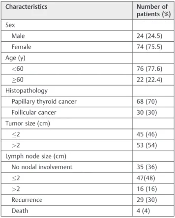
shown in ►Table 1. The median age at diagnosis was 43 years. The median follow-up was 21 months from the time of the index operation. Cervical recurrence was de-fined as metastatic involvement of any cervical LN devel-oping 6 months after surgery. Cases of cervical recurrences were mainly isolated LNs. Of the 98 patients, 71 (72%) patients underwent preoperative US, and 35 (36%) underwent preoperative CT; 8 patients underwent both studies. Fifty-six patients had postoperative US, and 42 had postoperative CT scans.
An analysis of factors predicting local recurrence is shown in ►Table 2. Positive LN recurrence was higher in men compared with women, although the difference did not reach statistical significance (p¼0.784). In addition, age, tumor size, and LN size were not significant risk factors for local recurrence (p¼0.608,p¼0.385,p¼0.875, respectively).
Positive LN status on US was not correlated with the prediction of recurrence. In contrast, positive or negative LN status as detected by CT was significantly correlated with postoperative locoregional recurrence (p¼0.01;►Table 3). The US and CT analyses demonstrated similar efficacy at detecting thyroid nodules, but differed significantly in their ability to accurately evaluate cervical LN involvement in recurrence, with CT outperforming US (►Table 4).
Discussion
PTC is the most common endocrine neoplasia with a tendency for local and regional metastasis.15The prognostic impact of
cervical LN involvement has been investigated in many studies, but remains a controversial issue.16–18Initial studies suggested that the presence of nodal metastasis had no effect in overall or disease-free survival, but this notion has been recently refuted.19Patients with PTC with cervical LN metas-tasis at initial presentation are more susceptible to recurrence than those without LN involvement.16–18The preoperative LN metastasis rate in our series was 64%, which was higher than the published 23%.19The recurrence rate in our series (30%) was higher than that previously reported in published studies (10 to 20%),18,20 which can mostly likely be attributed to differences in surgical techniques and treatments between different surgeons. However, the surgical management of Table 2 Risk factors correlated with recurrence
Risk factors No. of patients with recurrence (%)
pValue
Sex
Male (n¼18) 8 (44.4) 0.784
Female (n¼56) 22 (39.3)
Age (y)
60 (n¼58) 22 (37.9) 0.538
>60 (n¼16) 8 (50)
Tumor size (cm)
2 (n¼38) 18 (47.4) 0.385
>2 (n¼36) 12 (33.3)
Lymph node size (cm)
No nodal involvement (n¼26)
10 (38.5) 0.875
2 (n¼36) 14 (38.9)
>2 (n¼12) 6 (50)
aThe chi-square test was used to measure
pvalues (p<0.05 was considered significant by Fisher’s exact tests).
Table 1 Patient demographics
Characteristics Number of
patients (%)
Sex
Male 24 (24.5)
Female 74 (75.5)
Age (y)
<60 76 (77.6)
60 22 (22.4)
Histopathology
Papillary thyroid cancer 68 (70)
Follicular cancer 30 (30)
Tumor size (cm)
2 45 (46)
>2 53 (54)
Lymph node size (cm)
No nodal involvement 35 (36)
2 47(48)
>2 16 (16)
Recurrence 29 (30)
Death 4 (4)
Table 3 Correlation of the preoperative lymph node status with recurrence risk
Preoperative radiologic investigation
No. of patients with recurrence (%)
pValue
Negative LN status on US (n¼20)
6 (30) 0.778
Positive LN status on US (n¼34)
12 (35.3)
Negative LN status on CT (n¼15)
1 (6.67) 0.01
Positive LN status on CT (n¼14)
10 (62.5)
Abbreviations: CT, computed tomography; LN, lymph node; US, ultrasonography.
aThe chi-square test was used to measure
malignant thyroid nodule was based on FNAC and frozen section evaluation, with the FNAC being more sensitive and cost-effective.21
In this study, we evaluated age, sex, tumor size, LN size, and US and CTfindings preoperatively to determine the relation-ship of these factors with nodal recurrence. Univariate analy-sis has been used, due to small sample, although it was insufficient in comparison with multivariate analysis. Age and sex are considered prognostic factors (2010 AJCC staging system),17,22 but these factors were not associated with cervical recurrence in our study.
In addition, tumor size and LN size were not predictive of cervical recurrence. Preoperative LN metastasis was associat-ed with locoregional recurrence, as has been describassociat-ed for other studies.18,23 In 560 Japanese patients with thyroid cancer who underwent total thyroidectomy, those with US-detectable metastasis had a significantly worse relapse-free survival than those with negative USfindings.24Hay et al25 and Spires et al26showed that LN metastasis was related to a higher recurrence rate but did not adversely influence survival.
Our study and those mentioned previously would indicate that positive preoperative USfindings for LN metastasis could indicate a more aggressive disease course and strongly pre-dict the need for additional surgery in the future, whereas negative preoperative US LN findings indicate a lower risk with patients less likely to need future surgery.26However, most importantly, we determined that preoperative detec-tion of LN metastases with CT-based detecdetec-tion was signifi -cantly more specific, sensitive, and accurate than US-based detection; all patients who had negative preoperative CT LN metastasisfindings remained recurrence-free, whereas 30% of the patients with negative US LN metastasesfindings went on to develop locoregional recurrence. This strongly suggests that preoperative CT detection of LN metastases is a better modality for predicting locoregional recurrence in patients with DTC. On the other hand, the sensitivity and specificity of positive LN status on US in this study were less than those in other published studies.15,27,28
Conclusions
According to this study, patients with thyroid cancer with positive radiologic investigations (either US or CT) for LN metastasis have an increased risk of regional node recur-rence after total thyroidectomy. CT was superior to US at detecting metastatic LN involvement, and, therefore, on the basis of our data, we suggest that CT should be further studied and considered as a more suitable alternative to US
for preoperative evaluation and postoperative follow-up investigations.
References
1 Davies L, Welch HG. Increasing incidence of thyroid cancer in the United States, 1973–2002. JAMA 2006;295(18):2164–2167 2 Gilliland FD, Hunt WC, Morris DM, Key CR. Prognostic factors for
thyroid carcinoma. A population-based study of 15,698 cases from the Surveillance, Epidemiology and End Results (SEER) program 1973–1991. Cancer 1997;79(3):564–573
3 Mazzaferri EL, Kloos RT. Clinical review 128: current approaches to primary therapy for papillary and follicular thyroid cancer. J Clin Endocrinol Metab 2001;86(4):1447–1463
4 Johnson NA, LeBeau SO, Tublin ME. Imaging surveillance of differentiated thyroid cancer. Radiol Clin North Am 2011;49(3): 473–487, vi
5 Cady B, Rossi R. An expanded view of risk-group definition in differentiated thyroid carcinoma. Surgery 1988;104(6):947–953 6 Hay ID, Thompson GB, Grant CS, et al. Papillary thyroid carcinoma
managed at the Mayo Clinic during six decades (1940–1999): temporal trends in initial therapy and long-term outcome in 2444 consecutively treated patients. World J Surg 2002;26(8):879–885 7 Simon D, Goretzki PE, Witte J, Röher HD. Incidence of regional recurrence guiding radicality in differentiated thyroid carcinoma. World J Surg 1996;20(7):860–866, discussion 866
8 Mazzaferri EL, Jhiang SM. Long-term impact of initial surgical and medical therapy on papillary and follicular thyroid cancer. Am J Med 1994;97(5):418–428.Erratum in: Am J Med 1995;98:215 9 Pacini F, Molinaro E, Castagna MG, et al. Recombinant human
thyrotropin-stimulated serum thyroglobulin combined with neck ultrasonography has the highest sensitivity in monitoring differ-entiated thyroid carcinoma. J Clin Endocrinol Metab 2003;88(8): 3668–3673
10 Schlumberger M, Berg G, Cohen O, et al. Follow-up of low-risk patients with differentiated thyroid carcinoma: a European per-spective. Eur J Endocrinol 2004;150(2):105–112
11 Torlontano M, Crocetti U, Augello G, et al. Comparative evaluation of recombinant human thyrotropin-stimulated thyroglobulin lev-els, 131I whole-body scintigraphy, and neck ultrasonography in the follow-up of patients with papillary thyroid microcarcinoma who have not undergone radioiodine therapy. J Clin Endocrinol Metab 2006;91(1):60–63
12 Saggiorato E, Aversa S, Deandreis D, et al. Galectin-3: presurgical marker of thyroid follicular epithelial cell-derived carcinomas. J Endocrinol Invest 2004;27(4):311–317
13 Marshall CL, Lee JE, Xing Y, et al. Routine pre-operative ultraso-nography for papillary thyroid cancer: effects on cervical recur-rence. Surgery 2009;146(6):1063–1072
14 Greene FL, Page DL, Fleming ID, et al , eds. American Joint Committee on Cancer; American Cancer Society. AJCC Cancer Staging Manual. 6th ed. New York, NY: Springer-Verlag; 2002 15 González HE, Cruz F, O’Brien A, et al. Impact of preoperative
ultrasonographic staging of the neck in papillary thyroid carci-noma. Arch Otolaryngol Head Neck Surg 2007;133(12): 1258–1262
Table 4 A comparison of the prognostic accuracy of radiologic modalities for predicting recurrence
Modality Sensitivity (%) Specificity (%) Accuracy (%) PPV (%) NPV (%)
US 66.67 77.78 74 60 82.35
CT 80 90 86.6 80 90
16 Shaha AR. Prognostic factors in papillary thyroid carcinoma and implications of large nodal metastasis. Surgery 2004;135(2):237–239 17 Shaha A. Treatment of thyroid cancer based on risk groups. J Surg
Oncol 2006;94(8):683–691
18 Pacini F, Schlumberger M, Dralle H, Ilisea R, Smith Y, Viersinga V. [European consensus on the management of patients with differ-entiated carcinoma of the thyroid from follicular epithelium]. Vestn Khir Im I I Grek 2008;167(1):52–56[Article in Russian] 19 Londero SC, Krogdahl A, Bastholt L, et al; Danish Thyroid Cancer
Group. Papillary thyroid microcarcinoma in Denmark 1996–2008: a national study of epidemiology and clinical significance. Thyroid 2013;23(9):1159–1164
20 Dionigi G, Dionigi R, Bartalena L, Boni L, Rovera F, Villa F. Surgery of lymph nodes in papillary thyroid cancer. Expert Rev Anticancer Ther 2006;6(9):1217–1229
21 Caraci P, Aversa S, Mussa A, Pancani G, Ondolo C, Conticello S. Role offine-needle aspiration biopsy and frozen-section evaluation in the surgical management of thyroid nodules. Br J Surg 2002;89(6): 797–801
22 Schlumberger M, Pacini F, Wiersinga WM, et al. Follow-up and management of differentiated thyroid carcinoma: a European perspective in clinical practice. Eur J Endocrinol 2004;151(5): 539–548
23 Wada N, Masudo K, Nakayama H, et al. Clinical outcomes in older or younger patients with papillary thyroid carcinoma: impact of lymphadenopathy and patient age. Eur J Surg Oncol 2008;34(2): 202–207
24 Ito Y, Tomoda C, Uruno T, et al. Ultrasonographically and anato-mopathologically detectable node metastases in the lateral com-partment as indicators of worse relapse-free survival in patients with papillary thyroid carcinoma. World J Surg 2005;29(7): 917–920
25 Hay ID, Grant CS, van Heerden JA, Goellner JR, Ebersold JR, Bergstralh EJ. Papillary thyroid microcarcinoma: a study of 535 cases observed in a 50-year period. Surgery 1992;112(6): 1139–1146, discussion 1146–1147
26 Spires JR, Robbins KT, Luna MA, Byers RM. Metastatic papillary carcinoma of the thyroid: the significance of extranodal extension. Head Neck 1989;11(3):242–246
27 Poehls JL, Chen H, Sippel RS. Preoperative ultrasonographyfi nd-ings predict the need for repeated surgery in papillary thyroid cancer. Endocr Pract 2012;18(3):403–409