Instituto do Coração do Hospital das Clínicas - FMUSP
Correspondência: Paulo Sampaio Gutierrez - InCor - Av. Dr. Eneas C. Aguiar, 44 Cep 05403-000 - São Paulo, SP - E-mail: [email protected]
Received: 1/10/03 Accepted: 3/31/03
Arq Bras Cardiol, volume 82 (nº 2), 134-8, 2004
Paulo Sampaio Gutierrez, Maria Adelaide Albergaria Pereira, Regina Célia Martins O liveira, N oedir A ntonio Groppo Stolf, Maria de Lourdes Higuchi
São Paulo, SP - Brazil
Thyroid Hormone Levels in Patients with Aortic Dissection.
Comparison with Controls and Correlation with the
Percentage of the Aortic Media Composed of Myxoid Deposits
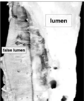
Aortic dissection (dissecting aneurysm) is characteri-zed by the separation of the aortic wall into 2 sheets at the medial layer, along the longitudinal axis of the artery, thus forming a false channel for the blood flow 1 (fig. 1). It is a
life-threatening but relatively rare disease, whose incidence has been evaluated in 5-29 cases/million persons/year 2,3.
Seve-ral conditions are associated with it: systemic arterial hy-pertension, the most common (70-90% of the cases); Marfan’s syndrome; Hurler’s syndrome; and traumatism, in-cluding surgical management of the aorta 1. A small
percen-tage of patients have no associated conditions. The patho-genesis of this disease remains unclear. Although most pa-tients with aortic dissection have systemic arterial hyper-tension, they correspond only to a very small percentage of people with this last condition. Weakness of the wall is pre-sumed to be present, but the actual factors underlying it ha-ve not been discoha-vered 4. Histological analyses targeting
this objective describe 3 main findings in the media of aortas with dissection: fragmentation of elastic fibers, a decrease in the number of smooth muscle cells, and an increase in mu-coid, basophilic material 5, a pattern frequently called
“mediocystic necrosis” (fig. 2). In spite of being more promi-nent in such cases, these alterations are not specific to the dissection; they can also appear with aging, as secondary phenomena in many aortic diseases, and in patients with hy-pertension but not dissection 5.
The mucoid material, increased in aortic dissections, is one of the more important components of the medial layer. Proteoglycans, composed of a protein core and lateral chains of glycosaminoglycans, a family of long-chained sugars; or glycosaminoglycans such as hyaluronan (hyaluronic acid) by themselves are the molecules that give the tissues this his-tological characteristic 6, also called myxoid (myxos=mucus).
This material is similar to pretibial or retroocular myxedema present in some patients with thyroid diseases.
In a necropsy series 7, more pathologic alterations were
found in thyroids from patients with aortic dissection than from controls paired by sex, age, systemic arterial hyperten-sion, and diabetes. The authors considered that part of the alterations (multiple nodular goiter, acinar atrophy and
fi-Objective - Deposits of myxoid material, similar to myxedema related to thyroid disease, are described in the medial layer of aortas with dissection. We analyzed the cli-nical or subclicli-nical thyroid dysfunction of patients with this disease and analyzed whether a correlation exists bet-ween serum levels of thyroid-related hormones and the my-xoid content of the aortic media.
Methods - We measured, with standard methods, se-rum levels of triiodothyronine (T3), thyroxine (T4), and thyroid stimulating hormone (TSH) in 28 patients who un-derwent aortic dissection and free T4 in 20 of them. The sa-me hormones were quantified in 20 control patients mat-ched by sex and age. Results were compared by using the Mann-Whitney test. We also measured the percentage of the aortic media occupied by myxoid material in the surgi-cal specimens of 25 of the patients with aortic dissection and analyzed its correlation with hormone levels by using the Pearson test.
Results - In the 20 pairs in which the amount of hormo-nes was compared, the mean values for T3, T4, free T4, and TSH were 1.22ng/mL, 9.89mcg/dL, 1.18ng/dL, and 5.45 mi-croIU/mL in study patients and 1.15ng/mL, 8.57mcg/dL, 1.32ng/dL, and 2.15 microIU/mL in controls. Neither these differences nor the correlation between the percentage of myxoid content (mean=30%) and the values for T3, T4, free T4, and TSH (mean 1.22ng/mL, 9.44mcg/dL, 1.20ng/dL, and 5.08 microIU/mL, respectively; n= 25) were significant.
Conclusion - Our data suggest that serum levels of thyroid hormones have no relation with the myxoid con-tent in the aortic media in cases of aortic dissection.
brosis, thyroiditis including Hashimoto’s disease) could cause hypothyroidism, but this condition was documented with clinical and biochemical evaluations of thyroid func-tion in only one of their cases.
Thus, aortic dissection could occur preferentially in persons with systemic arterial hypertension and thyroid di-sorders. The objective of the present study was to verify whether patients with aortic disease have an association with clinical or subclinical thyroid dysfunction, both by checking the relation between mucoid content in the aortic media and serum levels of thyroid hormones and by compa-ring these levels with those of control patients without aor-tic disease.
Methods
Twenty-eight patients (21 males) who had undergone surgical correction of aortic dissection in the ascending aorta (dissection type A according to the Stanford classifi-cation 8 or types I or II according to De Bakey’s
classifica-tion 9) were included in the study. Only persons who had
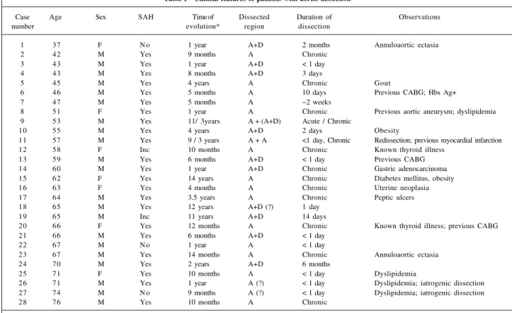
been operated on were selected in order to to make the group homogenous, because in most patients surgery is in-dicated as soon as possible after the diagnosis is establis-hed, considering the life-threatening nature of the disease. Age ranged from 37 to 76 years (mean 58, median 59). The main clinical data of these patients are presented in table I.
Patients who had undergone coronary artery revascu-larization with aortotomy were enrolled as controls. They were matched with the aortic dissection patients with regard to sex and age.
Triiodothyronine (T3), thyroxine (T4), free T4, and thyroid stimulating hormone (TSH) were quantified in the sera of the study patients and of 20 control patients by stan-dard methods (radioimmunoassay, chemoluminescence, or IRMA). In 8 patients, free T4 was not measured.
Three-micrometer sections of the aortas of 25 patients, sampled during surgery, were stained with Alcian blue (a dye that stains in blue glycosaminoglycans, including tho-se that take part in the composition of proteoglycans). To in-crease contrast, the sections were counterstained with he-matoxylin (fig. 2). The slides were examined under a Leica mi-croscope coupled to a Quantimet image analysis system, using a 40x objective. Blue areas were quantified by detec-tion, and the total area of the aortic media was measured. The medial layer of each sample was scanned perpendicu-larly to the long axis of the aortic wall. The quantifications were performed in at least 20 screen fields. Additional fields were also measured after the 20th, until a total length of the media was completed. Mean percentage of area occupied by myxoid material was then calculated in each aorta.
SPSS for Windows 6.0 was used for the statistical study. The amount of thyroid hormone in case and control groups was compared using the Mann-Whitney rank sum test, and the presence of systemic arterial hypertension in these 2 groups was compared using Fisher’s exact test. Due to the absence of control cases, only data of 20 patients (14 con-cerning free T4) were included in these analyses. Correlation between the percentage of mucoid content and hormone levels was verified by the Pearson test in 25 of the study patients, because the aorta was not sampled in the remaining 3 cases. Tests with P≤5% were considered significant.
Results
Sixteen of 20 patients with aortic dissection (80.0%) and 10 of 14 controls (71.4%; information not found about 6 of them) had systemic arterial hypertension. Therefore, the groups were not different concerning the proportion of hy-pertensive patients (P=0.69).
Two of the study patients and one control were known to have thyroid disease and had been treated pharma-cologically; one of them had a low level of TSH.
Fig. 2 - Histologic section of the medial layer of an aorta with dissection, stained with Alcian-blue to display proteoglycans, and counterstained with hematoxylin (pink). Objective magnification: 40x; bar (lower right corner)=20µm.
The amounts of T3, T4, free T4, and TSH in the serum are shown in Table 2. Besides the 3 patients mentioned above, hormonal alterations compatible with hyperthyroi-dism (a high level of free T4 and low TSH) were present in 2 study patients and 3 controls. High TSH, suggestive of hy-pothyroidism, either clinical (low free T4) or subclinical (normal free T4), was found in 2 patients in each group and in 1 aortic dissection patient in whom the free T4 level was not measured.
No significant difference was found between study patients and controls concerning hormone levels.
Table 3 and figure 3 present the percentage of area oc-cupied by mucoid material in the first group. No correlation was found between the hormone levels and the percentage of area of myxoid material.
Discussion
Although controversial 10, some evidence exists that
thyroid-disease associated deposits may not be restricted to pretibial or retroocular myxedema, but rather somehow are generalized 11. Thus, since the decades following the
description by Gsell and Erdhein in the 1920s of an increa-sed amount of basophilic substance in the aortas with dis-section, studies have tried to verify a possible relationship between thyroid dysfunction and this arterial disease.
Kountz and Hempelmann 12 found a great percentage of
ca-ses of aortic dissection in patients undergoing thyroidecto-my as a treatment for hypertension, but no association bet-ween the 2 pathologic conditions was found by Burchell 13.
More recently, a case was reported 14 in which an iatrogenic
dissection followed coronary angioplasty in a patient with myxedema, but a series 15 analyzing 48 patients with
subcli-nical or overt hypothyroidism suggested that the outcome after this procedure was not different from that in euthyroid patients. In 1994, Rosenmann and Yarom 7 reported an
in-creased frequency of thyroid pathologic lesions in patients with aortic dissection, although without correlation with ei-ther clinical or biochemical evaluation of thyroid function. On the other hand, glycosaminoglycans, composing or not proteoglycan chains, probably give the myxoid his-tological aspect to both thyroid disease-related and aortic dissection-related deposits, but the types of sugar chains involved may be different. Indications exist that either hya-luronan or decorin, a small sulfated proteoglycan, may be linked to thyroid-related deposits 16-19, whereas in a
pre-vious study we analyzed by immunohistochemistry the mu-coid-increased areas in aortas with dissection, and neither decorin nor hyaluronan was marked 20. Despite this
discre-pancy, the actual nature of each of these deposits is not fully elucidated, and it is not possible to rule out that a link bet-ween them might exist.
Evidence against the link between thyroid
dysfunc-Table I - Clinical features of patients with aortic dissection
Case Age Sex SAH Time of Dissected Duration of Observations
number evolution* region dissection
1 37 F N o 1 year A+D 2 months Annuloaortic ectasia
2 42 M Yes 9 months A Chronic
3 43 M Yes 1 year A+D < 1 day
4 43 M Yes 8 months A+D 3 days
5 45 M Yes 4 years A Chronic Gout
6 46 M Yes 5 months A 10 days Previous CABG; Hbs Ag+
7 47 M Yes 5 months A ~2 weeks
8 51 F Yes 1 year A Chronic Previous aortic aneurysm; dyslipidemia
9 53 M Yes 11/ 3years A + (A+D) Acute / Chronic
10 55 M Yes 4 years A+D 2 days Obesity
11 57 M Yes 9 / 3 years A + A <1 day, Chronic Redissection; previous myocardial infarction
12 58 F Inc 10 months A Chronic Known thyroid illness
13 59 M Yes 6 months A+D < 1 day Previous CABG
14 60 M Yes 1 year A+D Chronic Gastric adenocarcinoma
15 62 F Yes 14 years A Chronic Diabetes mellitus, obesity
16 63 F Yes 4 months A Chronic Uterine neoplasia
17 64 M Yes 3.5 years A Chronic Peptic ulcers
18 65 M Yes 12 years A+D (?) 1 day
19 65 M Inc 11 years A+D 14 days
20 66 F Yes 12 months A Chronic Known thyroid illness; previous CABG
21 66 M Yes 6 months A+D < 1 day
22 67 M N o 1 year A < 1 day
23 67 M Yes 14 months A Chronic Annuloaortic ectasia
24 70 M Yes 2 years A+D 6 months
25 71 F Yes 10 months A < 1 day Dyslipidemia
26 71 M Yes 1 year A (?) < 1 day Dyslipidemia; iatrogenic dissection
27 74 M N o 9 months A (?) < 1 day Dyslipidemia; iatrogenic dissection
28 76 M Yes 10 months A Chronic
Table III - Thyroid hormone levels and percentage of aortic medial layer occupied by mucoid substance
Case T3 T4 Free T4 TSH % mucoid
(ng/ml) (mcg/dl) (ng/dl) (microUi/ml)
1 1.5 11.4 1.5 3.0 37
2 1.3 9.2 1.4 0.5 28
3 0.8 13.7 - 4.3 35
4 1.5 10.0 1.3 1.9 25
5 1.5 11.0 1.6 2.3 15
6 0.9 7.3 1.4 2.4 32
7 0.9 6.5 0.9 2.8 26
8 1.1 6.7 - 3.5 28
9 1.4 12.1 0.9 1.4 40
11 1.9 11.3 1.1 1.8 29
12 1.1 7.4 - 2.2 37
13 0.9 8.1 0.6 6.3 22
14 0.7 9.3 1.3 2.1 23
16 1.7 8.9 1.2 1.4 23
18 1.6 18.5 - 1.0 29
19 1.1 7.6 1.2 2.8 24
20 1.9 10.8 1.6 0.2 31
21 1.8 12.4 1.3 1.3 25
22 1.2 10.2 - < 0.1§ 28
23 0.9 9.1 - 3.1 22
24 1.0 9.1 1.6 3.9 31
25 0.6 6.7 1.2 3 31
26 1.1 7.7 - 2.0 57
27 0.7 <2.5* 0.27 72.8 40
28 1.0 7.5 - 1.5 33
R -0.15 -0.14 -0.27 0.23
P 0.47 0.49 0.30 0.27
T3 - triiodotyronine; T4 - tirosine; TSH - thyroid stimulating hormone; - - free T4 not measured; * - for statistical purposes, value was computed as 2.4; § -for statistical purposes, value was computed as 0.09; r - correlation coefficients between % mucoid and each thyroid hormone levels.
% m y x o id t is s u
e7060 T3
50 40 30 20 10 0
0 0.5 1 1.5 2
T3 (ng/ml) % m y x o id t is s u e 70 60 50 40 30 20 10 0
0 10 20
T4 (mcg/dl) T4 % m y x o id t is s u e % m y x o id t is s u e 70 60 50 40 30 20 10 0 70 60 50 40 30 20 10 0 0
0 0.5 1 1.5 2
Free T4 (ng/dl)
Free T4
2 4 6 8
TSH (microUI/ml)
TSH*
* - For graphical purpuses, case 27 expurged
Fig. 3 - Relation between thyroid hormone levels and percent area of myxoid tissue in the aortic medial layer.
tion and aortic dissections indicates that the first disease is more common in women, while most cases of the arterial illness occur in men (3:1 male/female ratio 21, approximately
the same present in our study patients).
Thus, considering not only the presence of myxoid material in both conditions, but also the controversy con-cerning a possible association between them, we carried out the present study aiming to determine whether clinically evi-dent or subclinical thyroid dysfunction was present in pa-tients with aortic dissection. Only papa-tients in the late, stable postoperative period of aortic dissection were selected to avoid biasing the findings with alterations that could be
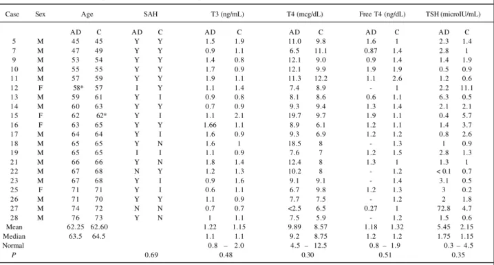
re-Table II - Age, sex, presence of systemic arterial hypertension and hormone levels in 20 patients with aortic dissection and in controls
Case Sex Age SAH T3 (ng/mL) T4 (mcg/dL) Free T4 (ng/dL) TSH (microIU/mL)
AD C AD C AD C AD C AD C AD C
5 M 45 45 Y Y 1.5 1.9 11.0 9.8 1.6 1 2.3 1.4
7 M 47 49 Y Y 0.9 1.1 6.5 11.1 0.87 1.4 2.8 1
9 M 53 54 Y Y 1.4 0.8 12.1 9.0 0.9 1.4 1.4 1.9
10 M 55 55 Y Y 1.7 0.9 12.1 9.9 1.9 1.9 0.5 0.9
11 M 57 59 Y Y 1.9 1.1 11.3 12.2 1.1 2.6 1.2 0.6
12 F 58* 57 I Y 1.1 1.4 7.4 8.9 - 1 2.2 11.1
13 M 59 61 Y I 0.9 0.8 8.1 8.6 0.6 1.1 6.3 0.5
14 M 60 63 Y Y 0.7 0.9 9.3 9.4 1.3 1.4 2.1 2.1
15 F 62 62* Y I 1.1 2.1 19.7 9.7 1.9 1.1 0.4 5.7
16 F 63 65 Y Y 1.66 1.1 8.9 6.1 1.2 1.1 1.4 3.7
17 M 64 64 Y I 1.6 0.9 9.3 6.9 1.2 1.2 0.8 2.6
18 M 65 65 Y N 1.6 1 18.5 8 - 1.3 1 0.9
19 M 65 65 I I 1.1 0.9 7.6 7 1.2 1.5 2.8 1.3
21 M 66 66 Y N 1.8 1.4 12.4 8 1.3 1 1.3 1
22 M 67 68 N Y 1.2 1.3 10.2 8 - 1.2 < 0.1 0.7
23 M 67 68 Y I 0.9 1.6 9.1 9.1 - 1.4 3.1 0.5
25 F 71 71 Y I 0.6 1.1 6.7 9.8 1.2 1.3 3 0.2
26 M 71 70 Y Y 1.1 0.9 7.7 7.5 - 1.2 2 1.8
27 M 74 72 N N 0.7 0.7 <2.5 6.5 0.27 1 72.8 4.7
28 M 76 73 Y N 1 1.1 7.5 5.9 - 1.2 1.5 0.6
Mean 62.25 62.60 1.22 1.15 9.89 8.57 1.18 1.32 5.45 2.15
Median 63.5 64.5 1.1 1.1 9.2 8.75 1.2 1.2 1.75 1.15
Normal 0.8 – 2.0 4.5 – 12.5 0.8 – 1.9 0.3 – 4.5
P 0.69 0.48 0.30 0.51 0.35
other hand, some patients were in a very late postsurgical period (up to many years). Modifications in thyroid func-tion could have occurred after the dissecfunc-tion. These possi-bilities would have greater implications if the results had shown either differences between the 2 groups or that the myxoid content was related to the hormone levels.
Acknowledgments
This research was funded by a grant from the Funda-ção de Amparo à Pesquisa do Estado de São Paulo
(FAPESP) - 97/02923-4. The authors are grateful to Adriana
Psota and Solange A. Consorte for their technical support, and to Dr Fabio Fernandes, Dr Paulo M. P. Fernandes, Roberto A. P. Mota, and Débora S. Valejo for their help in en-rolling patients in the study.
1. Roberts WC. Aortic dissection: anatomy, consequences, and causes. Am Heart J 1982; 101: 195-214.
2. Fuster V, Halperin JL. Aortic dissection: a medical perspective. J Card Surg 1994; 9: 713-28.
3. Meszaros I, Morocz J, Szlavi J, Schmidt J, Tornoci L, Nagy L, Szep L. Epidemiolo-gy and clinicopatholoEpidemiolo-gy of aortic dissection. Chest 2000; 117:1271-8. 4. Wilson SK, Hutchins GM. Aortic dissecting aneurysms. Causative factors in
204 subjects. Arch Pathol Lab Med 1982; 106: 175-80.
5. Schlatmann TJM, Becker AE. Pathogenesis of dissecting aneurysm of aorta: com-parative histopathologic study of significance of medial changes. Am J Cardiol 1977; 39: 21-6.
6. Scott J. Proteoglycan histochemistry – a valuable tool for connective tissue bio-chemists. Coll Relat Res 1985; 5: 541-75.
7. Rosenmann E, Yarom R. Dissecting aneurysm of the aorta and hypothyroidism. Isr J Med Sci 1994; 30: 510-3.
8. Daily PO, Trueblood W, Stinson EB, Wuerflein RD, Shumway NE (1970) Ma-nagement of acute aortic dissections. Ann Thorac Surg 10: 237-47. 9. DeBakey ME, Henly WS, Cooley DA, Morris GC, Crawford FS, Beall AC Jr.
Sur-gical management of dissecting aneurysms of the aorta. J Thorac Cardiovasc Surg 1965; 49: 130-48.
10. Peacey SR, Flemming L, Messenger A, Weetman AP. Is Graves’ dermopathy a ge-neralized disorder? Thyroid 1996; 6: 41-5.
11. Wortsman J, Dietrich J, Traycoff RB, Stone S. Preradial myxedema in thyroid disea-se. Arch Dermatol 1981; 117: 635-8.
12. Kountz WB, Hempelmann LH. Chromatrophic degeneration and rupture of the aorta following thyroidectomy in cases of hypertension. Am Heart J 1940; 20: 599-610.
References
13. Burchell HB. Aortic dissection (dissecting hematoma; dissecting aneurysm of the aorta. Circulation 1955; 12: 1068-74.
14. Okamoto R, Makino K, Saito K, et al. Aorto-coronary dissection during angio-plasty in a patient with myxedema. Jpn Circ J 2000; 64: 316-20.
15. Mantzoros CS, Evagelopoulou K, Moses AC. Outcome of percutaneous translu-minal coronary angioplasty in patients with subclinical hypothyroidism. Thy-roid 1995; 5: 383-7.
16. Shishiba Y, Yanagishita M, Hascall VC. Effect of thyroid hormone deficiency on proteoglycan synthesis by human skin fibroblast cultures. Connect Tissue Res 1988; 17: 119-35.
17. Shishiba Y, Takeuchi Y, Yokoi N, Ozawa Y, Shimizu T. Thyroid hormone excess stimulates the synthesis of proteoglycan in human skin fibroblasts in culture. Acta Endocrinol (Copenh) 1990; 123: 541-9.
18. Sisson JC. Hyaluronic acid in localized myxedema. J Clin Endocrinol Metab 1968; 28: 433-6.
19. Imai Y, Odajima R, Inoue Y, Shishiba Y. Effect of growth factors on hyaluronan and proteoglycan synthesis by retroocular tissue fibroblasts of Graves’ oph-thalmopathy in culture. Acta Endocrinol (Copenh) 1992; 126: 541-52. 20. Gutierrez PS, Reis MM, Aiello VD, Higuchi ML, Stolf NAG, Lopes EA.
Distri-bution of hyaluronan and dermatan/chondroitin sulphate proteoglycans in hu-man aortic dissection. Connect Tis Res 1998; 37(3-4): 151-61.
21. DeSanctis RW, Doroghazi RM, Austen WG, Buckley MJ. Aortic dissection. N Engl J Med 1987; 317: 1060-7.
22. Weetman AP. Hypothyroidism: screening and subclinical disease. Br Med J 1997; 314 (7088): 1175-8.
lated to critical status. In accordance with accepted crite-ria22, thyroid function was evaluated by the serum level of
T3, T4, free T4, and mostly TSH.