Nocturnal desaturation: predictors and the effect on sleep
patterns in patients with chronic obstructive pulmonary
disease and concomitant mild daytime hypoxemia*
RENATA CLAUDIA ZANCHET1, CARLOS ALBERTO DE ASSIS VIEGAS2
*Study carried out at the Hospital Universitário de Brasília (HUB, University Hospital of Brasília) and at the Universidade Católica de Brasília (UCB, Catholic University of Brasília) - Brasília, Distrito Federal, Brazil.
1. Adjunct Professor of the Physical Therapy Course of the Universidade Católica de Brasília (UCB, Catholic University of Brasília) - Brasília, Distrito Federal, Brazil
2. Adjunct Professor at the Universidade de Brasília (UnB, University of Brasília) School of Medicine - Brasília, Distrito Federal, Brazil
Correspondence to: Renata Claudia Zanchet. QS 07. Lote 01, Núcleo de Reabilitação Pulmonar, Sala A-04, Águas Claras - CEP: 71966-700, Taguatinga, DF, Brazil.
Phone: 55 61 3356 9005. E-mail: [email protected]
Submitted: 18 March 2005. Accepted, after review: 20 September 2005.
ABSTRACT
Objective: To determine the nocturnal oximetry pattern in chronic obstructive pulmonary disease patients having no sleep apnea and presenting mild daytime hypoxemia, to identify probable daytime parameters capable of predicting nocturnal desaturation, and to evaluate the influence of nocturnal desaturation on the sleep pattern of these patients.
Methods: Twenty-five patients were divided into two groups: those with nocturnal desaturation and those without.
Results: Comparing the first group (52%) with the second, we found the following: age, 63 + 5 years versus 63 + 6 years; forced expiratory volume in the first second = 53 + 31% versus 56 + 19% predicted; ratio of forced expiratory volume in the first second to forced vital capacity, 49 + 14% versus 52 + 10%; arterial oxygen tension, 68 + 8 mmHg versus 72 + 68 mmHg; and arterial oxygen saturation, 93 + 2% versus 94 + 1%. Patients in the nocturnal desaturation group presented lower daytime arterial oxygen saturation and nocturnal arterial oxygen saturation by pulse oximetry. There was no difference between the two groups in terms of the sleep patterns observed. The ratio of forced expiratory volume in the first second to forced vital capacity was found to correlate with forced vital capacity, daytime arterial oxygen tension and daytime arterial oxygen saturation. In addition, arterial oxygen saturation by pulse oximetry during exercise was found to correlate with nocturnal arterial oxygen saturation by pulse oximetry. However, only daytime arterial oxygen saturation was predictive of nocturnal desaturation. Conclusion: The only variable capable of predicting nocturnal
desaturation was daytime arterial oxygen saturation. Nocturnal desaturation did not influence the sleep patterns of patients with chronic obstructive pulmonary disease accompanied by mild daytime hypoxemia.
INTRODUCTION
In patients with chronic obstructive pulmonary disease (COPD), the quality of sleep can suffer, and such patients can present a drop in nocturnal levels of blood gases.(1-3)
There is a consensus that the greatest nocturnal desaturation occurs during rapid eye movement (REM) sleep(4-5) and that patients with COPD are
more hypoxic during sleep than they are when at rest during the day.(6-7) Hypoxemia is also greater
during sleep than during maximum exercise.(8)
The principal causes of nocturnal hypoxemia are alveolar hypoventilation(9) and altered
ventilation/perfusion ratio.(10) In addition, there is
a correlation between nocturnal hypoxemia and daytime parameters, such as arterial oxygen saturation (SaO2), arterial oxygen tension (PaO2)(6,11)
and decreased ventilatory response to hypercapnia.(12)
However, in addition to the differences in methodology, various authors have failed to evaluate the predictive value of each variable,(11,13) making
it difficult to interpret and extrapolate the results presented.
Furthermore, the influence that nocturnal desaturation has on the sleep patterns, pulmonary hemodynamics and life expectancy of patients with COPD (without hypoxemia or with mild daytime hypoxemia) has yet to be clarified in the literature.(14)
In view of this, the objectives of this study were as follows: to determine the nocturnal oximetry pattern in patients with COPD with mild daytime hypoxemia and without sleep apnea; to identify probable daytime parameters capable of predicting nocturnal desaturation; and to evaluate the influence of nocturnal desaturation on the sleep patterns of these patients.
METHODS
A cross-sectional study involving patients with COPD was carried out from August of 2003 to April of 2004. The COPD was diagnosed in accordance with the criteria defined by The Global Initiative for Chronic Obstructive Lung Disease.(15)
This study included patients admitted to the Pulmonary Rehabilitation Program of the University Hospital of Brasília and the Catholic University of Brasília. All of the patients with COPD were former smokers, smoke-free for at least six months, and
clinically stable for the past four weeks at least, with daytime PaO2 greater than 60 mmHg and SaO2 greater than 90%. Patients with sleep apnea syndrome (apnea-hypopnea index 5 events/hour), orthopedic problems or any other problems that might result in sleep disturbance were excluded from the study. All of the patients were using bronchodilators and oral theophylline, and none were being treated with continuous oxygen therapy or corticosteroids.
The patients evaluated were divided into two groups: those who presented nocturnal desaturation (group ND) and those who did not (group NND).
Patients who presented oxygen saturation by pulse oximetry (SpO2) 90% during 30% of their total sleep time were designated ND group patients.(16)
The study was approved by the Ethics Committee of the University Hospital of Brasília. All patients gave written informed consent.
The patients were submitted to the tests described below over a maximum period of one week.
Absolute values of forced vital capacity (FVC), forced expiratory volume in one second (FEV1) and FEV1/FVC ratio (%) were determined using a Vmax-22 series spirometer (SensorMedics, Yorba Linda, CA, USA), and the relative values predicted for gender, age and height were calculated based on the values described by Knudson et al.(17)
Spirometry was carried out according to the norms established by the American Thoracic Society.(18)
Regarding arterial blood gas analysis during wakefulness, values were determined for PaO2, arterial carbon dioxide tension (PaCO2) and SaO2 using a Ciba Corning 278 Gas System (Ciba-Corning, Diagnostics Corp., Medfield, MA, USA).
A 6-minute walk test was administered, immediately after which SpO2 values were measured with a model 920M pulse oximeter (Healthdyne Technologies, Marietta, GA, USA).
Body mass index, calculated using the formula weight in kilograms/height in square meters, was evaluated.
computerized polysomnography system (Healthdyne Technologies, Marietta, GA, USA) Traditional polysomnography variables were evaluated in accordance with Rechtschaffen and Kales.(19)
Values of the studied variables are presented as means standard deviation. Student's t-test for independent samples was used in the comparative analysis between ND and NND group values. Pearson's correlation test was used to determine the level of correlation between the variables studied during daytime and those evaluated during sleep. (For this analysis, we considered the groups as a whole, regardless of the nocturnal desaturation.) Logistic regression was used to calculate the odds ratios and identify the independent variables that were predictive of nocturnal desaturation. In accordance with this mathematical model the following variables were tested: FVC, FEV1, FEV1/ FVC, PaCO2, PaO2, SaO2 at rest and SpO2 during exercise. Values of p 0.05 were considered statistically significant.
RESULTS
Among the 25 patients studied, 13 (52%) presented nocturnal desaturation (ND group).
Of the 13 ND group patients, two were females, and the mean age was 63 ± 5 years. Of the 12 NND patients, three were females and the mean age was 63 ± 6 years.
Values relating to anthropometry, arterial blood gas, spirometry, and SpO2 during exercise are presented in Table 1. There were no statistically significant differences between the two groups regarding these values (p < 0.05).
We observed that the ND group patients presented lower SpO2 values during REM and non-REM (Nnon-REM) sleep, as well as lower minimum SpO2 during sleep and a greater percentage of sleep time with SpO2 90%, than did the NND group patients (p < 0.05) (Table 1).
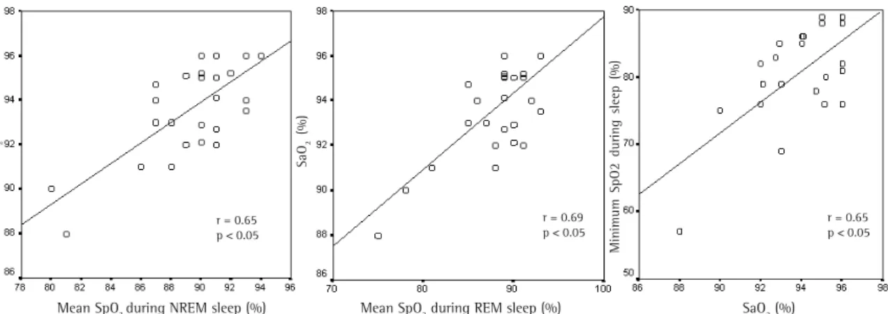
In the analysis of the study sample as a whole, daytime SaO2 and PaO2 correlated positively and significantly with SpO2 during nocturnal wakefulness, with mean SpO2 during sleep and with minimum SpO2 during sleep (Figure 1). Daytime SaO2 and PaO2 correlated negatively and significantly with the percentage of sleep time with SpO2 90%. However, there was a positive and significant correlation between the FEV1/FVC ratio (%) and SpO2
Variable ND NND (n = 13) (n = 12) BMI (kg/m2) 25 + 4 25 + 5 FVC (% of predicted) 84 + 33 86 + 20 FEV1/FVC (%) 49 + 14 52 + 10 FEV1 (% of predicted) 53 + 31 56 + 19 PaO2 (mmHg) 68 + 8 72 + 6 PaCO2 (mmHg) 33 + 5 36 + 3 SaO2 (%) 93 + 2 94 + 1 SpO2 during exercise (%) 90 + 4 90 + 3 SpO2 time < 90% (%) 73.0 + 27.8 9.5 + 6.8* SpO2 (%)- during wakefulness 88.4 + 3.0 91.6 + 1.7* SpO2 (%) - in NREM sleep 87.4 + 3.4 91.3 + 1.7* SpO2 (%) - in REM sleep 85.7 + 4.8 90.1 + 2.1* SpO2 (%) - minimum 77.2 + 8.6 83.6 + 4.3* during sleep
Data expressed as mean standard deviation; *p 0.05; ND: patients with nocturnal desaturation; NND: patients with no nocturnal desaturation; BMI: body mass index; FVC: forced vital capacity; FEV1: forced expiratory volume in one second; PaO2: arterial oxygen tension; PaCO2: arterial carbon dioxide tension; SaO2: arterial oxygen saturation; SpO2: arterial oxygen saturation by pulse oximetry
TABLE 1
Data relating to anthropometry, spirometry, arterial
blood gas and SpO2 - during exercise and during
polysomnography - of the groups studied
Variable ND NND (n = 13) (n = 12) Apnea-hypopnea index 3.1 + 5.3 1.5 + 1.7 (events/hour)
Total sleep time 334.0 +68.8 309.5 +82.6 (minutes)
Total sleep time/ 73.5 +13.3 72.2 +14.9 Total time in bed (%)
Sleep latency 29.0 +30.7 28.6 +27.5 (minutes)
REM sleep latency 87.9 +33.0 138.0 +91.4 (minutes)
Periodic lower limb movement 1.0 + 2.0 1.5 + 4.5 Stage changes 134.5 +42.6 119.6 +26.2 Micro-arousals index 31.1 +11.9 29.6 + 8.3 (events/hour)
Stage 1 (%) 20.6 + 6.2 20.7 + 9.0 Stage 2 (%) 47.8 + 6.9 50.2 + 9.6 Delta sleep (%) 9.2 + 2.7 8.3 + 3.3 REM sleep (%) 20.7 + 5.2 19.1 + 7.4
TABLE 2
Polysomnographic variables of the groups studied
during sleep. In addition, SpO2 during exercise correlated positively and significantly with SpO2 during sleep (Figure 2).
Polysomnographic data are presented in Table 2, showing that there were no statistically significant differences between the two groups studied in terms of sleep patterns.
Based on the odds ratios obtained, the only independent predictor of nocturnal desaturation was daytime SaO2. We observed that each percentage point increase in daytime SaO2 reduces the chance of nocturnal desaturation by 49%.
DISCUSSION
Of the 25 patients studied, 13 (52%) presented nocturnal desaturation, according to the definition
adopted,(16) during 30% to 90% of their sleep time.
These results are very close to those obtained in another study of patients with COPD with mild hypoxemia,(16) 45% of which were found to present
nocturnal desaturation. The results of this study refute the findings of some other authors(6) who
reported that daytime SaO2 values lower than or equal to 93% always result in nocturnal desaturation, since two of our NND group patients presented daytime SaO2 values of 91% and 92%, respectively.
The literature presents controversial results regarding the impact of nocturnal desaturation, in isolation, on patients with COPD. It has been reported that nocturnal desaturation promotes an increase in pulmonary arterial pressure.(7) However,
other authors have stated that, among patients with mild daytime hypoxemia, pulmonary arterial
Figure 1 - Correlation between daytime SaO2 and mean SpO2 during sleep, and between daytime SaO2 and minimum SpO2 during sleep in the 25 patients studied. SaO2: arterial oxygen saturation; SpO2: arterial oxygen saturation by pulse oximetry
SaO
2
(%)
SaO
2
(%)
Minimum SpO2 during sleep (%)
Mean SpO2 during NREM sleep (%) Mean SpO2 during REM sleep (%) SaO2 (%) r = 0.69
p < 0.05 r = 0.65p < 0.05
r = 0.65 p < 0.05
Figure 2 - Correlation between daytime SaO2 and sleep time with SpO2 < 90%, between FEV1/FVC ratio and mean SpO2 during sleep, and between SpO2 during exercise and mean SpO2 during sleep in the 25 patients studied.
SaO2: arterial oxygen saturation; SpO2: arterial oxygen saturation by pulse oximetry; FEV1: forced expiratory volume in one second; FVC: forced vital capacity
SaO
2
(%)
SpO2 time 90 FEV1/FVC (%) SpO2 during exercise (%)
Mean SpO
2
during sleep
Mean SpO
2
during sleep (%)
r =- 0.59 p < 0.05
r = 0.45
pressure was the same for those presenting nocturnal desaturation as for those not presenting such desaturation.(20) The survival of these patients
is also an object of controversy. A recent review of the literature(14) indicates that there is no scientific
evidence of the deleterious effect of nocturnal hypoxemia, in isolation, on the survival of these patients.
The present study demonstrated that, although SpO2 during sleep correlates with the FEV1/FVC ratio (%), daytime SaO2, daytime PaO2 and SpO2 during exercise, only daytime SaO2 was a predictor of nocturnal desaturation. This finding corroborates those of other authors.(6,21)
The literature presents different results on the predictive parameters of nocturnal desaturation. Some authors found that SpO2 during sleep correlates positively with daytime FEV1(21), SaO
2 and
PaO2.(6,11) In addition, it has been observed that the
greater the nocturnal desaturation, the lower the ventilatory response to hypercapnia and hypoxemia.(12) Regarding exercise, a situation of
ventilatory and cardiac stress, the lower the SaO2 during exercise, the lower the SpO2 during sleep.(6,8,13) Furthermore, we found nocturnal SpO
2
to correlate negatively with functional residual capacity,(6) PaCO
2(6,13) and daytime sleepiness.(12)
However, of all the factors that correlated with nocturnal SpO2, only SaO2,(6,21) daytime sleepiness(12)
and daytime PaO2(8,12) were independent predictors
of desaturation during sleep.
The discrepancy in the results presented in the literature can result from the differences in the methodology used, including the innumerable definitions of nocturnal desaturation used in the studies, such as a drop in SpO2 greater than 4% in relation to baseline for a minimum of five minutes(6)
and 30% or more of total sleep time with an SpO2 below 90%.(13,16) In this study, we adopted the
definition proposed by Levi-Valensi et al.(16) due
to its greater clinical importance.
As a response to hypoxemia and/or hypercapnia, patients with COPD present increased ventilation and respiratory effort, often resulting in awakening.(22) According to some authors,(23)
desaturation is accompanied by awakenings and/ or sleep stage changes.
In this study, there were no statistically significant differences between the ND group and the NND group in any polysomnography parameters
except for oximetry. Therefore, the number of awakenings, sleep stage changes, or movements during sleep, was not altered by nocturnal desaturation. This result corroborates those of other authors,(12,14,24) Some of whom(24) stated that
nocturnal desaturation is not the only factor that can cause sleep alterations in patients with COPD. In conclusion, 52% of the patients studied presented nocturnal desaturation, and the only variable capable of predicting such desaturation was daytime SaO2. Furthermore, nocturnal desaturation does not influence the sleep pattern of patients with COPD accompanied by mild daytime hypoxemia.
REFERENCES
1. Fleetham JA. Is chronic obstructive pulmonary disease related to sleep apnea-hypopnea syndrome? Am J Respir Crit Care Med. 2003;167(1):3-4.
2. Sanders MH, Newman AB, Haggerty CL, Redline S, Lebowitz M, Samet J, et al. Sleep Heart Health Study. Sleep and sleep-disordered breathing in adults with predominantly mild obstructive airway disease. Am J Respir Crit Care Med. 2003;167(1):7-14.
3. Zanchet RC, Vigas CAA, Lima TSM. Influência da reabilitação pulmonar sobre o padrão de sono de pacientes portadores de doença pulmonar obstrutiva crônica. J Bras Pneumol. 2004;30(5):439-44. 4. Douglas NJ. Sleep in patients with chronic obstructive
pulmonary disease. Clin Chest Méd. 1998;19(1):115-25. 5. Sergi M, Rizzi M, Andreoli A, Pecis M, Bruschi C, Fanfulla F. Are COPD patients with nocturnal REM sleep-related desaturation more prone to developing chronic respiratory failure requiring long-term oxygen therapy? Respiration. 2002;69(2):117-22. Comment in: Respiration. 2002;69(2):115-6.
6. Little SA, Elkholy MM, Chalmers GW, Farouk A, Patel KR, Thomson NC. Predictors of nocturnal oxygen desaturation in patients with COPD. Respir Med. 1999;93(3):202-7.
7. McNicholas WT. Impact of sleep in COPD. Chest. 2000;117(2 Suppl):48S-53S.
8. Mulloy E, McNicholas WT. Ventilation and gas exchange during sleep and exercise in severe COPD. Chest. 1996;109(2):387-94.
9. Douglas NJ, Flenley DC. Breathing during sleep in patients with obstructive lung disease. Am Rev Respir Dis. 1990;141(4 Pt 1):1055-70.
10. Fletcher EC, Gray BA, Levin DC. Nonapneic mechanisms of arterial oxygen desaturation during rapid-eye-movement sleep. J Appl Physiol. 1983;54(3):632-9. 11. Brijker F, van der Elshout FJJ, Heijdra YF, Folgering HT.
Underestimation of nocturnal hypoxemia due to monitoring conditions in patients with COPD. Chest. 2001;119(6):1820-6.
and mildly hypoxic patients with COPD. Eur Respir J. 1995;8(1):74-7.
13. Chaouat A, Weitzenblum E, Kessler R, Schott R, Charpentier C, Levi-Valensi P, et al. Outcome of COPD patients with mild daytime hypoxaemia with or without sleep-related oxygen desaturation. Eur Respir J. 2001;17(5):848-55.
14. Weitzenblum E, Chaouat A. Sleep and chronic obstructive pulmonary disease. Sleep Med Rev. 2004;8(4):281-94. 15. Pauwels RA, Buist AS, Calverley PM, Jenkins CR, Hurd
SS; GOLD Scientific Committee. Global strategy for the diagnosis, management, and prevention of chronic obstructive pulmonary disease. NHLBI/WHO Global Initiative for Chronic Obstructive Lung Disease (GOLD) Workshop summary. Am J Respir Crit Care Med. 2001;163(5):1256-76. Comment in: Am J Respir Crit Care Med. 2001;163(5):1047-8.
16. Levi-Valensi P, Weitzenblum E, Rida Z, Aubry P, Braghiroli A, Donner C, et al. Sleep-related oxygen desaturation and daytime pulmonary hemodynamics in COPD patients. Eur Respir J. 1992;5(3):301-7. Erratum in: Eur Respir J. 1992;5(5):645.
17. Knudson RJ, Lebowitz MD, Holberg CJ, Burrows B. Changes in the normal maximal expiration flow-volume curve with growth and aging. Am Rev Respir Dis. 1983;127(6):725-34.
1 8 . A m e r i c a n T h o ra c i c S o c i e t y. S t a n d a rd i z a t i o n o f
spirometry - 1994 update. Am J Respir Crit Care Med. 1995;152(3):1107-36.
19. Rechtschaffen A, Kales A, editors. A manual of standardized terminology, techniques and scoring system for sleep stages of human subjects. Washington, U.S. Government Printing Office, 1968. (Publication nº 204). 20. Chaouat A, Weitzenblum E, Kessler R, Charpentier C, Ehrhart M, Levi-Valensi P, et al. Sleep-related O2 desaturation and daytime pulmonary haemodynamics in COPD patients with mild hypoxaemia. Eur Respir J. 1997;10(8):1730-5. 21. Heijdra YF, Dekhuijzen PN, van Herwaarden CL,
Folgering HT. Nocturnal saturation and respiratory muscle function in patients with chronic obstructive pulmonary disease. Thorax. 1995;50(6):610-2. 22. Tatsumi K, Kimura H, Kunitomo K, Kuriyama T,
Watanabe S, Honda Y. Sleep arterial oxygen desaturation and chemical control of breathing during wakefulness in COPD. Chest. 1986;90(1):68-73.
23. Arand DL, McGinty DJ, Littner MD. Respiratory patterns associated with hemoglobin desaturation during sleep in chronic obstructive pulmonary disease. Chest. 1981;80(2):183-90.