Abstract
Objective: To investigate the use of nasal intermittent positive pressure ventilation (NIPPV) in level three neonatal intensive care units (NICU) in northeastern Brazil.
Methods: This observational cross-sectional survey was conducted from March 2009 to January 2010 in all level three NICUs in northeastern Brazil that are registered in the Brazilian Registry of Health Establishments (Cadastro Nacional de Estabelecimentos de Saúde, CNES) of the Ministry of Health. Questionnaires about the use of NIPPV were sent to the NICU directors in each institution. Statistical analysis was conducted using the software Epi-Info 6.04 and double data entry. A chi-square test was used to compare variables, and the level of statistical signiicance was set at p ≤ 0.05.
Results: This study identiied 93 level three NICUs in northeastern Brazil registered in CNES, and 87% answered the study questionnaire. Most classiied themselves as private institutions (30.7%); 98.7% used NIPPV;92.8 % adapted mechanical ventilators for NIPPV and used short binasal prongs as the interface (94.2%). Only 17.3% of the units had a protocol for the use of NIPPV. Mean positive inspiratory pressure and positive end-expiratory pressure were 20.0 cmH2O (standard deviation [SD]: 4.47) and 5.0 cmH2O (SD: 0.84).
Conclusion: NICUs in northeastern Brazil use nasal intermittent positive pressure ventilation, but indications and ventilation settings are not the same in the different institutions.
J Pediatr (Rio J). 2012;88(1):48-53: Ventilation, neonatology, intensive care units, infant.
O
riginala
rticleCopyright © by Sociedade Brasileira de Pediatria
48 Introduction
The use of mechanical ventilation (MV) ensures higher survival rates among patients with respiratory failure due to several diseases, particularly in newborns that, due to their lung immaturity, are more susceptible to respiratory distress and failure. Despite its importance, it may lead to complications, and its use is one of the main causes of lung lesions, particularly bronchopulmonary dysplasia, in premature infants.1-3 Although the importance of reducing
time receiving invasive MV is well recognized, this procedure is complex and affected by several factors, such as the different stages of lung development, underlying disease, secondary complications, cardiorespiratory interactions and associations between central respiratory control and respiratory muscles.4
Noninvasive respiratory support is an important alternative to reduce MV duration and to progress from
Practices of use of nasal intermittent positive pressure
ventilation (NIPPV) in neonatology in northeastern Brazil
Sara Karla F. de Medeiros,1 Werther Brunow de Carvalho,2 Cláudio F. R. Soriano3
1. Professora, Centro de Ensinos Superiores de Maceió, Maceió, AL, Brazil. Fisioterapeuta, Universidade Estadual de Ciências da Saúde de Alagoas (UNCISAL) e Hospital Geral do Estado de Alagoas, Maceió, AL, Brazil.
2. Professor titular, Neonatologia e Terapia Intensiva, Instituto da Criança, Hospital das Clínicas, Faculdade de Medicina, Universidade de São Paulo (USP), São Paulo, SP, Brazil.
3. Professor adjunto, Medicina em Urgência e Emergência, Terapia Intensiva e Neonatologia, Universidade Federal de Alagoas (UFAL) e UNCISAL, Maceió, AL, Brazil.
No conflicts of interest declared concerning the publication of this article.
Suggested citation: de Medeiros SK, Carvalho WB, Soriano CF. Practices of use of nasal intermittent positive pressure ventilation (NIPPV) in neonatology in northeastern Brazil. J Pediatr (Rio J). 2012;88(1):48-53.
Manuscript submitted Aug 4 2011, accepted for publication Oct 3 2011.
MV to spontaneous breathing. Current scientiic and clinical interest in a noninvasive type of support, the nasal intermittent positive pressure ventilation (NIPPV), has increased.5-7 This type of ventilation is deined as the provision of positive pressure without using an intratracheal tube or tracheotomy. It ensures intermittent and noninvasive inspiratory support at a positive inspiratory pressure greater than expiratory pressure.8
Although NIPPV has been widely used, reports recommend further studies to investigate the real function of this ventilation mode and to accurately deine the conditions and methods to achieve optimal results when using it. Several aspects of the use of NIPPV in neonates remain unclear. Its actual beneits, indications, mode of use and complications have not been deined in the literature,7,9,10 and no uniform technique for its use has been established. In addition, it is known that socioeconomic development is directly associated with healthcare promotion and instruments in any geographic region. Therefore, great differences in NIPPV methods may be expected between the practices adopted and discussed worldwide and those put into practice in Brazil, as well as between the uses of this ventilation mode in different Brazilian regions.
This study conducted a survey of the different uses of NIPPV in third level neonatal intensive care units (NICUs) in northeastern Brazil.
Methods
Study design
This observational cross-sectional survey included all third level NICUs in northeastern Brazil that were registered in the Brazilian Registry of Health Establishments (Cadastro Nacional de Estabelecimentos de Saúde, CNES) of the Ministry of Health. A third level NICU was deined as a place where there are high complexity services and qualiied human resources in different areas.11 NICUs were excluded from the study if their directors or representatives refused to answer the questionnaire or sign the informed consent term, if access was denied, and if the units were closed or had no beds available for neonatal care.
Data were collected from March 2009 to January 2010 using questionnaires that were mailed to the directors of the third level NICUs. The material mailed was divided into three parts: (1) guidelines to ill out the questionnaire and mail it back; (2) a list of terms for their uniform use in the study, in which the following terms were described: NIPPV, continuous positive airway pressure, hood, short binasal prong, nasopharyngeal prong, short single prong, face mask, nasal mask; and (3) questionnaire with 20 multiple choice questions about the use of NIPPV. Each question was followed by a blank space for comments.
First, the CNES was reviewed to ind out the number, distribution by county and contact person in each NICU in northeastern Brazil. In the irst phase of data collection, the questionnaires were mailed to the directors or technical directors of the NICU of the institutions registered in the CNES. The answers were returned by mail in 30 days. After that, in the second phase of the study, phone calls were made to the NICU directors or representatives that did not send back the questionnaire in the irst phase. They were told that another envelope would be sent to them and were asked to return the questionnaire in it. The second envelope was expected to be returned in another 30 days after the date when it was posted. In the irst phase of data collection, one of the authors visited the institutions that did not return the questionnaire. During the visits, the questionnaire was handed to the participants, but no additional information about how to respond to it was provided. When the ICU director could not be reached, another neonatologist in the same institution answered the questionnaire.
The variables under analysis were: number of NICUs in northeastern Brazil; number of institutions by state; type of institution; number of ICU beds. The variables analyzed to investigate knowledge of the use of NIPPV were: when NIPPV started being used in the institution; pressure source; interface used; nasal septum protection; use of nasogastric tube; newborn positioning during NIPPV; use of synchronized ventilation; frequency of synchronized ventilation use; NIPPV indications; NIPPV protocol; mean parameters of use; associated complications; weaning modes; use of support after NIPPV.
Data analysis and ethical aspects
The Epi-Info 6.04 and double data entry were used for tabulation and statistical analysis. Data were analyzed using descriptive statistics: means, standard deviations and percentiles. A chi-square test was used for comparisons between variables, and the level of signiicance was set at ≤ 0.05. This study was approved by the Ethics in Research Committees of Universidade Federal de São Paulo (UNIFESP) and Universidade Estadual de Ciências da Saúde de Alagoas (UNCISAL).
Results
Variables n %
Pressure source
Ventilator used for noninvasive ventilation* 3 4.3
Adapted ventilator 64 92.8
Inter 3 50 72.4
Inter 5 34 49.2
Sechrist IV 100B 9 13
Takaoka Smart 3 4.3
Dixtal 3010 1 1.4
Type of mechanical ventilator not informed 9 13
No data 2 2.9
Synchronized ventilation (type)
No synchronized ventilation 30 43.5
Synchronized ventilation using abdominal sensor 1 1.4
Synchronized ventilation using MV trigger sensitivity 30 43.4
Synchronized ventilation not informed 8 11.5
Interface†
Short binasal prong 65 94.2
Nasopharyngeal prong‡ 8 11.5
Nasal face 5 7.3
Facial mask 5 7.3
Short single prong -
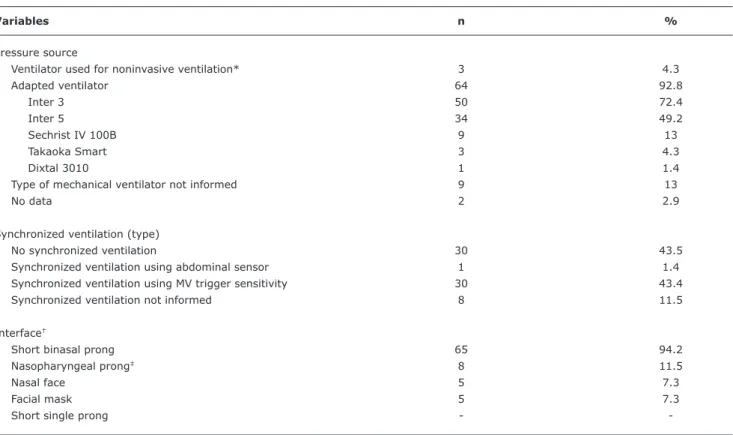
-Table 1 - Distribution of 69 NICUs according to pressure source, use of synchronized ventilation and type of interface in nasal intermittent positive pressure ventilation
MV = mechanical ventilator; NICU = neonatal intensive care unit. * Type of mechanical ventilator not informed.
† This variable accepted more than one option. ‡ Adapted prong.
questionnaires were answered by the ICU director in 47/78 (60.2%) of the institution and by a neonatologist on duty in 31/78 (39.8%). The institutions were classiied as private in 24/78 (30.7%) of the cases; as public, state-run institutions in 23/78 (29.4%); as public, city-run in 12/78 (15.3%); as federal units in 7/78 (8.9%); as non-proit organizations in 8/78 (10.2%); and as mixed units in only 3/78 (3.8%), in which case two or more classiications applied. One of the institutions did not deine its nature.
NIPPV was used in 69/78 (88.4%) of the NICUs in northeastern Brazil. The lowest rates of NIPPV use were found in the states of Ceará (50%) and Maranhão (66.7%). The states of Bahia, Sergipe, Alagoas, Paraíba and Rio Grande do Norte had a 100% rate of NIPPV use. There were no statistically signiicant differences in NIPPV use between the states in northeastern Brazil. Only 2.9% of the units that used NIPPV reported having used it for over ive years, whereas 40.6% had started using it 1 to 2 years before. A protocol for its use was followed in 12 (17.4%) institutions, but their protocols were not described.
The ICU distribution according to pressure source, use of synchronized ventilation and type of interface to provide NIPPV is shown in Table 1.
During the application of NIPPV with nasal prongs, 61/69 (88.4%) of the units reported using nasal septum protection. Sixteen (26.2%) used hydrocolloid dressings, 9/61 (14.7%), adhesive tape, 5/61 (8.2%), special adhesive tape, such as Micropore™ tape, and 31/61 (50.8%) did not inform the type of material used. The shape of the septum protector was informed only by 5/61 (8.2%) institutions, which described it as a “pig snout.”
The main indications of NIPPV reported by the institutions were after programmed weaning (79.7%), to avoid apneia in preterm infants (78.3%) and to avoid tracheal intubation when there were signs of respiratory failure (69.6%). The least frequent indication was its use for preterm infants after the administration of exogenous surfactant (24.6%).
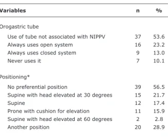
Table 2 - Distribution of NICUs according to use of orogastric tube and ideal infant positioning in nasal intermittent positive pressure ventilation
Variables n %
Orogastric tube
Use of tube not associated with NIPPV 37 53.6
Always uses open system 16 23.2
Always uses closed system 9 13.0
Never uses it 7 10.1
Positioning*
No preferential position 39 56.5
Supine with head elevated at 30 degrees 15 21.7
Supine 12 17.4
Prone with cushion for elevation 11 15.9 Supine with head elevated at 60 degrees 2 2.8
Another position 20 28.9
NICU = neonatal intensive care unit; NIPPV = nasal intermittent positive pressure ventilation.
* This variable accepted more than one option.
Table 3 - Maximum peak inspiratory pressure, positive end-expiratory pressure, inspiratory low, inspiratory time and respiratory rate during nasal intermittent positive pressure ventilation in the 69 institutions included in the study
Mean SD
PIP (cmH2O) 20.0 4.47
PEEP (cmH2O) 5.0 0.84
RR (ipm) 20.0 8.27
Tinsp (s) 0.45 0.06
Flow (l/min) 10.8 3.21
Table 4 - Distribution of NICUs according to complications associated with use of nasal intermittent positive pressure ventilation in neonates
Complications* n %
Nasal septum lesion 59 85.5
Abdominal distension 41 59.4
Epistaxis 29 42.0
Increased gastric residues 21 30.4
Gastrointestinal perforation 2 2.9
Pneumothorax 1 1.4
PEEP = positive end-expiratory pressure; PIP = positive inspiratory pressure; RR = respiratory rate cycle; SD = standard deviation; Tinsp = inspiratory time.
NICU = neonatal intensive care unit. * This variable accepted more than one option. Table 3 shows mean maximum positive end-expiratory
pressure, peak inspiratory pressure (PIP), inspiratory low, respiratory rate and inspiratory time during NIPPV as reported by the institutions.
Complications during the use of NIPPV in neonates in the institutions in northeastern Brazil are shown in Table 4.
The parameters most frequently used for NIPPV weaning were respiratory rate per cycle and fraction of inspired O2. The values of respiratory rate per cycle reported as adequate for weaning by most units ranged from 10 to 12 per minute, and the fraction of inspired O2 described by most institutions was 21%.
After NIPPV weaning, 60 (87.0%) of the institutions used continuous positive airway pressure, 25 (36.2%) used oxyhoods, and 7 (10.1%) had patients breathe room air. Of all the institutions under study, 26 (37.6%) reported using the 3 types of support and deining the choice according to the clinical conditions of the newborn.
Discussion
Currently, there are no published studies that show how third level NICUs in Brazil act in relation to the use of NIPPV. This is the irst study to investigate that use, and our focus was on the northeastern region of Brazil because its distance from larger centers and the precariousness of resources assigned to health care may contribute to speciic conditions during the care provided to neonates.12
This study about NIPPV in northeastern Brazil showed that 88.4% of the units use this ventilation mode. This percentage is higher than those found in similar studies, such as the one conducted by Ryan et al.,13 who found that, of 17 units studied in Canada, only 9 (53%) used NIPPV, and by Owen et al.,9 who found that only 48% of the British neonatal units used NIPPV. However, no inference should be made for current users because of the growing number of publications about this topic and their relections on the increase of the use of this ventilation mode by several national and international institutions.
about the use of NIPPV in infants and children aged 15 days to 17 years in Spanish institutions found that nonspeciic noninvasive mechanical ventilators were used in only 2% of the cases.8,14 Studies in the literature do not clarify whether this inding may affect the inal results of NIPPV, but suggest that non-adapted ventilators demand higher pressures because air leaks cannot be compensated.15
In addition, a high number (56.3%) of the institutions included in this study reported that they used synchronized ventilation in NIPPV. Despite that, only one of the units reported using abdominal sensors to detect the inspiratory effort made by neonates; the others used the sensitivity trigger available in conventional ventilators to achieve synchronization. This synchronization mechanism has been classiied as inacceptable for newborns, particularly when using NIPPV, because of the dificulty in detecting the respiratory effort in consequence of the substantial leaks at the interface.16-18 Therefore, most of the institutions in northeastern Brazil seem to face dificulties in the provision of NIPPV that is truly synchronized with the inspiratory effort of newborns, and the results of studies about the eficacy of NIPPV cannot be safely applied because most studies were conducted using synchronized ventilation.8,19,20
Almost all NICUs in northeastern Brazil included in this study (94.2%) used short binasal prongs to provide NIPPV, which is in agreement with data in current literature.7,19,21,22 A systematic review of the literature in this area showed that short binasal prongs, although not free of complications, were easier to use, comparatively less invasive, had lower resistance and were clinically more appropriate.23 A relatively high number of NICUs in northeastern Brazil (11.5%) also reported using nasopharyngeal prongs. Six of these units (8.7%) described this interface as an adaptation of an aspiration tube inserted from the nostril to the pharynx of the newborn. This type of adaptation was necessary because of the lack of appropriate interfaces available in the NICU. There are no details in the literature about this practice, and future studies about its use should be conducted to deine the rate of complications or of success and failure during NIPPV.
Only two NICUs in northeastern Brazil described no complications associated with the use of NIPPV, and 50% reported more than 1 complication. These data contradict those reported in similar studies, which found few complications of NIPPV use.9,14 The most important complication described by the NICUs in northeastern Brazil were nasal septum lesions, followed by abdominal distension. The institutions included in this study have sought alternatives to prevent nasal septum lesions: 88.4% use hydrocolloid dressings, adhesive tape or Micropore™ tapes to protect the nostrils. The use of non-synchronized NIPPV is associated with the provision of low into the stomach when the glottis is closed, which increases the amount of air that lows into the abdomen.18,24 This may explain
the high rate of abdominal distension found in this study. One of the units included in this study associated the use of NIPPV with the occurrence of pneumothorax, and two, with gastrointestinal perforation, also reported in similar studies.18,25
Currently, the best scientiic evidence in favor of the use of NIPPV is the support to reduce intubation rates and avoid apneia.24,26,27 The NIPPV indications most often mentioned by NICUs in northeastern Brazil were use after extubation (79.7%) and during apneic episodes (78.3%). Less frequent indications suggested its use as a primary support mode to treat respiratory distress syndrome (24.6%). Owen et al. found that only 59% of the English units used NIPPV to avoid extubation failure, whereas 80% indicated this type of ventilation to avoid apneic episodes after the failure in using continuous positive airway pressure, and 16%, as the irst ventilation mode.9
Despite the high incidence of accidental extubation in neonates,5,28 no study has been found in the literature about the use of NIPPV in this situation. Our study found that a high percentage of NICUs in northeastern Brazil (53.1%) consider NIPPV a safe procedure after accidental extubation. Further studies should investigate its chances of success in this case.
In the literature, there is no consensus about what are the optimal parameters to keep newborns well adapted or to wean them gradually from NIPPV. Studies about the beneits of NIPPV use many different parameters, which makes it dificult to reproduce their positive results in clinical practice. This study about the use of NIPPV in northeastern Brazil relected this condition. Although some institution in northeastern Brazil (43.5%) reported using pressures ranging from 16 to 20 mmH2O, a relatively high number of units use extreme PIP pressures, ranging from 5 to 10 H2O or from 26 to 30 H2O. Another reason for the great variation associated with PIP descriptions in this study may be the lack of speciication of the questionnaires about the pathology that affected the patient during NIPPV.
About 50% of the NICUs included in this study classiied the reduction of respiratory rate and the fraction of inspired oxygen for the gradual weaning from NIPPV as important parameters. Owen et al.9 found that the English NICUs used PIP reduction and positive end-expiratory pressure as primary weaning parameters.
Data in this study cannot be used as guidelines for NIPPV, but our indings describe what NICUs in northeastern Brazil do when using this noninvasive ventilation mode for newborns. Based on these data, studies with other levels of evidence may be conducted to investigate how aspects of the use of NIPPV in northeastern Brazil affect the end results of this type of ventilation.
References
1. Hutchison AA, Bignall S. Non-invasive positive pressure ventilation
in the preterm neonate: reducing endotrauma and the incidence
of bronchopulmonary dysplasia. Arch Dis Child Fetal Neonatal Ed. 2008;93:64-8.
2. Gonzaga AD, Duque Figueira BB, Sousa JM, de Carvalho WB. Tempo de ventilação mecânica e desenvolvimento de displasia broncopulmonar. Rev Assoc Med Bras. 2007;53:64-7.
3. Kulkarni A, Ehrenkranz RA, Bhandari V. Effect of introduction of synchronized nasal intermittent positive-pressure ventilation in a neonatal intensive care unit on bronchopulmonary dysplasia and growth in preterm infants. Am J Perinatol. 2006;23:233-40. 4. Donn SM, Sinha SK. Invasive and noninvasive neonatal mechanical
ventilation. Respir Care. 2003;48:426-39.
5. Bhandari V. Accidental extubations - are the infants trying to tell us something? J Pediatr (Rio J). 2010;86:167-9.
6. Bhandari V. Nasal intermitent positive pressure ventilation in the
newborn: review of literature and evidence-based guidelines. J Perinatol. 2009;30:505-12.
7. Owen LS, Morley CJ, Davis PG. Neonatal nasal intermittent positive
pressure ventilation: what do we know in 2007? Arch Dis Child Fetal Neonatal Ed. 2007;92:F414-8.
8. Barbosa AP, Johnston C, Carvalho WB. Ventilação não-invasiva em neonatologia e pediatria. São Paulo: Atheneu; 2007. 9. Owen LS, Morley CJ, Davis PG. Neonatal nasal intermittent positive
pressure ventilation: a survey of practice in England. Arch Dis Child Fetal Neonatal Ed. 2008;93:F148-50.
10. Migliori C, Motta M, Angeli A, Chirico G. Nasal bilevel vs. continuous positive airway pressure in preterm infants. Pediatr Pulmonol. 2005;40:426-30.
11. Brasil, Ministério da Saúde, Secretaria Executiva. Gestante de alto risco: sistemas estaduais de referência hospitalar à gestante de alto risco. Brasília: Ministério da Saúde; 2001. 32 p.
12. Rede Norte-Nordeste de Saúde Perinatal. Fortaleza: Seminário de lançamento da rede Norte-Nordeste de saúde perinatal; 2006.
http://www.renospe.org. Acess: 26/05/2009
13. Ryan CA, Finer NN, Peters KL. Nasal intermittent positive-pressure ventilation offers no advantages over nasal continuous positive airway pressure in apnea of prematurity. Am J Dis Child. 1989;143:1196-8.
14. Magin EC, Ódena MP, Cid JL, Torres FM, Garcia Teresa MA, Villanueva AM, et al. Estudio epidemiológico de la ventilación no invasiva en las UCIP en España [resumo]. An Pediatr. 2007;67:98. 15. Medina Villanueva A, Prieto Espuñes S, Los Arcos Solas M, Rey
Galán C, Concha Torre A, Menéndez Cuervo S, et al. Aplicación de ventilación no invasiva en una unidad de cuidados intensivos pediátricos. An Pediatr. 2005;62:13-9.
16. Alvo M, Hirschheimer MR. Instalações de gasoterapia para ventilação pulmonar mecânica. In: Carvalho WB, Freddi NA, Hirschheimer MR, Proença JO, Ribeiro R. Ventilação pulmonar mecânica em pediatria. São Paulo: Atheneu; 1993. p. 21-30. 17. John J, Björklund LJ, Svenningsen NW, Jonson B. Airway and
body surface sensors for triggering in neonatal ventilation. Acta Paediatr. 1994;83:903-9.
18. Courtney SE, Barrington KJ. Continuous positive airway pressure and noninvasive ventilation. Clin Perinatol. 2007;34:73-92. 19. Davis PG, Morley CJ, Owen LS. Non-invasive respiratory support
of preterm neonates with respiratory distress: continuous
positive airway pressure and nasal intermittent positive pressure ventilation. Semin Fetal Neonatal Med. 2009;14:14-20. 20. Ali N, Claure N, Alegria X, D’Ugard C, Organero R, Bancalari E.
Effects of non-invasive pressure support ventilation (NI-PSV) on ventilation and respiratory effort in very low birth weight infants.
Pediatr Pulmonol. 2007;42:704-10.
21. Lamy F. Avaliação da ventilação pulmonar mecânica em recém-nascidos no Instituto Fernandes Figueira [dissertação]. Rio de Janeiro: Pós-Graduação em Saúde da Criança e da Mulher, Instituto Fernandes Figueira, Fundação Oswaldo Cruz; 1995.
22. Kolpelman AE, Holbert D. Use of oxygen cannulas in extremely low birthweight infants is associated with mucosal trauma and bleeding, and possibly with coagulase-negative staphylococcal sepsis. J Perinatol. 2003;23:94-7.
23. De Paoli AG, Davis PG, Faber B, Morley CJ. Devices and pressure
sources for administration of nasal continuous positive airway pressure (NCPAP) in preterm neonates. Cochrane Database Syst Rev. 2008;(1):CD002977.
24. Moretti C, Giannini L, Fassi C, Gizzi C, Papoff P, Colarizi P. Nasal
low-synchronized intermittent positive pressure ventilation to facilitate weaning in very low-birthweight infants: unmasked
randomized controlled trial. Pediatr Int. 2008;50:85-91.
25. Garland JS, Nelson DB, Rice T, Neu J. Increased risk of gastrointestinal perforations in neonates mechanically ventilated with either face mask or nasal prongs. Pediatrics. 1985;76:406-10.
26. Lemyre B, Davis PG, de Paoli AG. Nasal intermittent positive pressure ventilation (NIPPV) versus nasal continuous positive airway pressure (NCPAP) for apnea of prematurity. Cochrane Database Syst Rev. 2002;(1):CD002272.
27. Davis PG, Lemyre B, de Paoli AG. Nasal intermittent positive pressure ventilation (NIPPV) nasal continuous positive airway pressure (NCPAP) for preterm neonates after extubation. Cochrane Database Syst Rev. 2001;(3):CD003212.
28. Carvalho FL, Mezzacappa MA, Calil R, Machado H da C. Incidence and risk factors of accidental extubation in a neonatal intensive care unit. J Pediatr (Rio J). 2010;86:189-95.
study, as well as the differences between the application methods in northeastern Brazil and other parts of the world, reinforce the need to conduct further investigations to clarify these issues. Further studies should be conducted for that purpose, as well as to ind out how the institutions use NIPPV for newborns at speciic ages, weights and pathologies, in the search for a safer NIPPV protocol adapted to the conditions and resources available in each place where it is used.
Acknowledgments
The authors thank all the institutions and professionals that participated in this study.
Correspondence:
Sara Karla Ferreira de Medeiros
Av. Pedro Américo, 240, apto. 701, Genoa, Pajuçara CEP 57025-890 - Maceió, AL - Brazil
Tel.: +55 (82) 9928.3467