Autores
Fellype Carvalho Barreto1 Andréa Emilia Marques Stinghen2
Rodrigo Bueno de Oliveira3,6 Ana Tereza Barufi Franco5 Andréa Novais Moreno1
Daniela Veit Barreto1 Roberto Pecoits-Filho1 Tilman B. Drüeke4 Ziad A. Massy4
1 Pontifical Catholic University
of Paraná.
2 Federal University of Paraná. 3 University of São Paulo. 4 Université de Picardie Jules
Verne.
5 Nove de Julho University, Uninove.
6 Faculty of Medical Sciences,
State University of Campinas (UNICAMP).
Data de submissão: 18/02/2014. Data de aprovação: 07/03/2014.
Correspondência para:
Fellype Carvalho Barreto. School of Medicine, Pontificia Universidade Católica do Paraná. Rua Imaculada Conceição, nº 1155. Curitiba, PR, Brazil. CEP: 80215-901.
E-mail: [email protected] Tel: (41) 271-1657.
The quest for a better understanding of chronic kidney
disease complications: an update on uremic toxins
Em busca de uma melhor compreensão da doença renal crônica:
uma atualização em toxinas urêmicas
A doença renal crônica (DRC) caracteriza-se pela redução progressiva da filtração glome-rular e/ou presença de proteinúria, e subse-quente retenção progressiva de compostos orgânicos, denominados toxinas urêmicas. Nas últimas décadas, um grande número destes compostos foi identificado, assim como seus efeitos adversos no organis-mo, sobretudo no sistema cardiovascular. Nesta revisão, apresentamos a classificação das toxinas urêmicas, proposta pelo gru-po europeu de estudo em toxinas urêmicas (EUTox), e discutiremos os efeitos de algu-mas das principais toxinas, como ADMA, fosfato, FGF-23, PTH, AGEs, indoxil sulfa-to e para-cresil sulfasulfa-to. Além disso, aborda-remos as principais estratégias terapêuticas para aumentar a remoção das toxinas urê-micas por métodos convectivos e/ou adsorti-vos; e para diminuir a produção e absorção intestinal dessas toxinas por meio de inter-venções dietéticas e farmacológicas, respec-tivamente. A compreensão de que múltiplas toxinas contribuem para a uremia expõe a necessidade de novos alvos-terapêuticos, com potencial impacto positivo na progres-são da DRC e na sobrevida dos pacientes.
R
ESUMOPalavras-chave: diálise; doenças cardiovas-culares; falência renal crônica; uremia.
Chronic kidney disease is characterized by a progressive reduction of glomeru-lar filtration rate and/or the appearance of proteinuria, and subsequently the progressive retention of organic waste compounds called uremic toxins (UT). Over the last decades, a large number of such compounds have been identified and their effects on organs and tissues, especially the cardiovascular system, has been demonstrated. In this review, we present the current classification of UT, as proposed by the EUTox Group, and the effects of some of the probably most important UTs, such as phosphate, FGF-23, PTH, AGEs, indoxyl sulfate and para-cresyl sulfate. We provide an over-view on therapeutic approaches aimed to increase their extracorporeal removal via convective and/or adsorptive strategies and to lower their intestinal production/ absorption via dietetic and pharmaco-logical interventions. The recognition that multiple toxins contribute to the uremia supports the need for new therapeutic tar-gets, with a potentially positive impact on CKD progression and survival.
A
BSTRACTKeywords: cardiovascular diseases; dialysis; kidney failure, chronic; uremia.
I
NTRODUCTIONIn the last two decades, renewed interest has emerged about the uremic syndrome and its negative impact on chronic kidney disease (CKD) patient outcomes. The syndrome is primarily caused by the progressive decline in kidney function that leads to an accumulation of organic waste products.1 These waste products, not all identified as yet, are called “uremic toxins” or “uremic retention solutes”. Under
normal conditions, they are excreted by the kidneys. Thus, their concentrations increase progressively with the progression of CKD, interacting negatively with various biological functions. The clinical spectrum of this pathophysiological phenomenon is generally known as uremia (literally, ‘‘urine in the blood’’) and at present more frequently designed as the uremic state.2,3
The first publication in this field is from 1877,4 and little was known regar-ding the nature of uremic toxins before the
interventions directed to modulate the total body/ plasma levels of uremic toxins.
Uremic toxins can be classified according to their physico-chemical characteristics and removal by dialysis into the following 3 classes:
I. Small water-soluble: compounds with a maximum molecular weight (MW) of 500 Daltons (Da). The main molecules in this group include urea, creatinine and guanidines, which are easily removed by dialysis. They do not necessarily have toxic effects.
II. Middle molecules: Compounds of moderately elevated MW (> 500 Da), with β2-microglobulin and leptin as prototypes. They can only be removed by dialysis membranes with pores large enough to allow their passage. Many of these compounds are peptides. They affect a large number of organs and systems.
III. Protein-bound compounds: They are generally of low MW. The prototypes of this group are phenols and indoles. They exert a variety of toxic effects and are difficult to remove by dialysis.
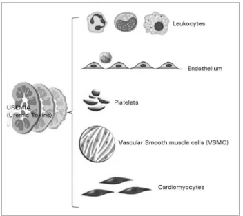
A large number of uremic toxins have deleterious effects on various organs and tissues in the body, mainly the cardiovascular system (Figure 1). The pathophysiological mechanisms involved are complex and far from being completely understood. They include reactive oxidative stress, inflammation, protein glycation and cellular transdifferentiation. In this review, we present the main toxin prototype groups, based on physico-chemical characteristics, clinical effects, and current or potential interventions aimed to modulate their concentration through extracorporeal removal methods and/or pharmacological approaches capable of opposing their toxic effects.
SMALLWATER-SOLUBLECOMPOUNDS GUANIDINES
An important group of small water-soluble compounds consists of guanidine compounds, metabolites of L-arginine that have long been known for their neurotoxic effects.8 There are three types of methylated arginine residues: monomethyl arginine (MMA), symmetric dimethylarginine (SDMA) and asymmetric dimethylarginine (ADMA), the latter being the most abundant one. Several studies have shown an increase in serum ADMA levels in CKD, which may be particularly relevant to the presence of TABLE 1 REQUIREMENTSFORAGIVENCOMPOUNDBE
CONSIDEREDASAUREMICTOXIN4
Chemically identified and accurately measured; Total body/plasma levels should be higher than in non-uremic subjects;
High concentrations should be related whith specific dysfunctions/symptoms, which disappear when concentrations are reduced;
Biological activity of this compound should be proven in
ex vivo, in vivo or in vitro studies, and finally, experimental concentrations of this molecule in those studies should match those found in body fluids or tissue from uremic patients.
introduction of hemodialysis (HD) into clinical prac-tice in the 1960s. The concept of uremic toxins has thus been developed at that time, and their identifica-tion and biological activities were actively investigated subsequently by a small number of research groups (Man and Funck-Brentano in France, Bergström in Sweden, etc). These research activities led to the
“middle molecule’’ hypothesis.5,6 However, studies in this field ceased subsequently because no convincing demonstration of their role in the uremic syndrome could be made at that time. The topic was resuscitated later on, in the early 1990ies, by the nephrology group in Gent, Belgium (Ringoir, Vanholder). They were able to provide new, scientifically valid evidence in favor of the biological activity of several uremic toxins. Finally, in 1999 Vanholder’s group, together with other researchers in Europe, launched the European Uremic Toxins (EUTox) Work Group, which contributed substantially to the acceptance of the concept and the demonstration of the role of uremic toxins. Subsequently, the idea disseminated worldwide, as reflected by a remarkable increase in published reports devoted to this field.
According to EUTox, a compound can be considered as a uremic toxin if it fits a postulate, similar to Koch’s postulate, as modified by Massry et al. in 1977 (Table 1).3,7 At present, there are 152 solutes listed in the EUTox database (http://eutoxdb. odeesoft.com/index.php) and certainly an increase in this number in the following years can be expected.
nephrotic proteinuria, although its correlation with glomerular filtration rate (GFR) has been found to be poor.9,10 Initially, it was assumed that this increase was the result of lower renal clearance. Later studies, however, suggested that the increase was due to both augmented synthesis and reduced catabolism of these compounds.11 Of note, even though guanidi-nes and urea are structurally similar, the distribution volume of the former is significantly greater, which results in a decrease in removal efficiency by dialysis procedures.12
ADMA has long been associated with endothelial dysfunction. In 1992, Vallence et al.13 identified MMA and ADMA as endogenous inhibitors of nitric oxide synthase (eNOS). However, the intracellular concentration of MMA is very small, whereas ADMA is the most abundant eNOS inhibitor. ADMA has been further linked to cardiovascular disease (CVD) and mortality in both the general and CKD population ever since.14,15
SDMA, the structural counterpart of ADMA, was considered inert until recently. However, it is currently recognized as a pro-inflammatory and a pro-oxidant agent.16,17 Unlike ADMA, SDMA was demonstrated to be capable of increasing TNF-α and interleukine-6 (IL-6) expression, and enhancing NFkB activation, in human monocytic cell line THP-1. In line with this in vitro data, SDMA has been associated with inflammatory markers, including TNF-α and IL-6, in pre-dialysis patients at different stages of CKD.17
URICACID
Uric acid (MW: 168 Da) is considered a uremic toxin since a considerable body of evidence suggests that supraphysiologic uric acid concentrations have deleterious effects on the kidney and on the cardiovascular system. In CKD, factors like reduced GFR, increased renal vascular resistance and co-existent insulin resistance as well as the use of diuretics can lead to hyperuricemia, defined as the accumulation of serum uric acid beyond its solubility point in water (6.8 mg/dL).18 Other factors such as high purine/protein diet, alcohol consumption and high cell turnover can also contribute to increase its serum concentration.
Classically, hyperuricemia could lead to hyperuricosuria and deposition of uric acid crystals in the kidney, increasing the risk of nephrolithiasis, renal inflammation and decrease of GFR by intraluminal obstruction and increased concentration of urate anion in renal tissue.19,20 On the other hand, non-crystal dependent effects of uric acid have been proposed, but this remains a matter of debate. Though urate anion, the predominant form of uric acid under physiologic pH, has long been considered a powerful antioxidant at physiologic concentrations, experimental data have shown that at higher concentrations this anion can lead to endothelial dysfunction, increased systemic cytokine production, activation of the renin-angiotensin-aldosterone system and last, not least, glomerular lesions.21
In the clinical setting, observational studies have reported conflicting results. In the Cardiovascular Health Study, no association was observed between serum uric acid level and the incidence of CKD.22 Similarly, in the patient cohort of the MDRD Study, uric acid was not found to be an independent risk factor for CKD progression.23 In contrast, Hsu et al. evaluated a cohort of 177,570 participants followed over 25-years and found that increased serum uric acid levels were independently associated with an increased risk of end-stage kidney disease (ESKD).24 A hypothesis to explain these apparent inconsistencies is that uric acid clearance is impaired in CKD, which in turn can act as a confounding factor.21 Taking into account interventional studies aimed to decrease serum uric acid levels in CKD, the results, although encouraging,25 are not strong enough to systematically recommend allopurinol as a therapy for halting or decreasing CKD progression.
INORGANICPHOSPHATE (PI)
The regulatory mechanisms responsible for Pi homeostasis, such as 1,25(OH)2-vitamin D, parathyroid hormone (PTH) and fibroblast growth factor-23 (FGF23), together with Klotho,26 are disrupted in the CKD setting. Pi overload may occur early on in the course of CKD, while hyperphosphatemia appears only when GFR falls below 30 ml/min/1.73 m2 of body surface.27 When CKD progresses and dialysis treatment is required, the positive balance of Pi becomes more pronounced due to (i) loss of kidney function and (ii) insufficient removal by conventional hemodialysis (HD) or peritoneal dialysis (PD) (Table 2). A detailed discussion of Pi homeostasis and hyperphosphatemia treatment can be found elsewhere.26,28
More recently, the role of Pi as a potential cardiovascular toxin has received further support. Several observational studies have suggested that high Pi levels, even within the normal range, may be associated with an increased risk of incident CVD,37 left ventricular hypertrophy (LVH) and increased risk of heart failure,38 coronary disease,39 and higher mortality40 in the general population.
Experimental studies have shed some light on the mechanisms by which Pi acts as a cardiovascular toxin, above and beyond the promotion of VC. Apolipoprotein E knockout mice fed an atherogenic diet with high (1.6%) Pi content had significantly more atheroma at the site of the aortic sinus than mice fed a low (0.2%) or standard (0.6%) Pi diet, while lipid profile and blood pressure were not affected.41 Furthermore, it has been demonstrated in two different mouse models of CKD that lowering serum Pi levels with sevelamer, a phosphate-binding agent, improved VC and atherosclerotic lesions,34 aortic stiffness, diastolic dysfunction and prevented ventricular hypertrophy.42 In vivo studies have demonstrated that Pi overload may lead to endothelial dysfunction, by inducing apoptosis,43 impaired acethylcholine-induced vasodilation,44,45 inhibition of nitric oxide production44 and downregulation of annexin II.46 Interestingly, lowering serum Pi levels (at least in animal models of CKD) by either dietary Pi restriction or sevelamer administration ameliorates endothelial dysfunction.43,47
A major limitation in the interpretation of Pi effects in vivo is the difficulty to know whether they are the result of a direct action of Pi or rather due to indirect mechanisms, e.g. via an increase in serum PTH levels. In two studies using a rat model of CKD, in which the animals underwent parathyroidectomy and continuous controlled infusion of PTH, the authors were able to demonstrate a direct Pi effect on the heart, inducing myocardial hypertrophy, cardiomyocyte hyperplasia and interstitial fibrosis, and vessels in the absence of changes in serum PTH concentration.48-50
Recent clinical evidence attributing Pi an important role as a culprit in CVD in both CKD and general population has brought this compound to the forefront of uremic toxins. It is also worth mentioning that the amount of Pi in Western diet, especially due to its use as a food additive, is probably much higher than the one recommended by health authorities, resulting in the excessive exposure to a continuous TABLE 2 ESTIMATEDWEEKLYREMOVALOFPHOSPHATE
BYDIALYTICMETHODS
Dialytic methods Duration/ frequency
Phosphate removal (mg/wk) Conventional HD* 4h; 3x/wk 2.356 ± 864
Short daily HD* 2-3h; 6x/wk 2.452 ± 720 Nocturnal HD* 6-8h; 6x/wk 8.000 ± 2.800
Hemodiafiltration 4h; 3x/wk 3.570 ± 270
Peritoneal dialysis
APD 2.739 ± 1.042
CAPD 2.790 ± 1.022
HD: Hemodialysis; APD: Automated peritoneal dialysis; CAPD: Continuous ambulatory peritoneal dialysis. * Using high-flux membranes.
Pi overload. Therefore, given the increased CVD and mortality risk associated with hyperphosphatemia, the proposition has been made to label food products with high Pi content in the interest of public health, similarly to what is currently recommended for food NaCl content.
MIDDLEMOLECULES
FIBROBLASTGROWTHFACTOR-23
FGF23 was described more than 10 years ago by two independent groups.51,52 This hormone, secreted by osteocytes and osteoblasts, is a protein constituted by 251-amino-acids (MW: 30,000 Da). Its major function is the control of mineral metabolism, particularly serum Pi and calcitriol levels, through inhibition of renal tubular Pi reabsorption and 1α-hydroxylase activity. FGF23 also acts on the parathyroid gland inhibiting PTH secretion. All these actions are mediated by FGF23 binding to FGF receptors (FGFR) and its co-receptor Klotho, which greatly increases the binding affinity of FGF23 for FGFR.53,54
The serum FGF23 level increases early in the course of CKD, probably due to a state of primary FGF23 excess, resulting from a combination of increased production and decreased degradation.55 As GFR declines, FGF23 increases and can reach levels > 1,000-fold above normal in patients with CKD stage 5D. Furthermore, residual clearance by the kidney and removal by dialysis do not appear to modify serum FGF23 to a significant extent.56
Numerous reports showed a positive association between elevated FGF23 levels and adverse clinical outcomes, such as CKD progression,57,58 LVH,59 VC60 and mortality in pre-dialysis and dialysis CKD populations.61,62 Considering the above effects of FGF23 on biological functions, one might expect to see evidence in support of a causal relationship in this respect. However, available experimental findings do not allow a clear conclusion. For example, complete neutralization of FGF23 action by a specific antibody has been shown to be harmful since it aggravated hyperphosphatemia, VC, and mortality in a rat model of CKD and mineral bone disorder (MBD).63 This observation suggests that FGF23, like PTH, exerts beneficial effects in early stages of CKD but that its excessive rise with the progression of CKD to ESKD is a maladaptation syndrome with highly deleterious consequences.
Regarding LVH, convincing experimental evidence has recently been provided by Faul et al. in favor of a direct noxious effect of FGF23.64 The authors showed that the intramyocardial or intravenous injection of FGF23
in wild-type mice induced LVH, independent of Klotho which is not expressed in the myocardium.64 In addition, the authors reported that elevated FGF23 levels were independently associated with LVH in a large, racially diverse CKD cohort, in line with findings by others in the general population.65 However, not all authors have been able to find an association of high FGF23 levels with vascular calcification66 or mortality59,67 Apparent inconsistencies can be explained by small sample size, different approaches to adjust for confounding factors, imaging of different arterial beds, lack of prospective data, and finally, different FGF23 mechanisms of toxicity.
Therefore, it is not yet clear if FGF23 is only a surrogate marker of CKD-MBD, CKD progression and cardiovascular disease or whether it is also an active player and hence a potential therapeutic target. For a more detailed review of FGF23 the reader should refer to recent comprehensive reviews.68,69
LEPTIN
The hormone leptin is a 167-amino-acid secreted protein (MW: 16,000 Da), expressed mainly in adipose tissue. It was originally proposed to be an antiobesity factor. However, the majority of obese healthy individuals have elevated serum leptin levels. Therefore, the hypothesis of leptin resistance in human obesity emerged.70 The classical actions of leptin include the control of feeding behavior, energy balance, fertility and immune function. Recently, other actions of leptin have been described, such as its influence on bone mass and the cardiovascular system.71 At the cellular level, leptin stimulates alkaline phosphatase activity, as well as the proliferation, migration and calcification of VSMCs.72,73
Although clinical studies in the general population have shown a link between leptin and CVD,74,75 leptin’s role in CKD seems to be more complex. A group of authors have suggested that dialysis patients with high, rather that low, serum leptin levels have better outcomes,76 However, there was no association between serum leptin and body mass index (BMI). Clearly, the relation between serum leptin and BMI is complex in ESKD, and other group did not find any survival advantage in obese HD patients as compared to lean HD patients.77
independent predictor of metabolic syndrome but not of clinical outcomes. Interestingly, PTH was an independent predictor of plasma leptin levels.78 Taking into account these two findings, one could speculate that leptin is related to factors that exert a more close impact on the clinical outcomes of CKD patients, such as mineral disturbances and metabolic factors. This hypothesis remains to be proven in future clinical long-term studies.
PARATHYROIDHORMONE
Secondary hyperparathyroidism, characterized by an increase in PTH (MW: 9.4 kDa) synthesis and secretion and by parathyroid gland hyperplasia, is a common complication of CKD. Elevated PTH levels are generally found since the early stages of CKD (GFR > 60 ml/min/1.73 m2), in any case much sooner than hyperphosphatemia.27 As one of the most important regulators of bone metabolism, PTH in excess induces significant changes in bone structure and function, leading to the development of high turnover bone disease which, in association with CKD, is called osteitis fibrosa.29,79 It is characterized by increased bone fragility, which may explain, at least in part, the association between sHPT and increased fracture risk.80 In addition, sHPT may cause bone marrow fibrosis and impair erythropoiesis. It has been suggested that very high PTH levels contribute to the polyneuropathy, glucose intolerance, dyslipidemia and inflammation of the uremic state.81
It is important to recognize that toxic PTH levels may lead to deleterious effects on many other organs and tissues due to the ubiquitous expression of its main receptor, PTH1R, including the cardiovascular system. Observational studies have found associations between high PTH levels and CVD in the setting of CKD, such as VC,82 disturbed left ventricular function83 and mortality.84 Observational studies have pointed toward an association between PTH and cardiovascular mortality in the general population as well.85 CKD patients with severe sHPT often develop severe VC, which are particularly located in the arterial media such as the ones observed in digitial arteries of the hands and the feet, and which can entirely regress after surgical parathyroidectomy.86
The effect of PTH on the CV system is an issue under continuous investigation. Experimental studies using a rat model of CKD (5/6 nephrectomy) in which the animals underwent parathyroidectomy, the continuous infusion of supraphysiological rates of
synthetic PTH was associated with the development of extensive VC - independently of serum Pi levels or the presence of uremia.48 In another study using the same CKD rat model, higher PTH levels were associated with myocardial hypertrophy and fibrosis along with higher myocardial expression of inflammation markers and oxidative stress.49 Furthermore, studies have reported the interplay between PTH on the one hand and aldosterone and norepinephrine release on the other, suggesting additional pathophysiologic pathways by which sHPT may lead to cardiovascular damage.87,88
Finally, there is increasing evidence that circulating fragments of PTH, which are elevated in the uremic state, are hormonally active, exerting partially opposing effects to those of intact PTH. Thus, it has been suggested that 7-84 PTH fragments may be involved in the skeletal resistance to PTH observed in CKD.89
There is an intricate relationship between PTH and other factors involved in the disturbances of mineral metabolism in CKD, which may fog the importance of PTH per se as a risk factor for uremia-related complications. The inconclusive, non-definitive results of the recent EVOLVE trial have further contributed to this uncertainty, since in intent-to-treat analysis there was no significant advantage of cinacalcet treatment over best presently available standard treatment in the combined primary endpoint (cardiovascular events plus death) despite a marked decrease in serum PTH.90 However, when performing prespecified corrections of major confounders such as age and study drug discontinuation the better control of hyperparathyroidism was associated with a nominally significant superiority in hard outcomes. Of note, numerous previous clinical and experimental studies support the hypothesis that PTH acts as a systemic uremic toxin, with direct and indirect effects on a variety of tissues and organs, and severe sHPT is a major threat to CKD patient outcomes. Therefore, PTH remains an important therapeutic target to avoid bone and cardiovascular complications in such patients.
ADVANCED GLYCATION END PRODUCTS (AGES) AND
ADVANCEDOXIDIZEDPROTEINPRODUCTS (AOPPS)
main sources of AGEs. The exogenous forms come from diet whereas the endogenous ones are formed in the body. AGE transformation of nutrients occurs when foods are processed at high temperature. In contrast, endogenous transformation results from exposure to high glucose levels such as in diabetic patients, from aging, and from uremia.91,92 AGEs result in a first step from non-enzymatic glycation of proteins by aldehyde and ketones to form a Schiff base. After rearrangement of the these structures, the intermediary products are formed (Amadori products), and finally, AGEs in a second step.93 There are more than 20 AGE compounds, including 1,2-dicarbonyl precursor compounds glyoxal, methylglyoxal, and the end products, N-carboxymethyl-lysine, pentosidine, and hydroimidazolone as the best characterized compounds, which serve as markers of AGE accumulation in a range of tissues.94 AGEs main pathway of elimination from the body is through the urine, or through dialysis in case of renal function replacement.95 Patients under PD are more susceptible than HD patients to systemic and local formation of AGEs, not only as a result of the uremic state but also of the constant exposure of the peritoneum to high levels of glucose and glucose degradation products generated during sterilization of dialysis fluid by heat.95 Moreover, AGEs accumulate progressively in the peritoneal mesothelial layer with increasing dialysate dwell time.96 Another cause of increased AGE formation in the uremic state is oxidati-ve stress, generated by an imbalance between pro-oxi-dant forces (such as an increase in the ratio of oxidized glutathione to reduced glutathione) and anti-oxidant defense system (such as reduced superoxide dismutase/ peroxidase activity).
AGEs exert several potentially deleterious effects in the body. In the cardiovascular system, their accumulation contributes to myocardial changes, endothelial dysfunction, arterial stiffness, and atherosclerotic plaque formation. When these molecules bind to collagen and elastin, they accumulate in the matrix of blood vessels in a non functional and disorderly way, changing endothelial vasomotor tone modulation, platelet adhesion, and cell proliferation.97 AGEs exert their actions in the vascular system by binding to a specific receptor, called RAGE. Activation of RAGE induces an inflammatory response, leading to the effects described above and also increasing the production of adhesion molecules, increasing proliferation of the vessel intimal layer, angiogenesis and oxidative stress.98
RAGE is expressed in all cells involved in atherogenesis, including monocytes, macrophages, endothelial cells and VSMC. Interestingly, these cells do not express significant amounts of RAGE under physiological conditions, but can be induced to express RAGE more vigorously in conditions where their ligands and/or transcription factors accumulate, like in uremia.99
Advanced oxidation protein products (AOPPs) are a class of dityrosine-containing protein products, which arise from the reaction between chlorinated oxidants and plasma proteins. Increased levels of AOPPs are detected in uremic serum since predialysis stage. AOPPs have been suggested to be not only reliable markers of oxidative stress but also mediators of inflammation, by triggering monocyte activation.100 They have been associated with podocyte injury,101 incident thrombo-occlusive atherosclerotic CV events in the pre-dialysis setting102 and carotid atherosclerosis in ESKD patients.103 These findings support the hypothesis that AOPPs should be considered as uremic toxins.
PROTEIN-BOUNDUREMICTOXINS INDOXYLSULFATE
Indoxyl sulfate (IS) is a low-MW (213.21 Da) uremic toxin derivated from dietary protein. It is an indol derived from the amino acid tryptophan by action of intestinal bacteria.104 It is normally excreted into the urine, with its mean urinary excretion rate ranging from 50-70 mg/day in healthy persons. In CKD patients, as renal function declines it accumulates in serum due to its reduced renal clearance.105 The main part of IS in the blood of CKD patients is bound to serum albumin, which means that its urinary excretion occurs mainly by tubular secretion, mainly by proximal tubular cells, and secondarily by glomerular filtration. Organic anion transporter (OAT) 1, the prototypic para-amino hippurate transporter, is localized in renal tubular cells and interacts with a wide range of small endogenous substances, including IS. IS is taken up from the blood by OAT1 and OAT3 at the basolateral membrane of tubular cells and accumulates in the cells at high concentrations.106
in endothelial damage and triggers the production of pro-inflammatory molecules, inhibition of endothelial regeneration and repair, and endothelial free radical
production.108 In addition, the participation of IS in
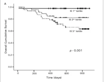
VC, endothelial microparticle release, disruption of adherent junctions of endothelial cells, proliferation of VSMCs, renal and cardiac fibrosis, and impairement of osteoclast differentiation and function has been reported as well.109-112 It has been proposed that IS may affect the remnant nephrons, especially proximal tubular cells, and stimulate tubulointerstitial fibrosis, glomerular sclerosis, and the progression of CKD by increasing the gene expression of TGF-β1, TIMP-1, and pro-α1collagen, leading to a further loss of nephrons, completing the vicious circle of progressive renal injury.104 Several clinical studies have shown that high IS levels are associated with high IL-6 levels, coronary artery disease, vascular damage, progression of CKD and mortality (Figure 2).112-116
compounds involved ρ-cresol; however, it was discovered subsequently that this compound is found in only tiny concentrations in the body since it is rapidly metabolized to its conjugates by the intestinal microbiota. Of note, it has been shown that the biochemical impact of ρ-cresol is not necessarily the same as the impact of its conjugates.118 Previous studies have demonstrated that these uremic toxins are first taken up by the kidneys, blood vessels, bones, and across the blood-brain barrier via OATs and then induce the production of oxygen free radicals and inflammatory cytokines in the respective organs.119-122 ρ-cresol affects the inflammatory response, interfering with the activation of polymorphonuclear leukocytes and endothelial response to cytokines.123 However, neither in uremic patients nor in normal people this molecule can be detected in the circulation in its unconjugated form, in contrast to PCS.124 Meijers et al. demonstrated that PCS induces the release of endothelial microparticles, even in the absence of endothelial injury, suggesting that this toxin is involved in endothelial dysfunction.125 Furthermore, Schepers et al. observed a pro-inflammatory effect of PCS, measured by increased formation of free radicals produced by leukocytes, contributing to vascular damage in patients with CKD.126 Importantly, high PCS levels have been associated with mortality in CKD.124 In addition, Koppe et al. observed that normal mice treated with PCS for 4 weeks, developed insulin resistance, loss of fat mass, and ectopic redistribution of lipid in muscle and liver, mimicking features associated with CKD.127
T
HERAPEUTIC STRATEGIESAs discussed above, the uremic syndrome is a complex condition resulting from the effects of wide variety of toxins. Current strategies aimed to decrease their serum concentration are dietary and pharmacological interventions, mainly via modulation of intestinal absorption capacity through binding effects and/or reduction of ingested amounts of the toxins or their precursors, and extra-corporeal removal via PD and HD.
Notably, removal of middle and protein-bound uremic
toxins by conventional HD and PD is insufficient. The main therapeutic strategies aimed to reduce the level of these compounds and possible advantages are briefly discussed below.
DIETARYINTERVENTION
Protein-bound uremic toxins are generally produced from the metabolism of amino acids in the intestine. Thus, low-protein diet is often considered as a possible
Figure 2. Kaplan-Meyer estimates of several survival probability for CKD patients as a function of tertiles of serum indoxyl sulfate levels. Footnote: reprinted with permission from Barreto FC (ref.115).
PARA-CRESOL (P-CRESOL) ANDP-CRESYLSULFATE (PCS)
dietary approach to reduce the serum concentration of these toxins. Animal experiments have shown that rats fed a low-protein diet had lower concentration of protein-bound solutes.128 Recently, it was demonstrated that a reduced protein and Pi intakes associated with a very low protein diet, supplemented with ketoanalogues and essential amino acids, significantly lowered IS in CKD patients.129 However, nephrologists should be aware of the fact that reducing protein intake excessively may deteriorate the nutritional status of CKD patients and that continuous nutritional evaluations by an expert dietitian are required. Another interesting issue that needs further investigation is the influence of vegetarian diets on the production of uremic toxins. The urinary excretion of PCS and IS was 62% and 59% lower, respectively, in vegetarians than in participants consuming an unrestricted diet, which was associated with higher fiber intake and lower protein intake.130 Finally, the use of pre and probiotics has been suggested as interesting approach to reduce IS and PCS serum levels as well.131,132
PHARMACOLOGICALINTERVENTION
The rationale for using oral sorbents to achieve a reduction of protein-bound toxins in the circulation is that they are derived from the intestinal metabolism of amino acids. Clinical studies ha-ve demonstrated that the oral sorbent AST-120 (Kremezin®) is associated with lower levels of IS,133 slower progression of CKD134 and improved survi-val after dialysis was started.135 Experimental stu-dies had also shown potentially useful therapeutic effects of AST-120 in preventing IS-induced pa-thological alterations, such as VC.136,137 Results of two EPPIC trials, that evaluated the effectiveness of AST-120 added to standard-of-care therapy in moderate to severe CKD, did not support effi-cacy of this drug in slowing CKD progression. However, a subgroup analysis indicated a trend for AST-120 associated reduction of CKD progression in compliant patients with rapid decline of renal function.138 Further trials are required to confirm the reality of this trend.
The use of specific types of phosphate binders has also been proposed as a possible means to decrease the concentrations of several uremic toxins, apart from their main therapeutic utilization in the control of hyperphosphatemia. Recently, two small clinical studies pointed to the possibility of achieving a
pharmacologic modulation of leptin139 and FGF23140 levels in a cohort of early stage CKD patients via the use of sevelamer. Moreover, Vlassara et al. demonstrated in patients with diabetes and early CKD that sevelamer reduced markers of inflammantion and oxidative stress, independently of changes in Pi, possibly due to sevelamer’s capacity to bind AGEs in the intestinal lumen.141 In a recent observational cross-sectional study, in PD patients, sevelamer use was associated with lower levels of ρ-cresol as well.142 In a pilot study in healthy volunteers treatment with acarbose, an alpha-glucosidase inhibitor, decreased the generation and serum concentrations of the protein bound uremic solute ρ-cresol.143 Whether these actions may lead to an improvement of specific uremia-related complications and overall outcome of patients with CKD has yet to be shown.
HEMODIALYSISWITHHIGH-FLUXMEMBRANES
Several randomized controlled trials (RCT) aimed to demonstrate that an increase in the clearance of higher weight molecules via the use of high-flux dialysis membranes led to a survival benefit as compared to standard low-flux membranes. The first large-scale RCT was the HEMO study.144 It failed to find a difference in mortality between the two groups. It also was unable to demonstrate a benefit of high dialysis dose as compared to standard dialysis dose. Post hoc secondary analyses of the HEMO study pointed to a benefit of using high-flux membranes in terms of cardiac outcomes,145 and decreased cerebro-vascular disease mortality in patient subcategories,145 possibly as the result of better middle molecule removal due to the larger pore size of this type of membrane. It is worth mentioning that high-flux HD has no effect on protein-bound uremic toxin levels. Similarly to the HEMO trial, the subsequently done MPO trial also failed to demonstrate a survival advantage with the use of high-flux as compared to low-flux dialysis membranes.146 It must be pointed out that positive results obtained by secondary analyses may strengthen, but do not prove, the hypothesis that high-flux treatment may improve cardiovascular events and survival in certain subpopulations of HD patients.
CONVECTIVESTRATEGIES (HEMOFILTRATIONORHEMODIAFILTRATION)
protein-bound uremic toxins.147,148 By comparing
three main convective strategies in parallel, named pre-dilution hemodiafiltration (HDF), post-dilution
HDF and pre-hemofiltration (HF), Meert et al.
reported that under similar convective volumes,
post- and pre-dilution HDF had similar effects on protein-bound molecules removal, though the former appeared to be better for small water soluble compounds and β2-microgobulin. Pre-HF was superior to pre-HDF only for β2-microgobulin removal and not at all superior to post-HDF.148 In summary, post-HDF was found to be the most effective convective strategy for solute removal. Mid-dilution HDF, a new strategy that allows simultaneous infusion in a pre- and post-dilution fashion, appears to be as efficient as post-dilution HDF for small water-soluble and protein-bound solutes removal.149
A beneficial impact of convective therapies on hard clinical outcomes is however far from being firmly established. Although benefit has been suggested by
observational studies,150,151 the results obtained in
recent RCTs are less clear.152,153 Thus, Grooteman
et al. found in a recent multicenter RCT that dialysis patients assigned to high-efficiency post-dilution on-line HDF had less cardiovascular events but there was no difference between the two groups in the
primary endpoint, namely all-cause mortality.152 Ok
et al. set up an RCT comparing the effect of on-line
HDF to that of high-flux HD.153 The primary outcome
(composite of death from any cause and nonfatal cardiovascular events) was not different between the
two groups. However, in adjusted Cox-regression
analysis treatment with high-efficiency on-line HD
was associated with a 46% risk reduction for overall mortality. In a very recent RCT, Asci et al. failed to find an advantage in fatal or non-fatal cardiovascular event-free survival with high flux HD as compared
to standard HD treatment.154 Only Maduell et al.
were able to demonstrate that dialysis patients randomized to on-line HDF had a better survival
than those randomized to standard HD.155 Whether
the superiority of HDF was due to a better clearance of uremic toxins in the middle-to-large MW range remains to be demonstrated.
ADSORPTION
Other possible therapeutic strategies that could be used to improve dialysis removal of protein-bound
uremic toxins are: addition of activated charcoal or 5% albumin to the dialysate; use of highly permeable dialysis membranes to promote albumin leakage; fractionated plasma separation and adsorption.156 They have not yet been used in large clinical trials, though. The risk of aggravating malnutrition, limited effectiveness due to saturation of adsorption sites and thrombotic complications are potential limitations of these therapies.157 Additional studies are required before recommending these strategies in routine clinical use.
PERITONEALDIALYSIS
Data on uremic toxins in PD is scarce. In spite of the serum concentration of protein-bound uremic toxins being lower in PD compared to HD patients, their removal is worse in the former group.158 These observations suggest that factors beyond the dialysis method, such as intestinal generation and/or metabolism, may play a role to determine their serum levels.
Although several studies have suggested a benefit of therapeutic intervention on middle and protein-bound uremic toxins levels, most of these data came from pos hoc analysis or small studies. Larger, prospective, well-designed RCT aimed to identify the impact of reducing the levels of different uremic toxins on hard outcomes, such as overall and cardiovascular mortality, are required.
G
ENERALCONCLUSIONSThe study of uremic toxins is of growing interest in ne-phrology. Evidence from clinical and experimental stu-dies have demonstrated the impact of these compounds on a variety of organs and systems. Notably, uremic toxins have recently been claimed as new cardiovas-cular risk factors, attracting interest from other fields of medicine.159 Understanding the effects of uremic toxins may provide a more comprehensive view of CKD and its complications and, most likely, new therapeutic targets to retard the progression of CKD and to counteract its cardiovascular complications.
R
EFERENCES1. Meyer TW, Hostetter TH. Uremia. N Engl J Med 2007;357:1316-25. PMID: 17898101 DOI: http://dx.doi.org/10.1056/NEJMra071313 2. Vanholder R. Uremic toxins. Nephrologie 2003;24:373-6. 3. Glassock RJ. Uremic toxins: what are they? An integrated
overview of pathobiology and classification. J Ren Nutr 2008;18:2-6. DOI: http://dx.doi.org/10.1053/j.jrn.2007.10.003 4. Mahomed FA. On the Pathology of Uraemia and the Socalled
5. Man NK, Terlain B, Paris J, Werner G, Sausse A, Funck-Brentano JL. An approach to "middle molecules" identification in artificial kidney dialysate, with reference to neuropathy prevention. Trans Am Soc Artif Intern Organs 1973;19:320-4.
6. Babb AL, Johansen PJ, Strand MJ, Tenckhoff H, Scribner BH. Bi-directional permeability of the human peritoneum to middle molecules. Proc Eur Dial Transplant Assoc 1973;10:247-62.
7. Vanholder R, De Smet R, Glorieux G, Argilés A, Baurmeister U, Brunet P, et al.; European Uremic Toxin Work Group (EUTox). Review on uremic toxins: classification, concentration, and interindividual variability. Kidney Int 2003;63:1934-43. PMID: 12675874
8. D'Hooge R, Pei YQ, Marescau B, De Deyn PP. Convulsive action and toxicity of uremic guanidino compounds: behavioral assessment and relation to brain concentration in adult mice. J Neurol Sci 1992;112:96-105. PMID: 1469446 DOI: http:// dx.doi.org/10.1016/0022-510X(92)90138-B
9. Eloot S, Schepers E, Barreto DV, Barreto FC, Liabeuf S, Van Biesen W, et al. Estimated glomerular filtration rate is a poor predictor of concentration for a broad range of uremic toxins. Clin J Am Soc Nephrol 2011;6:1266-73. DOI: http://dx.doi. org/10.2215/CJN.09981110
10. Kielstein JT, Böger RH, Bode-Böger SM, Frölich JC, Haller H, Ritz E, et al. Marked increase of asymmetric dimethylarginine in patients with incipient primary chronic renal disease. J Am Soc Nephrol 2002;13:170-6.
11. Baylis C. Nitric oxide deficiency in chronic kidney disease. Am J Physiol Renal Physiol 2008;294:F1-9. PMID: 17928410 12. Eloot S, van Biesen W, Dhondt A, de Smet R, Marescau B, De
Deyn PP, et al. Impact of increasing haemodialysis frequency versus haemodialysis duration on removal of urea and guanidino compounds: a kinetic analysis. Nephrol Dial Transplant 2009;24:2225-32. DOI: http://dx.doi.org/10.1093/ndt/gfp059 13. Vallance P, Leone A, Calver A, Collier J, Moncada S.
Endogenous dimethylarginine as an inhibitor of nitric oxi-de synthesis. J Cardiovasc Pharmacol 1992;20:S60-2. PMID: 1282988 DOI: http://dx.doi.org/10.1097/00005344-199204002-00018
14. Böger RH, Bode-Böger SM, Szuba A, Tsao PS, Chan JR, Tangphao O, et al. Asymmetric dimethylarginine (ADMA): a novel risk fac-tor for endothelial dysfunction: its role in hypercholesterolemia. Circulation 1998;98:1842-7. PMID: 9799202
15. Zoccali C, Bode-Böger S, Mallamaci F, Benedetto F, Tripepi G, Malatino L, et al. Plasma concentration of asymmetrical dimethylarginine and mortality in patients with end-stage renal disease: a prospective study. Lancet 2001;358:2113-7. PMID: 11784625 DOI: http://dx.doi. org/10.1016/S0140-6736(01)07217-8
16. Bode-Böger SM, Scalera F, Kielstein JT, Martens-Lobenhoffer J, Breithardt G, Fobker M, et al. Symmetrical dimethylarginine: a new combined parameter for renal function and extent of coronary artery disease. J Am Soc Nephrol 2006;17:1128-34. DOI: http://dx.doi.org/10.1681/ASN.2005101119
17. Schepers E, Barreto DV, Liabeuf S, Glorieux G, Eloot S, Barreto FC, et al. Symmetric dimethylarginine as a proinflammatory agent in chronic kidney disease. Clin J Am Soc Nephrol 2011;6:2374-83. DOI: http://dx.doi.org/10.2215/ CJN.01720211
18. Jalal DI, Chonchol M, Chen W, Targher G. Uric acid as a target of therapy in CKD. Am J Kidney Dis 2013;61:134-46. DOI: http://dx.doi.org/10.1053/j.ajkd.2012.07.021
19. Umekawa T, Chegini N, Khan SR. Increased expression of monocyte chemoattractant protein-1 (MCP-1) by renal epithelial cells in culture on exposure to calcium oxalate, phosphate and uric acid crystals. Nephrol Dial Transplant 2003;18:664-9. DOI: http://dx.doi.org/10.1093/ndt/gfg140 20. Spencer HW, Yarger WE, Robinson RR. Alterations of renal
function during dietary-induced hyperuricemia in the rat. Kidney Int 1976;9:489-500. DOI: http://dx.doi.org/10.1038/ ki.1976.63
21. Johnson RJ, Kang DH, Feig D, Kivlighn S, Kanellis J, Watanabe S, et al. Is there a pathogenetic role for uric acid in hypertension and cardiovascular and renal disease? Hypertension 2003;41:1183-90. 22. Chonchol M, Shlipak MG, Katz R, Sarnak MJ, Newman AB,
Siscovick DS, et al. Relationship of uric acid with progression of kidney disease. Am J Kidney Dis 2007;50:239-47. PMID: 17660025 DOI: http://dx.doi.org/10.1053/j.ajkd.2007.05.013 23. Madero M, Sarnak MJ, Wang X, Greene T, Beck GJ, Kusek JW,
et al. Uric acid and long-term outcomes in CKD. Am J Kidney Dis 2009;53:796-803. DOI: http://dx.doi.org/10.1053/j. ajkd.2008.12.021
24. Hsu CY, Iribarren C, McCulloch CE, Darbinian J, Go AS. Risk factors for end-stage renal disease: 25-year follow-up. Arch Intern Med 2009;169:342-50. PMID: 19237717 DOI: http:// dx.doi.org/10.1001/archinternmed.2008.605
25. Goicoechea M, de Vinuesa SG, Verdalles U, Ruiz-Caro C, Ampuero J, Rincón A, et al. Effect of allopurinol in chronic kidney disease progression and cardiovascular risk. Clin J Am Soc Nephrol 2010;5:1388-93. DOI: http://dx.doi.org/10.2215/CJN.01580210 26. Uribarri J. Phosphorus homeostasis in normal health and
in chronic kidney disease patients with special emphasis on dietary phosphorus intake. Semin Dial 2007;20:295-301. DOI: http://dx.doi.org/10.1111/j.1525-139X.2007.00309.x 27. Levin A, Bakris GL, Molitch M, Smulders M, Tian J, Williams LA,
et al. Prevalence of abnormal serum vitamin D, PTH, calcium, and phosphorus in patients with chronic kidney disease: results of the study to evaluate early kidney disease. Kidney Int 2007;71:31-8. DOI: http://dx.doi.org/10.1038/sj.ki.5002009 28. Barreto FC, de Oliveira RA, Oliveira RB, Jorgetti V.
Pharmacotherapy of chronic kidney disease and mineral bone disorder. Expert Opin Pharmacother 2012;12:2627-40. DOI: http://dx.doi.org/10.1517/14656566.2011.626768
29. Sampaio EA, Lugon JR, Barreto FC. Fisopatologia do hiperparatireoidismo secundário J Bras Nefrol 2008;30:6-10. 30. Block GA, Hulbert-Shearon TE, Levin NW, Port FK.
Association of serum phosphorus and calcium x phosphate product with mortality risk in chronic hemodialysis patients: a national study. Am J Kidney Dis 1998;31:607-17. DOI: http:// dx.doi.org/10.1053/ajkd.1998.v31.pm9531176
31. Kestenbaum B, Sampson JN, Rudser KD, Patterson DJ, Seliger SL, Young B, et al. Serum phosphate levels and mortality risk among people with chronic kidney disease. J Am Soc Nephrol 2005;16:520-8. DOI: http://dx.doi.org/10.1681/ ASN.2004070602
32. Giachelli CM. Vascular calcification: in vitro evidence for the role of inorganic phosphate. J Am Soc Nephrol 2003;14:S300-4. DOI: http://dx.doi.org/10.1097/01.ASN.0000081663.52165.66 33. Phan O, Ivanovski O, Nguyen-Khoa T, Mothu N, Angulo
J, Westenfeld R, et al. Sevelamer prevents uremia-enhanced atherosclerosis progression in apolipoprotein E-deficient mice. Circulation 2005;112:2875-82. PMID: 16267260 DOI: http:// dx.doi.org/10.1161/CIRCULATIONAHA105.541854 34. Phan O, Ivanovski O, Nikolov IG, Joki N, Maizel J, Louvet L,
et al. Effect of oral calcium carbonate on aortic calcification in apolipoprotein E-deficient (apoE-/-) mice with chronic renal failure. Nephrol Dial Transplant 2008;23:82-90. DOI: http:// dx.doi.org/10.1093/ndt/gfm699
35. Barreto DV, Barreto Fde C, de Carvalho AB, Cuppari L, Draibe SA, Dalboni MA, et al. Phosphate binder impact on bone remodeling and coronary calcification-results from the BRiC study. Nephron Clin Pract 2008;110:c273-83. DOI: http://dx.doi.org/10.1159/000170783
36. Chertow GM, Burke SK, Raggi P; Treat to Goal Working Group. Sevelamer attenuates the progression of coronary and aortic calcification in hemodialysis patients. Kidney Int 2002;62:245-52. DOI: http://dx.doi.org/10.1046/j.1523-1755.2002.00434.x 37. Dhingra R, Sullivan LM, Fox CS, Wang TJ, D'Agostino RB
38. Dhingra R, Gona P, Benjamin EJ, Wang TJ, Aragam J, D'Agostino RB Sr, et al. Relations of serum phosphorus levels to echocardiographic left ventricular mass and incidence of heart failure in the community. Eur J Heart Fail 2010;12:812-8. DOI: http://dx.doi.org/10.1093/eurjhf/hfq106
39. Cancela AL, Santos RD, Titan SM, Goldenstein PT, Rochitte CE, Lemos PA, et al. Phosphorus is associated with coronary artery disease in patients with preserved renal function. PLoS One 2012;7:e36883. DOI: http://dx.doi.org/10.1371/journal. pone.0036883
40. Tonelli M, Sacks F, Pfeffer M, Gao Z, Curhan G; Cholesterol And Recurrent Events Trial Investigators. Relation between serum phosphate level and cardiovascular event rate in people with coronary disease. Circulation 2005;112:2627-33. PMID: 16246962
41. Ellam T, Wilkie M, Chamberlain J, Crossman D, Eastell R, Francis S, et al. Dietary phosphate modulates atherogenesis and insulin resistance in apolipoprotein E knockout mice-brief report. Arterioscler Thromb Vasc Biol 2011;31:1988-90. DOI: http://dx.doi.org/10.1161/ATVBAHA.111.231001
42. Maizel J, Six I, Dupont S, Secq E, Dehedin B, Barreto FC, et al. Effects of sevelamer treatment on cardiovascular abnormalities in mice with chronic renal failure. Kidney Int 2013;84:491-500. DOI: http://dx.doi.org/10.1038/ki.2013.110
43. Di Marco GS, Hausberg M, Hillebrand U, Rustemeyer P, Wittkowski W, Lang D, et al. Increased inorganic phosphate induces human endothelial cell apoptosis in vitro. Am J Physiol Renal Physiol 2008;294:F1381-7. PMID: 18385273 DOI: http://dx.doi.org/10.1152/ajprenal.00003.2008
44. Shuto E, Taketani Y, Tanaka R, Harada N, Isshiki M, Sato M, et al. Dietary phosphorus acutely impairs endothelial function. J Am Soc Nephrol 2009;20:1504-12. DOI: http://dx.doi. org/10.1681/ASN.2008101106
45. Six I, Maizel J, Barreto FC, Rangrez AY, Dupont S, Slama M, et al. Effects of phosphate on vascular function under normal conditions and influence of the uraemic state. Cardiovasc Res 2012;96:130-9. PMID: 22822101 DOI: http://dx.doi. org/10.1093/cvr/cvs240
46. Di Marco GS, König M, Stock C, Wiesinger A, Hillebrand U, Reiermann S, et al. High phosphate directly affects endothelial function by downregulating annexin II. Kidney Int 2013;83:213-22. DOI: http://dx.doi.org/10.1038/ ki.2012.300
47. Van TV, Watari E, Taketani Y, Kitamura T, Shiota A, Tanaka T, et al. Dietary phosphate restriction ameliorates endothelial dysfunction in adenine-induced kidney disease rats. J Clin Biochem Nutr 2012;51:27-32. DOI: http://dx.doi.org/10.3164/jcbn.11-96 48. Neves KR, Graciolli FG, dos Reis LM, Pasqualucci CA,
Moysés RM, Jorgetti V. Adverse effects of hyperphosphatemia on myocardial hypertrophy, renal function, and bone in rats with renal failure. Kidney Int 2004;66:2237-44. PMID: 15569312 DOI: http://dx.doi.org/10.1111/j.1523-1755.2004.66013.x
49. Custódio MR, Koike MK, Neves KR, dos Reis LM, Graciolli FG, Neves CL, et al. Parathyroid hormone and phosphorus overload in uremia: impact on cardiovascular system. Nephrol Dial Transplant 2012;27:1437-45. DOI: http://dx.doi.org/10.1093/ ndt/gfr447
50. Graciolli FG, Neves KR, dos Reis LM, Graciolli RG, Noronha IL, Moysés RM, et al. Phosphorus overload and PTH induce aortic expression of Runx2 in experimental uraemia. Nephrol Dial Transplant 2009;24:1416-21. DOI: http://dx.doi. org/10.1093/ndt/gfn686
51. Yamashita T, Yoshioka M, Itoh N. Identification of a novel fibroblast growth factor, FGF-23, preferentially expressed in the ventrolateral thalamic nucleus of the brain. Biochem Biophy Res Comm 2000;277:494-8. DOI: http://dx.doi.org/10.1006/ bbrc.2000.3696
52. ADHR Consortium. Autossomal dominant hypophosphataemic rickets is associated with mutations in FGF23. Nat Genet 2000; 26:345-8. DOI: http://dx.doi.org/10.1038/81664
53. Urakawa I, Yamazaki Y, Shimada T, Iijima K, Hasegawa H, Okawa K, et al. Klotho converts canonical FGF receptor into a specific receptor for FGF23. Nature 2006;444:770-4. PMID: 17086194 DOI: http://dx.doi.org/10.1038/nature05315 54. Ben-Dov IZ, Galitzer H, Lavi-Moshayoff V, Goetz R,
Kuro-o M, Mohammadi M, et al. The parathyroid is a target organ for FGF23 in rats. J Clin Invest 2007;117:4003-8. PMID: 17992255
55. Larsson T, Nisbeth U, Ljunggren O, Jüppner H, Jonsson KB. Circulating concentration of FGF-23 increases as renal function declines in patients with chronic kidney disease, but does not change in response to variation in phosphate intake in healthy volunteers. Kidney Int 2003;64:2272-9. PMID: 14633152 DOI: http://dx.doi.org/10.1046/j.1523-1755.2003.00328.x 56. Isakova T, Xie H, Barchi-Chung A, Vargas G, Sowden N,
Houston J, et al. Fibroblast growth factor 23 in patients undergoing peritoneal dialysis. Clin J Am Soc Nephrol 2011;6:2688-95. DOI: http://dx.doi.org/10.2215/CJN.04290511
57. Fliser D, Kollerits B, Neyer U, Ankerst DP, Lhotta K, Lingenhel A, et al. Fibroblast growth factor 23 (FGF23) predicts progression of chronic kidney disease: the Mild to Moderate Kidney Disease (MMKD) Study. J Am Soc Nephrol 2007;18:2600-8. DOI: http://dx.doi.org/10.1681/ASN.2006080936
58. Titan SM, Zatz R, Graciolli FG, dos Reis LM, Barros RT, Jorgetti V, et al. FGF-23 as a predictor of renal outcome in diabetic nephropathy. Clin J Am Soc Nephrol 2011;6:241-7. DOI: http://dx.doi.org/10.2215/CJN.04250510
59. Hsu HJ, Wu MS. Fibroblast growth factor 23: a possible cause of left ventricular hypertrophy in hemodialysis patients. Am J Med Sci 2009;337:116-22. PMID: 19214027 DOI: http:// dx.doi.org/10.1097/MAJ.0b013e3181815498
60. Desjardins L, Liabeuf S, Renard C, Lenglet A, Lemke HD, Choukroun G, et al.; European Uremic Toxin (EUTox) Work Group. FGF23 is independently associated with vascular calcification but not bone mineral density in patients at various CKD stages. Osteoporos Int 2012;23:2017-25. PMID: 22109743
61. Gutiérrez OM, Mannstadt M, Isakova T, Rauh-Hain JA, Tamez H, Shah A, et al. Fibroblast growth factor 23 and mortality among patients undergoing hemodialysis. N Engl J Med 2008;359:584-92. PMID: 18687639 DOI: http://dx.doi. org/10.1056/NEJMoa0706130
62. Isakova T, Xie H, Yang W, Xie D, Anderson AH, Scialla J, et al. Fibroblast growth factor 23 and risks of mortality and end-stage renal disease in patients with chronic kidney disease. JAMA 2011;305:2432-9. PMID: 21673295 DOI: http://dx.doi. org/10.1001/jama.2011.826
63. Shalhoub V, Shatzen EM, Ward SC, Davis J, Stevens J, Bi V, et al. FGF23 neutralization improves chronic kidney disease-associated hyperparathyroidism yet increases mortality. J Clin Invest 2012;122:2543-53. PMID: 22728934 DOI: http:// dx.doi.org/10.1172/JCI61405
64. Faul C, Amaral AP, Oskouei B, Hu MC, Sloan A, Isakova T, et al. FGF23 induces left ventricular hypertrophy. J Clin Invest 2011;121:4393-408. PMID: 21985788 DOI: http://dx.doi. org/10.1172/JCI46122
65. Mirza MA, Larsson A, Melhus H, Lind L, Larsson TE. Se-rum intact FGF23 associate with left ventricular mass, hypertrophy and geometry in an elderly population. Atherosclerosis 2009;207:546-1. DOI: http://dx.doi. org/10.1016/j.atherosclerosis.2009.05.013
66. Scialla JJ, Lau WL, Reilly MP, Isakova T, Yang HY, Crouthamel MH, et al. Fibroblast growth factor 23 is not associated with and does not induce arterial calcification. Kidney Int 2013;83:1159-68. DOI: http://dx.doi.org/10.1038/ ki.2013.3
68. Oliveira RB, Moysés RM. FGF-23: state of the art. J Bras Nefrol 2010;32:323-1. DOI: http://dx.doi.org/10.1590/S0101-28002010000300015
69. Wolf M. Update on fibroblast growth factor 23 in chronic kidney disease. Kidney Int 2012;82:737-47. PMID: 22622492 DOI: http://dx.doi.org/10.1038/ki.2012.176
70. Considine RV, Sinha MK, Heiman ML, Kriauciunas A, Stephens TW, Nyce MR, et al. Serum immunoreactive-leptin concentrations in normal-weight and obese humans. N Engl J Med 1996;334:292-5. PMID: 8532024 DOI: http://dx.doi. org/10.1056/NEJM199602013340503
71. Friedman JM, Halaas JL. Leptin and the regulation of body weight in mammals. Nature 1998;395:763-70. PMID: 9796811 DOI: http://dx.doi.org/10.1038/27376
72. Parhami F, Tintut Y, Ballard A, Fogelman AM, Demer LL. Leptin enhances the calcification of vascular cells: artery wall as a target of leptin. Circ Res 2001;88:954-60. PMID: 11349006 DOI: http://dx.doi.org/10.1161/hh0901.090975
73. Oda A, Taniguchi T, Yokoyama M. Leptin stimulates rat aortic smooth muscle cell proliferation and migration. Kobe J Med Sci 2001;47:141-50. PMID: 11729375
74. Singhal A, Farooqi IS, Cole TJ, O'Rahilly S, Fewtrell M, Kattenhorn M, et al. Influence of leptin on arterial distensibility: a novel link between obesity and cardiovascular disease? Circulation 2002;106:1919-24. PMID: 12370213
75. Wallace AM, McMahon AD, Packard CJ, Kelly A, Shepherd J, Gaw A, et al. Plasma leptin and the risk of cardiovascular disease in the west of Scotland coronary prevention study (WOSCOPS). Circulation 2001;104:3052-6. PMID: 11748099 DOI: http://dx.doi.org/10.1161/hc5001.101061
76. Scholze A, Rattensperger D, Zidek W, Tepel M. Low serum leptin predicts mortality in patients with chronic kidney disease stage 5. Obesity (Silver Spring) 2007;15:1617-22. DOI: http:// dx.doi.org/10.1038/oby.2007.191
77. de Mutsert R, Snijder MB, van der Sman-de Beer F, Seidell JC, Boeschoten EW, Krediet RT, et al. Association between body mass index and mortality is similar in the hemodialysis population and the general population at high age and equal duration of follow-up. J Am Soc Nephrol 2007;18:967-74. DOI: http://dx.doi.org/10.1681/ASN.2006091050
78. de Oliveira RB, Liabeuf S, Okazaki H, Lenglet A, Desjardins L, Lemke HD, et al. The clinical impact of plasma leptin levels in a cohort of chronic kidney disease patients. Clin Kidney J 2013;6:63-70. DOI: http://dx.doi.org/10.1093/ckj/sfs176 79. Mac Way F, Lessard M, Lafage-Proust MH. Pathophysiology
of chronic kidney disease-mineral and bone disorder. Joint Bone Spine 2012;79:544-9. PMID: 23177912 DOI: http:// dx.doi.org/10.1016/j.jbspin.2012.09.014
80. Jadoul M, Albert JM, Akiba T, Akizawa T, Arab L, Bragg-Gresham JL, et al. Incidence and risk factors for hip or other bone fractures among hemodialysis patients in the Dialysis Outcomes and Practice Patterns Study. Kidney Int 2006;70:1358-66. PMID: 16929251 DOI: http://dx.doi. org/10.1038/sj.ki.5001754
81. Rodriguez M, Lorenzo V. Parathyroid hormone, a uremic toxin. Semin Dial 2009;22:363-8. DOI: http://dx.doi.org/10.1111/ j.1525-139X.2009.00581.x
82. Hernandes FR, Barreto FC, Rocha LA, Draibe SA, Canziani ME, Carvalho AB. Evaluation of the role of severe hyperparathyroidism on coronary artery calcification in dialysis patients. Clin Nephrol 2007;67:89-95. PMID: 17338428 DOI: http://dx.doi.org/10.5414/CNP67089
83. Drüeke T, Fauchet M, Fleury J, Lesourd P, Toure Y, Le Pailleur C, et al. Effect of parathyroidectomy on left--ventricular function in haemodialysis patients. Lancet 1980;1:112-4. DOI: http://dx.doi.org/10.1016/S0140-6736(80)90602-9
84. Block GA, Klassen PS, Lazarus JM, Ofsthun N, Lowrie EG, Chertow GM. Mineral metabolism, mortality, and morbidity in maintenance hemodialysis. J Am Soc Nephrol 2004;15:2208-18. DOI: http://dx.doi.org/10.1097/01.ASN.0000133041.27682.A2
85. van Ballegooijen AJ, Reinders I, Visser M, Dekker JM, Nijpels G, Stehouwer CD, et al. Serum parathyroid hormone in relation to all-cause and cardiovascular mortality: the Hoorn study. J Clin Endocrinol Metab 2013;98:E638-45. DOI: http:// dx.doi.org/10.1210/jc.2012-4007
86. Rostand SG, Drüeke TB. Parathyroid hormone, vitamin D, and cardiovascular disease in chronic renal failure. Kidney Int 1999;56:383-92. PMID: 10432376 DOI: http://dx.doi. org/10.1046/j.1523-1755.1999.00575.x
87. Tomaschitz A, Ritz E, Pieske B, Fahrleitner-Pammer A, Kienreich K, Horina JH, et al. Aldosterone and parathyroid hormone: a precarious couple for cardiovascular disease. Cardiovasc Res 2012;94:10-9. PMID: 22334595 DOI: http:// dx.doi.org/10.1093/cvr/cvs092
88. Potthoff SA, Janus A, Hoch H, Frahnert M, Tossios P, Reber D, et al. PTH-receptors regulate norepinephrine release in human heart and kidney. Regul Pept 2011;171:35-42. PMID: 21756942 DOI: http://dx.doi.org/10.1016/j.regpep.2011.06.002
89. Iwasaki Y, Yamato H, Nii-Kono T, Fujieda A, Uchida M, Hosokawa A, et al. Insufficiency of PTH action on bone in uremia. Kidney Int Suppl 2006:S34-6. DOI: http://dx.doi. org/10.1038/sj.ki.5001600
90. EVOLVE Trial Investigators; Chertow GM, Block GA, Correa-Rotter R, Drüeke TB, Floege J, Goodman WG, et al. Effect of cinacalcet on cardiovascular disease in pa-tients undergoing dialysis. N Engl J Med 2012;367:2482-94. PMID: 23121374 DOI: http://dx.doi.org/10.1056/NEJ-Moa1205624
91. Semba RD, Nicklett EJ, Ferrucci L. Does accumulation of advanced glycation and products contribute to the aging phenotype? J Gerontol A Biol Sci Med Sci 2010;65:963-75. PMID: 20478906
92. Schwedler S, Schinzel R, Vaith P, Wanner C. Inflammation and advanced glycation end products in uremia: simple coexistence, potentiation or causal relationship? Kidney Int Suppl 2001;78:S32-6. PMID: 11168979
93. Piperi C, Adamopoulos C, Dalagiorgou G, Diamanti-Kandarakis E, Papavassiliou AG. Crosstalk between advanced glycation and endoplasmic reticulum stress: emerging therapeutic targeting for metabolic diseases. J Clin Endocrinol Metab 2012;97:2231-42. PMID: 22508704 DOI: http://dx.doi.org/10.1210/jc.2011-3408 94. Thornalley PJ, Rabbani N. Highlights and hotspots of protein
glycation in end-stage renal disease. Semin Dial 2009;22:400-4. DOI: http://dx.doi.org/10.1111/j.1525-139X.2009.00589.x 95. Alhamdani MS, Al-Azzawie HF, Abbas FK. Decreased formation
of advanced glycation end-products in peritoneal fluid by carnosine and related peptides. Perit Dial Int 2007;27:86-9. 96. Brunkhorst R, Kruse M, Ehlerding G, Mahiout A. Advanced
glycosylated end-products (AGEs) in non-diabetic patients undergoing dialysis. Clin Nephrol 1996;46:265-6.
97. Zieman S, Kass D. Advanced glycation end product cross-linking: pathophysiologic role and therapeutic target in cardiovascular disease. Congest Heart Fail 2004;10:144-9. DOI: http://dx.doi.org/10.1111/j.1527-5299.2004.03223.x 98. Xu Y, Wang S, Feng L, Zhu Q, Xiang P, He B. Blockade of
PKC-beta protects HUVEC from advanced glycation end products induced inflammation. Int Immunopharmacol 2010;10:1552-9. DOI: http://dx.doi.org/10.1016/j.intimp.2010.09.006
99. Figarola JL, Shanmugam N, Natarajan R, Rahbar S. Anti-inflammatory effects of the advanced glycation end product inhibitor LR-90 in human monocytes. Diabetes 2007;56:647-55. PMID: 17327432 DOI: http://dx.doi.org/10.2337/db06-0936 100. Witko-Sarsat V, Friedlander M, Capeillère-Blandin C,
Nguyen-Khoa T, Nguyen AT, Zingraff J, et al. Advanced oxidation protein products as a novel marker of oxidative stress in uremia. Kidney Int 1996;49:1304-13. PMID: 8731095 DOI: http://dx.doi.org/10.1038/ki.1996.186
102. Descamps-Latscha B, Witko-Sarsat V, Nguyen-Khoa T, Nguyen AT, Gausson V, Mothu N, et al. Advanced oxidation protein products as risk factors for atherosclerotic cardiovascular events in nondiabetic predialysis patients. Am J Kidney Dis 2005;45:39-47. PMID: 15696442 DOI: http:// dx.doi.org/10.1053/j.ajkd.2004.09.011
103. Drüeke T, Witko-Sarsat V, Massy Z, Descamps-Latscha B, Guerin AP, Marchais SJ, et al. Iron therapy, advanced oxidation protein products, and carotid artery intima-media thickness in end-stage renal disease. Circulation 2002;106:2212-7. PMID: 12390950 DOI: http://dx.doi.org/10.1161/01. CIR.0000035250.66458.67
104. Niwa T. Uremic toxicity of indoxyl sulfate. Nagoya J Med Sci 2010;72:1-11.
105. Bolati D, Shimizu H, Yisireyili M, Nishijima F, Niwa T. Indoxyl sulfate, a uremic toxin, downregulates renal expression of Nrf2 through activation of NF-κB. BMC Nephrol 2013;14:56. DOI: http://dx.doi.org/10.1186/1471-2369-14-56
106. Taki K, Nakamura S, Miglinas M, Enomoto A, Niwa T. Accumulation of indoxyl sulfate in OAT1/3-positive tubular cells in kidneys of patients with chronic renal failure. J Ren Nutr 2006;16:199-203. DOI: http://dx.doi.org/10.1053/j.jrn.2006.04.020 107. Lowenstein J. The anglerfish and uremic toxins. FASEB J
2011;25:1781-5. DOI: http://dx.doi.org/10.1096/fj.11-0602ufm 108. Dou L, Bertrand E, Cerini C, Faure V, Sampol J, Vanholder R,
et al. The uremic solutes p-cresol and indoxyl sulfate inhibit endothelial proliferation and wound repair. Kidney Int 2004;65:442-51. DOI: http://dx.doi.org/10.1111/j.1523-1755.2004.00399.x
109. Bammens B, Evenepoel P, Verbeke K, Vanrenterghem Y. Removal of middle molecules and protein-bound solutes by peritoneal dialysis and relation with uremic symptoms. Kidney Int 2003;64:2238-43. DOI: http://dx.doi.org/10.1046/j.1523-1755.2003.00310.x
110. Neirynck N, Vanholder R, Schepers E, Eloot S, Pletinck A, Glorieux G. An update on uremic toxins. Int Urol Nephrol 2013;45:139-50. DOI: http://dx.doi.org/10.1007/s11255-012-0258-1
111. Mozar A, Louvet L, Godin C, Mentaverri R, Brazier M, Kamel S, et al. Indoxyl sulphate inhibits osteoclast differentiation and function. Nephrol Dial Transplant 2012;27:2176-81. DOI: http://dx.doi.org/10.1093/ndt/gfr647
112. Mozar A, Louvet L, Morlière P, Godin C, Boudot C, Kamel S, et al. Uremic toxin indoxyl sulfate inhibits human vascular smooth muscle cell proliferation. Ther Apher Dial 2011;15:135-9. DOI: http://dx.doi.org/10.1111/j.1744-9987.2010.00885.x 113. Lee CT, Kuo CC, Chen YM, Hsu CY, Lee WC, Tsai YC, et al.
Factors associated with blood concentrations of indoxyl sulfate and p-cresol in patients undergoing peritoneal dialysis. Perit Dial Int 2010;30:456-63. DOI: http://dx.doi.org/10.3747/pdi.2009.00092 114. Chiu CA, Lu LF, Yu TH, Hung WC, Chung FM, Tsai IT, et al.
Increased levels of total P-Cresylsulphate and indoxyl sulphate are associated with coronary artery disease in patients with diabetic nephropathy. Rev Diabet Stud 2010;7:275-84. DOI: http://dx.doi.org/10.1900/RDS.2010.7.275
115. Barreto FC, Barreto DV, Liabeuf S, Meert N, Glorieux G, Temmar M, et al.; European Uremic Toxin Work Group (EUTox). Serum indoxyl sulfate is associated with vascular disease and mortality in chronic kidney disease patients. Clin J Am Soc Nephrol 2009;4:1551-8. DOI: http://dx.doi. org/10.2215/CJN.03980609
116. Wu IW, Hsu KH, Lee CC, Sun CY, Hsu HJ, Tsai CJ, et al. p-Cresyl sulphate and indoxyl sulphate predict progression of chronic kidney disease. Nephrol Dial Transplant 2010;26:938-47. 117. de Loor H, Bammens B, Evenepoel P, De Preter V,
Verbeke K. Gas chromatographic-mass spectrometric analysis for measurement of p-cresol and its conjugated metabolites in uremic and normal serum. Clin Chem 2005;51:1535-8. DOI: http://dx.doi.org/10.1373/clinchem.2005.050781
118. Vanholder R, Van Laecke S, Glorieux G. What is new in uremic toxicity? Pediatr Nephrol 2008;23:1211-21.
119. Deguchi T, Ohtsuki S, Otagiri M, Takanaga H, Asaba H, Mori S, et al. Major role of organic anion transporter 3 in the transport of indoxyl sulfate in the kidney. Kidney Int 2002;61:1760-8. PMID: 11967025 DOI: http://dx.doi.org/10.1046/j.1523-1755.2002.00318.x
120. Tsutsumi Y, Deguchi T, Takano M, Takadate A, Lindup WE, Otagiri M. Renal disposition of a furan dicarboxylic acid and other uremic toxins in the rat. J Pharmacol Exp Ther 2002;303:880-7. DOI: http://dx.doi.org/10.1124/ jpet.303.2.880
121. Enomoto A, Niwa T. Roles of organic anion transporters in the progression of chronic renal failure. Ther Apher Dial 2007;1:S27-31. DOI: http://dx.doi.org/10.1111/j.1744-9987.2007.00515.x
122. Miyamoto Y, Watanabe H, Noguchi T, Kotani S, Nakajima M, Kadowaki D, et al. Organic anion transporters play an important role in the uptake of p-cresyl sulfate, a uremic toxin, in the kidney. Nephrol Dial Transplant 2011;26:2498-502. DOI: http://dx.doi.org/10.1093/ndt/gfq785
123. Dou L, Cerini C, Brunet P, Guilianelli C, Moal V, Grau G, et al. P-cresol, a uremic toxin, decreases endothelial cell response to inflammatory cytokines. Kidney Int 2002;62:1999-2009. PMID: 12427124 DOI: http://dx.doi.org/10.1046/j.1523-1755.2002. t01-1-00651.x
124. Liabeuf S, Barreto DV, Barreto FC, Meert N, Glorieux G, Schepers E, et al.; European Uraemic Toxin Work Group (EUTox). Free p-cresylsulphate is a predictor of mortality in patients at different stages of chronic kidney disease. Nephrol Dial Transplant 2010;25:1183-91. DOI: http://dx.doi.org/10.1093/ndt/gfp592
125. Meijers BK, Van Kerckhoven S, Verbeke K, Dehaen W, Vanrenterghem Y, Hoylaerts MF, et al. The uremic retention solute p-cresyl sulfate and markers of endothelial damage. Am J Kidney Dis 2009;54:891-901. PMID: 19615803 DOI: http:// dx.doi.org/10.1053/j.ajkd.2009.04.022
126. Schepers E, Meert N, Glorieux G, Goeman J, Van der Eycken J, Vanholder R. P-cresylsulphate, the main in vivo metabolite of p-cresol, activates leucocyte free radical production. Nephrol Dial Transplant 2007;22:592-6. DOI: http://dx.doi. org/10.1093/ndt/gfl584
127. Koppe L, Pillon NJ, Vella RE, Croze ML, Pelletier CC, Chambert S, et al. p-Cresyl sulfate promotes insulin resistance associated with CKD. J Am Soc Nephrol 2013;24:88-99. DOI: http://dx.doi.org/10.1681/ASN.2012050503
128. Brunet P, Dou L, Cerini C, Berland Y. Protein-bound uremic retention solutes. Adv Ren Replace Ther 2003;10:310-20. DOI: http://dx.doi.org/10.1053/j.arrt.2003.08.002
129. Marzocco S, Dal Piaz F, Di Micco L, Torraca S, Sirico ML, Tartaglia D, et al. Very low protein diet reduces indoxyl sulfate levels in chronic kidney disease. Blood Purif 2013;35:196-201. DOI: http://dx.doi.org/10.1159/000346628
130. Patel KP, Luo FJ, Plummer NS, Hostetter TH, Meyer TW. The production of p-cresol sulfate and indoxyl sulfate in vegetarians versus omnivores. Clin J Am Soc Nephrol 2012;7:982-8. DOI: http://dx.doi.org/10.2215/CJN.12491211
131. Meijers BK, De Preter V, Verbeke K, Vanrenterghem Y, Evenepoel P. p-Cresyl sulfate serum concentrations in haemodialysis patients are reduced by the prebiotic oligofructose-enriched inulin. Nephrol Dial Transplant 2010;25:219-24. DOI: http://dx.doi.org/10.1093/ndt/ gfp414
132. Rossi M, Klein K, Johnson DW, Campbell KL. Pre-, pro-, and synbiotics: do they have a role in reducing uremic toxins? A systematic review and meta-analysis. Int J Nephrol 2012;2012:673631. PMID: 23316359