Histopathological study comparing native and
post-transplant recurrent chronic hepatitis C with emphasis
on confounders with acute cellular rejection
Estudo histopatológico comparativo da hepatite crônica C nativa e recorrente
pós-transplante com ênfase em achados semelhantes à rejeição celular aguda
Isabela R. C. Fraga; Adriana Caroli-Bottino; Vera Lucia N. Pannain
Universidade Federal do Rio de Janeiro (UFRJ), Rio de Janeiro, Brazil.
First submission on 18/03/16; last submission on 13/07/16; accepted for publication on 24/07/16; published on 20/10/16
ABSTRACT
Introduction: Histological analyses of post-transplant liver biopsies may be dificult in distinguishing recurrent chronic hepatitis C (CHC) from other causes of graft dysfunction, especially acute cellular rejection (ACR). Objective: The aim of this study was to compare the histological characteristics of liver biopsies with CHC in transplant and non-transplant patients with hepatitis C virus (HCV) infection and assess the occurrence of indings common to ACR. Methods: We studied 40 biopsies from non-transplant and 30 biopsies from post-transplant patients, according to the Ishak score for necroinlammatory activity grade and stage of ibrosis. We also assessed the inlammatory iniltrate, steatosis, ductal changes, portal endotheliitis and central perivenulitis. Results: We found predominance of mild grade and stage in both groups. The portal inlammatory iniltrate was also mild and mainly lymphocytic in the two groups. Ductal changes were more frequent in the non-transplant patients. Steatosis was also mild in both groups, but predominated in non-transplant CHC patients. Portal endotheliitis occurred in 42.5% and 40% in non-transplant and post-transplant CHC, respectively. The frequency of centrilobular endotheliitis was similar in both groups. Conclusion: Histological indings in chronic hepatitis C are similar in non-transplant and post-transplant patients. In addition, morphological features characteristic of ACR are also observed in HCV infection of native livers as well as in the graft of patients with recurrent infection after transplantation.
Key words: chronic hepatitis C; liver transplantation; graft rejection; pathology.
INTRODUCTION
Cirrhosis secondary to hepatitis C virus (HCV) infection is currently the main indication for liver transplantation in several countries(1). However, virtually all patients with chronic hepatitis C
(CHC) who undergo transplantation develop recurrent infection during graft reperfusion in which viral titers reach pre-transplant levels a few hours after the procedure(2-4). Infection recurrence
is the leading cause of graft dysfunction and loss and is often associated with a faster progression to cirrhosis in comparison with non-transplant patients(2, 5). Therefore, post-transplant monitoring
is crucial for early diagnosis of disease recurrence and progression, since it allows timely initiation of antiviral treatment(6).
One of the differential diagnoses of recurrent HCV infection after liver transplantation is acute cellular rejection (ACR), which is also a frequent cause of early graft dysfunction(2). In patients
with graft dysfunction, histological analysis of liver biopsy is used to identify the undergoing process. However, distinguishing between infection recurrence and immunological rejection is challenging since some histological indings are common to both conditions(3, 7). Nevertheless, timely identiication of ACR is essential:
it leads to important therapeutic decisions, such as change in the immunosuppressive regimen, which can itself exacerbate the recurrent HCV infection or promote opportunistic infections.
The histological diagnosis of HCV infection is based on the grade of necroinlammatory activity and ibrosis stage(8, 9). One of
the main systems currently used for this evaluation is the Ishak score(9). Other often analyzed histological parameters include
steatosis, portal lymphoid follicles or aggregates, bile ducts injury and
endotheliitis(10). The diagnosis of ACR is based on the presence of mixed
portal inlammatory iniltrate composed of activated lymphocytes, neutrophils and eosinophils, endotheliitis and ductal injury. HCV infection can also present with endotheliitis and biliary ductal
injury(11). In this case, the presence of lobular necroinlammatory
activity, interface hepatitis and steatosis help establish the diagnosis of HCV infection, since they are absent in cases of ACR(12).
Several studies have been conducted to determine factors that increase sensitivity of histological analysis of liver graft biopsy in identifying viral relapse(2). Anyway, we deemed it important that
others be developed to compare simultaneously the histological characteristics of the liver in HCV infection in transplant and non-transplant patients, in addition to analyzing the frequency with which histological changes common to ACR occur.
METHODS
In this retrospective study, we conducted a search at the computerized database of the University Hospital Clementino Fraga Filho (Universidade Federal do Rio de Janeiro [UFRJ]) using the key words “chronic hepatitis C”, “liver transplant” and “hepatitis C virus” to identify patients with HCV infection who underwent liver biopsy between 2002 and 2012. Liver biopsy was performed in patients with elevated aminotransferase levels.
We excluded from the analysis the biopsies of patients who had undergone transplantation less than six months before the study, and those with histological sections with less than six completely represented portal tracts, clinical and/or histological evidence of ACR, ibrosing cholestatic hepatitis, biliary and/or vascular diseases secondary or not to the transplant, and coinfected with the hepatitis B virus and/or human immunodeiciency virus.
After this exclusion, we selected for analysis 40 liver biopsies from patients with native livers with HCV infection (non-transplant) and 30 with recurrent HCV infection at least six months after liver transplantation (post-transplant CHC). The diagnosis of infection was conirmed in all patients by polymerase chain reaction. Information about patients’ gender, age and viral genotype were obtained when available.
For each biopsy we analyzed ive histological sections using a Nikon E200 microscope. Each of the sections was stained with hematoxylin and eosin (HE), Masson’s trichrome and Gomori’s reticulin.
The study was conducted after approval by the Medical Ethics Committee of the University Hospital Clementino Fraga Filho-UFRJ, under registry number 064/11.
Histological analysis
For histological analysis, we considered the following parameters: grade of necroinlammatory activity and stage of ibrosis according to the Ishak score(9), portal and periportal
changes, and ductal epithelium and parenchymal abnormalities.
Grade and stage
Based on the Ishak score(9), we classiied the grade of
necroinlammatory activity as G1-G6 = mild, G7-G12 = moderate, and G13-G18 = marked. We considered the stage of ibrosis in those cases classiied with E1 and E2 as minimum/ mild, in those with E3 and E4 as moderate, and in the ones with E5 and E6 as marked.
Portal and periportal findings
Based on the Ishak score(9), we classiied interface hepatitis as
absent (A0), minimum (A1), mild (A2), moderate (A3) and marked (A4), and the intensity of the inlammatory portal iniltrate as mild (D1 and D2), moderate (D3) or marked (D4). As for the cell types present in the inlammatory iniltrate, we analyzed the presence of lymphocytes, eosinophils and neutrophils, which were classiied as present or absent. The presence of at least three eosinophils in at least one portal tract by analyzed biopsy was determined as the cut-off point in the analysis of portal eosinophils. Portal endotheliitis was considered mild when presenting only a small focus of inlammatory iniltrate permeating the venular endothelium, moderate when affecting up to 50% of the venular circumference, and severe when more than 50% of the circumference of the portal vein was involved.
Ductal epithelium findings
Parenchymal findings
We analyzed the frequency of steatosis, central perivenulitis (with or without centrilobular vein endotheliitis) and apoptosis. Steatosis was considered mild when lipid vacuoles were present in 5%-30% of the hepatocytes, moderate when 31%-60%, and marked when occurring in more than 60%. Central perivenulitis and apoptosis were classiied as present or absent.
Additional analyses
We applied the Banff criteria (portal inlammation, bile duct damage and venous endothelial inlammation – endotheliitis) to evaluate the occurrence of ACR-like indings in both groups(13). In order to assess the rapidity with which
ibrosis develops in transplanted patients, we analyzed the stage of ibrosis in relation to the moment in which the biopsy was
obtained.
Statistical analysis
After descriptive analysis of each variable regarding frequency, we used the test of independence (Wilks’ G²) followed by the Z-test. We applied Student’s t test to verify the occurrence of statistically signiicant differences between groups. We adopted a signiicance level of 0.05.
RESULTS
In the group of non-transplant patients, there was a predominance of the female gender (62.5%), whereas in the group with post-transplant CHC, the male gender predominated (83.4%). Average ages were 53.7 years and 55.8 years, respectively.
We were able to obtain information about the genotype in 31 non-transplant patients and in 21 patients with post-transplant CHC. Among those non-post-transplant, there was a predominance of genotype 1a (n = 13; 42%), followed by 1b (n = 12; 38.7%), genotype 3 (n = 4; 13%) and genotype 1 unspecified (n = 2; 6.3%). In the post-transplant group, the prevalent genotype was 1b (n = 9; 42.8%), followed by genotype 1 unspecified (n = 4; 19%), genotype 3 (n = 5; 24%) and genotype 1a (n = 3; 14.2%).
In the group of transplant patients, the time elapsed between the transplant and the biopsy that diagnosed viral recurrence ranged between 6 months and 11 years.
TABLE 1 − Comparison of grade and stage in non-transplant and transplant patients with CHC
Ishak score
Non-transplant (n = 40)
Post-transplant CHC (n = 30)
p-value (Fischer’s exact test)
Grade
Mild: 32 (80%) Moderate: 8 (20%)
Marked: 0
Mild: 22 (73.4%) Moderate: 8 (26.6%)
Marked: 0
0.258
Stage
Minimum/mild: 26 (65%) Moderate: 12 (30%)
Marked: 2 (5%)
Minimum/mild: 22 (73.4%) Moderate: 8 (26.6%)
Marked: 0
0.085
CHC: chronic hepatitis C.
Histological analysis
Grade of necroinflammatory activity and stage of fibrosis
There was a predominance of mild grade and stage in both groups. A comparison between groups showed no difference between the frequencies of each of these indings (p = 0.258 and p = 0.085, respectively; Table 1). Cirrhosis was observed only in the group of non-transplant patients.
Portal and periportal findings (Table 2)
The frequencies of mild interface hepatitis were similar in both groups (60%; p = 0.162). Three non-transplant patients (7.5%) showed A3 interface hepatitis (moderate) that was not found in any patient in the group with post-transplant CHC.
The portal inlammatory iniltrate was predominantly lymphocytic and of mild intensity in both groups. As for the cell types in the inlammatory iniltrate, there was no signiicant difference between groups regarding the participation of neutrophils
(p = 0.223) or eosinophils (p = 0.156). Portal endotheliitis was identiied in 42.5% of the biopsies of native livers and 40% of the biopsies of transplant patients. Cases classiied as mild endotheliitis prevailed in both groups, 76.5% in non-transplanted and 75% in post-transplant CHC, whereas moderate endotheliitis was present in 25% and 23.5%, respectively (p = 0.417).
Ductal epithelium findings
Both groups showed comparable frequencies of degeneration
(Figure 1), atrophy and ductular proliferation (p = 0.202). However, the prevalence of exocytosis was higher in the non-transplant group (p = 0.049) (Table 3 and Figure 2). We did
Parenchymal findings
Steatosis was predominantly mild in both groups (Table 5).
Moderate steatosis was not observed in any non-transplant patient, but occurred in four patients with post-transplant CHC (p = 0.033). In contrast, central perivenulitis was more frequent in transplant patients (40%) compared with non-transplant patients (20%; p = 0.034) (Figure 3). There was no difference between groups in the frequencies of centrilobular vein endotheliitis or apoptosis
(p = 0.276 and p = 0.388, respectively).
ACR-like findings
In the non-transplant group we observed the presence of all Banff criteria(13) for ACR. Excluding the portal inlammatory iniltrate,
which was present in all cases, we observed that 13 cases (32.5%) had only one ACR-like inding, whereas 12 cases (30%) had two indings, totaling 25 biopsies (62.5%) of non-transplant patients in whom we observed at least one of the diagnostic criteria for ACR. Applying the same criteria for the transplant group, we observed that 14 cases TABLE 2 − Comparison of the frequency of portal and periportal findings
between non-transplant and transplant patients with CHC Portal and
periportal changes
Non-transplant (n = 40)
Post-transplant CHC (n = 30)
p-value (Fischer’s exact
test)
Interface hepatitis (Ishak)
A0: 0 A1: 13 (32.5%)
A2: 24 (60%) A3: 3 (7.5%)
A4: 0
A0: 1 (3.3%) A1: 11 (36.7%)
A2: 18 (60%) A3: 0 A4: 0
0.162
Presence of
neutrophils 21 (52.5%) 13 (43.3%) 0.223
Presence of
eosinophils 9 (22.5%) 4 (13.3%) 0.156
Portal inlammatory
iniltrate
Mild: 28 (70%) Moderate: 12
(30%)
Mild: 24(80%)
Moderate: 6 (20%) 0.339
Endotheliitis
Mild: 13 (76.5%) Moderate: 4
(23.5%)
Mild: 9 (75%)
Moderate: 3 (25%) 0.417
CHC: chronic hepatitis C.
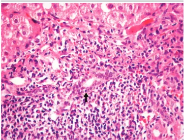
FIGURE 1 − Native liver with chronic hepatitis C: ductal degeneration – cytoplasmic
eosinophilia and loss of nuclear polarization (1,000×)
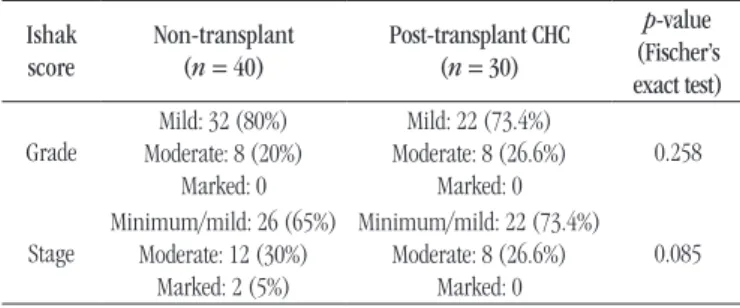
FIGURE 2 − Native liver with chronic hepatitis C: ductal exocytosis – arrow (400×)
TABLE 3 − Comparison of the frequency of ductal epithelium changes in non-transplant and non-transplant patients with CHC
Ductal epithelium changes
Non-transplant (n = 40)
Post-transplant CHC (n = 30)
p-value (Fischer’s exact
test)
Ductal degeneration 11 (27.5%) 6 (20%) 0.23
Ductal atrophy 8 (20%) 4 (13.3%) 0.226
Ductular proliferation 5 (12.5%) 6 (20%) 0.202
Ductal exocytosis 15 (37.5%) 6 (20%) 0.049
CHC: chronic hepatitis C.
TABLE 4 − Ratio of the number of portal tracts with bile ducts featuring exocytosis over the total number of portal tracts present in the analyzed biopsy in
non-transplant and transplant patients with CHC Ratio Non-transplant
(n = 40)
Post-transplant CHC (n = 30)
p-value (Fischer’s exact
test) Minimum
Maximum Median
Mean Standard deviation (n)
0.05 0.25 0.125 0.132 0.053
0.05 0.43 0.13 0.165 0.134
0.289
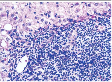
(46.6%) presented only one inding, and three cases (10%) showed two indings (Figure 4), totaling 17 (56.6%) transplant patients in whom we observed at least one of the ACR diagnostic criteria.
FIGURE 4 − Recurrent post-transplant chronic hepatitis C: ductal lesion and endotheliitis
(400×)
TABLE 5 − Comparison of the frequency of parenchymal findings in non-transplant and non-transplant patients with CHC
Parenchymal findings
Non-transplant (n = 40)
Post-transplant CHC (n = 30)
p-value (Fischer’s exact
test)
Apoptosis 16 (40%) 11 (36.6%) 0.388
Steatosis Mild: 22 (55%) Moderate: 0
Mild: 14 (46.6%) Moderate: 4
(13.4%)
0.033
Central perivenulitis 8 (20%) 12 (40%) 0.034
Endotheliitis of
centrilobular vein 7 (17.5%) 7 (23.3%) 0.276
CHC: chronic hepatitis C.
FIGURE 3 − Native liver with chronic hepatitis C: perivenulitis with centrilobular vein
endotheliitis (400×)
Analysis of the stage of fibrosis with respect to
time to obtain the biopsy after transplantation
Fibrosis was present in 12 of 13 patients biopsied within the irst two years after transplantation. Among these patients, nine were genotype 1. Information about the donors’ age was available in only six cases; in these, the average age was 49.2 years (range: 24-68 years).
DISCUSSION
In this comparison of histological sections of liver biopsies of non-transplant CHC patients and post-transplant CHC patients we observed that cirrhosis was present only in non-transplant patients, while moderate steatosis occurred only in transplant patients. Despite the presence of central perivenulitis in both groups, this inding was signiicantly more frequent in the post-transplant group. Regarding the other analyzed parameters, we found no signiicant differences between groups.
The absence of cirrhosis in transplant patients was probably due to the relatively short follow-up time after transplantation (up to four years in most patients). In studies evaluating the occurrence of cirrhosis in transplant patients, the average interval between transplantation and identiication of cirrhosis was 9.5 years(4).
In HCV infection, steatosis is associated with the same mechanisms of insulin resistance, except for patients infected with genotype 3, in whom the accumulation of fat is usually more prominent and a result of direct interference of the viral infection on lipid metabolism within the hepatocyte(7). Therefore,
steatosis is a frequent inding both in native and in grafted livers. Steatosis in HCV infection is characterized mainly by macrovesicles, mostly of mild to moderate intensity, without the characteristic centrilobular distribution of steatosis associated with non-alcoholic fatty liver disease(7). In our study, there
was a predominance of mild steatosis in both groups, whereas moderate steatosis was present only in transplant patients. We believe that genotype did not inluence this result, since among the ive patients with genotype 3 included in the study, only one of the transplant patients had moderate steatosis.
The expression “central perivenulitis” has been suggested to describe inlammatory changes and injuries affecting the centrilobular region(7). It is mainly found in ACR, but is also present
in other liver diseases(14). In fact, we found central perivenulitis
also in non-transplant patients and in a frequency greater than
patients with only CHC and excluded other liver diseases in which central perivenulitis can be present, we speculate that immunological phenomena occurring in this group could account for this inding. Another inding inconsistent with other studies was the presence of centrilobular vein endotheliitis, observed in 17.5% of the non-transplant patients, in contrast with the absence of identiication of this parameter by other
authors(3, 15).
In both groups, interface hepatitis was predominantly mild, unlike what was reported by some authors on recurrence of HCV infection(16). However, the time elapsed after transplantation
in most biopsies included in other studies was shorter than in
ours(16). It is possible that interface activity may increase as the
recurrence of viral infection progresses. In other studies, portal inlammation was absent or was classiied as minimal in half of the cases(16), in contrast to our indings of predominantly mild
portal inlammation in all patients after the transplant. The lobular indings in recurrent CHC are generally similar to those found in biopsies of native livers. However some authors report more marked lobular inlammation in the irst(17, 18).
Saxena et al. (2002)(19) described a signiicant increase in the
amount of apoptotic bodies in biopsies from transplant patients with recurrent hepatitis C when compared with biopsies from native livers. This was not observed in our study, since apoptosis was present in 40% of the biopsies of non-transplant patients and 36% of the transplant patients. The different methodologies of the two studies, both in the quantiication of apoptotic bodies, and in the time elapsed between transplantation and biopsy, prevent a comparative analysis of the results.
In addition to the parameters related to the grade of necroinlammatory activity, other portal changes have been evaluated in the comparison between CHC in native livers and CHC recurrence in transplanted livers, among them, bile duct lesions. Similarly to Poulsen and Christoffersen (1969)(20), we
morphologically classiied the injury to the bile duct epithelium as degeneration, proliferation, atrophy and exocytosis. All these indings were more frequent in the non-transplant group, with the exception of ductular proliferation, which showed no signiicant difference between the groups. Other authors also highlighted the presence of changes in bile ducts in 30% of non-transplant patients(3). This frequency was lower than the
one found in our study, in which ductal exocytosis was observed in 37.5% of the native livers. Considering that those authors studied ductal exocytosis along with other unspeciied biliary duct indings(3), we suggest that our results are superior to theirs
in relation to ductal exocytosis. Since there is evidence that the damage to the epithelium of bile ducts is associated with a higher degree of portal inflammatory activity(7), we also correlated
the intensity of portal inflammatory infiltrate with the several presentations of ductal injury. We did not observe correlation between these findings in any of the groups, even for exocytosis, which had a statistically significant difference between groups. Also regarding ductal exocytosis, we did not find a significant difference in the ratio between the number of portal spaces with affected bile ducts and the total number of portal tracts present in the analyzed biopsy. There is no uniformity in the literature on morphological characterization of ductal injury
in CHC(3, 7), which complicates the comparison of the results. In
addition to the challenges of morphological characterization, the pathogenesis of the ductal injury on the HCV infection is not established, and the possibilities of direct injury by the virus and/or immunological mechanisms should be considered(21),
which increases the number of variables that can determine ductal injury. These evidences point to a need for further studies to better clarify the interaction between HCV and the epithelium of the bile ducts, and consequently the ductal lesions observed
in CHC.
The time of ibrosis development in transplant and non-transplant patients infected by HCV is variable(11). Even though
the stage of ibrosis was mild in both groups, it is important to highlight that already in the irst two years after transplant, ibrosis was present in almost all samples. Considering that the time course of HCV infection is lower in this group, this result points to a greater progression of ibrosis in the post-transplantation period when compared with infection in native livers, as previously reported by other authors(4, 7).
In summary, our indings show that the histological indings in the liver of patients with post-transplant CHC resemble those of non-transplant patients. In addition, morphological indings characteristic of ACR are also observed in HCV infection of native livers as well as in the graft of patients with recurrent infection after transplantation. We conclude, based on these indings, that liver biopsy as an isolated diagnostic method in patients with recurrent CHC after liver transplant may be insuficient to accurately establish the differential diagnosis with ACR.
CONFLICT OF INTEREST
REFERENCES
1. Fernández-Yunquera A, Rincón D, Salcedo M, Bañares R. Update on the use of direct-acting antiviral agents for the treatment of chronic hepatitis C virus infection. Rev Esp Quimioter. 2013 Sep; 26(3): 189-92. PubMed PMID: 24080883.
2. Adeyi O, Fischer SE, Guindi M. Liver allograft pathology: approach to interpretation of needle biopsies with clinicopathological correlation. J Clin Pathol. 2010 Jan; 63(1): 47-74. PubMed PMID: 19847014. 3. Souza P, Prihoda TJ, Hoyumpa AM, Sharkey FE. Morphologic features resembling transplant rejection in core biopsies of native livers from patients with hepatitis C. Hum Pathol. 2009 Jan; 40(1): 92-7. PubMed PMID: 18790517.
4. Gane EJ. The natural history of recurrent hepatitis C and what inluences this. Liver Transpl. 2008 Oct; 14 Suppl 2: S36-44. PubMed PMID: 18825724.
5. Berenguer M. Natural history of recurrent hepatitis C. Liver Transpl. 2002 Oct; 8(10 Suppl 1): S14-8. PubMed PMID: 12362293.
6. Gawrieh S, Papouchado BG, Burgart LJ, Kobayashi S, Charlton MR, Gores GJ. Early hepatic stellate cell activation predicts severe hepatitis C recurrence after liver transplantation. Liver Transpl. 2005 Oct; 11(10): 1207-13. PubMed PMID: 16184568.
7. Moreira RK. Recurrent hepatitis C and acute allograft rejection: clinicopathologic features with emphasis on the differential diagnosis between these entities. Adv Anat Pathol. 2011 Sep; 18(5): 393-405. PubMed PMID: 21841407.
8. Goodman ZD. Grading and staging systems for inlammation and ibrosis in chronic liver diseases. J Hepatol. 2007 Oct; 47(4): 598-607. PubMed PMID: 17692984.
9. Ishak K, Baptista A, Bianchi L, et al. Histological grading and staging of chronic hepatitis. J Hepatol. 1995 Jun; 22(6): 696-9. PubMed PMID: 7560864.
10. Freitas NR, Teles SA, Matos MA, et al. Hepatitis C virus infection in Brazilian long-distance truck drivers. Virol J. 2010 Aug 27; 7: 205. PubMed PMID: 20799961.
11. Meriden Z, Forde KA, Pasha TL, et al. Histologic predictors of ibrosis progression in liver allografts in patients with hepatitis C virus infection. Clin Gastroenterol Hepatol. 2010 Mar; 8(3): 289-96, 296.e1-8. PubMed PMID: 19913638.
12. Petrovic LM, Villamil FG, Vierling JM, Makowka L, Geller SA. Comparison of histopathology in acute allograft rejection and recurrent hepatitis C infection after liver transplantation. Liver Transpl Surg. 1997 Jul; 3(4): 398-406. PubMed PMID: 9346770.
13. Demetris AJ, Kenneth PB, Dhillon AP, et al. Banff schema for grading liver allograft rejection: an international consensus document. Hepatology. 1997 Mar; 25(3): 658-63. PubMed PMID: 9049215. 14. Demetris AJ, Adeyi O, Bellamy CO, et al. Liver biopsy interpretation for causes of late liver allograft dysfunction. Hepatology. 2006 Aug; 44(2): 489-501. PubMed PMID: 16871565.
15. Hubscher SG. Central perivenulitis: a common and potentially important inding in late posttransplant liver biopsies. Liver Transpl. 2008 May; 14(5): 596-600. PubMed PMID: 18433067.
16. Ziarkiewicz-Wroblewska B, Wroblewski T, Ziolkowski J, et al. Evaluation of chronic HCV infection in transplanted livers using a modiied histological activity index. Ann Transplant. 2011 Jan-Mar; 16(1): 26-33. PubMed PMID: 21436771.
17. Gane EJ, Portmann BC, Naoumov NV, et al. Long-term outcome of hepatitis C infection after liver transplantation. N Engl J Med. 1996 Mar 28; 334(13): 815-20. PubMed PMID: 8596547.
RESUMO
Introdução: A análise histológica de biópsias hepáticas pós-transplante pode trazer dificuldades na distinção entre hepatite crônica C (HCC) recorrente e outras causas de disfunção do enxerto, sobretudo rejeição celular aguda (RCA). Objetivo: Comparar as características histológicas de biópsias hepáticas de pacientes transplantados e não transplantados portadores de HCC, além de avaliar a presença de achados comuns à RCA. Métodos: Foram estudadas 40 biópsias de pacientes não transplantados e 30 de transplantados, de acordo com o escore de Ishak para grau de atividade necroinflamatória e estágio de fibrose. Foram ainda avaliadas as características do infiltrado inflamatório, da esteatose, das alterações ductais e da endotelite portal e da perivenulite central. Resultados: Em ambos os grupos, houve predomínio de leve grau de atividade necroinflamatória e leve fibrose. O infiltrado inflamatório portal também foi leve e predominantemente linfocítico em ambos os grupos. Alterações ductais foram mais frequentes em pacientes não transplantados. Esteatose também foi leve em ambos os grupos, mas predominou nos pacientes não transplantados. Endotelite portal ocorreu em 42,5% e 40% em HCC não transplantada e HCC pós-transplante, respectivamente. A frequência de endotelite centrolobular foi semelhante nos dois grupos. Conclusão: Os achados histológicos na HCC são semelhantes em pacientes transplantados e não transplantados. Além disso, características morfológicas da RCA estão presentes na HCC, tanto em fígados nativos como em enxertos de pacientes com infecção recorrente após transplante.
18. Guido M, Fagiuoli S, Tessari G, et al. Histology predicts cirrhotic evolution of post transplant hepatitis C. Gut. 2002 May; 50(5): 697-700. PubMed PMID: 11950819.
19. Saxena R, Crawford JM, Navarro VJ, Friedman AL, Robert ME. Utilization of acidophil bodies in the diagnosis of recurrent hepatitis C infection after orthotopic liver transplantation. Mod Pathol. 2002 Sep; 15(9): 897-903. PubMed PMID: 12218206.
20. Poulsen H, Christoffersen P. Abnormal bile duct epithelium in liver biopsies with histological signs of viral hepatitis. Acta Pathol Microbiol Scand. 1969; 76(3): 383-90. PubMed PMID: 5823358.
21. Fillipowicz EA, Xiao Sy, Sower LE, Weems J, Payne DA. Detection of HCV in bile duct epithelium by laser capture microdissection (LCM). In Vivo. 2005 Jul-Aug; 19(4): 737-9. PubMed PMID: 15999543.
CORRESPONDING AUTHOR
Adriana Marques Caroli de Freitas Bottino