J Bras Pneumol. 2009;35(7):709-712
Case report
A 74-year-old white male who was a retired farmer, a nonsmoker and a former drinker presented with a one-year history of chest pain in the suprasternal notch, accompanied by edema and local erythema. His condition had worsened two weeks prior, aggravated by drainage of purulent material from a fistulous lesion. In addition, he reported evening fever, night sweats and weight loss (25 kg) in the last 12 months. The patient was under outpatient follow-up treatment in various sectors of the Santa Maria University Hospital, due to multiple comorbidities: systemic arterial hypertension; heart failure; mitral valve disease; pulmonary arterial hypertension; hypothyroidism; chronic renal failure; nonspecific colitis; and benign prostatic hyperplasia. He was being treated with furosemide, simvastatin, omeprazole,
levothy-Introduction
The extrapulmonary form of TB involving the sternum is quite rare. The most signifi-cant risk factor for the disease is open heart surgery.(1) Other risk factors include intrave-nous drug abuse, blunt thoracic trauma, closed cardiopulmonary resuscitation, subclavian vein catheterization, diabetes mellitus, HIV infec-tion, alcoholism and BCG vaccination.(1) Sternal TB usually affects young adults living in areas where TB is endemic. Since the advent of modern antituberculous therapy, the number of cases of sternal TB has dramatically decreased, there having been fewer than 20 cases reported in the literature since that time.(2) We report the case of a male patient without active pulmonary disease who was under follow-up treatment at a clinical medicine outpatient clinic when he developed sternal TB.
Sternal osteomyelitis caused by infection
with
Mycobacterium tuberculosis
*
Osteomielite esternal por Mycobacterium tuberculosis
Diego Michelon De Carli, Mateus Dornelles Severo, Carlos Jesus Pereira Haygert, Marcelo Guollo, Alex Omairi, Vinícius Dallagasperina Pedro,
Eduardo Pedrolo Silva, Arnaldo Teixeira Rodrigues
Abstract
We report the case of a 74-year-old male patient with a one-year history of chest pain in the suprasternal notch associated with erythema, edema and drainage of purulent material from a fistulous lesion. The patient was HIV-negative with no history of TB. A CT scan of the chest showed an osteolytic lesion in the sternum, and a biopsy revealed caseous granuloma, which, in the microbiological evaluation, was negative for fungi and acid-fast bacilli. The diagnosis of sternal osteomyelitis caused by Mycobacterium tuberculosis was confirmed using PCR.
Keywords: Thorax; Mycobacterium infections; Tuberculosis, osteoarticular; Sternum; Diagnosis.
Resumo
Descrevemos o caso de um paciente de 74 anos, masculino, com dor torácica na porção superior do esterno com um ano de evolução associada a eritema, edema e fístula com drenagem de material purulento. Paciente HIV negativo e sem história prévia de contato com TB. A TC de tórax evidenciou lesão osteolítica esternal, e o material de biópsia revelou granuloma caseoso negativo para fungos e bacilos álcool-ácido resistentes no exame microbio-lógico direto. O diagnóstico de osteomielite esternal por Mycobacterium tuberculosis foi realizado por PCR. Descritores: Tórax; Infecções por Mycobacterium; Tuberculose osteoarticular; Esterno; Diagnóstico.
* Study carried out at the Santa Maria University Hospital, Federal University of Santa Maria, Santa Maria, Brazil.
Correspondence to: Arnaldo Teixeira Rodrigues. Universidade Federal de Santa Maria, Av. Roraima, s/n, Camobi, CEP 98100-000, Santa Maria, RS, Brasil.
Tel 55 55 3025-7661. E-mail: [email protected] Financial support: None.
Submitted: 3 June 2008. Accepted, after review: 12 January 2009.
710 De Carli DM, Severo MD, Haygert CJP, Guollo M, Omairi A, Pedro VD et al.
J Bras Pneumol. 2009;35(7):709-712
Discussion
Brazil ranks 14th among the 23 coun-tries that, together, account for 80% of all TB cases worldwide, with a prevalence of 58 cases/100,000 population and an incidence of 47.2 cases/100,000 population.(3) Bone and joint involvement accounts for 1-3% of TB cases, and involvement of the sternum accounts for less than 1%.(4,5) Sternal TB predominantly roxine, hydralazine, clonazepam, sertraline,
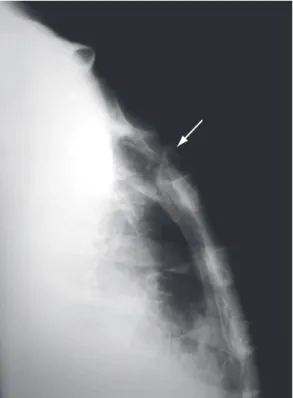
finasteride and doxazosin. Physical examination at admission revealed satisfactory general heath, extremely pale mucosae, an arterial pressure of 130/80 mmHg, a heart rate of 80 bpm, an axillary temperature of 36°C and a respiratory rate of 35 breaths/min. The patient presented a grade 3/6 systolic ejection murmur at the apex, radiating to the axillary region, and diminished breath sounds at both lung bases. In the region of the suprasternal notch, there was a poorly delineated, hyperemic edematous area (approxi-mately 10 cm in diameter) that was painful upon palpation and presented drainage of puru-lent secretion from a fistulous lesion (Figure 1). A swab specimen of the lesion was collected, and an open sternum biopsy was performed for histopathological analysis. Treatment with ciprofloxacin and clindamycin was initiated. The blood workup revealed normochromic normocytic anemia, the PPD test was positive (induration = 15 mm), HIV testing (ELISA) was negative, and erythrocyte sedimentation rate (ESR) was 48 mm/h. Imaging studies (anterior and lateral chest X-rays) revealed no pleuropul-monary findings suggestive of previous or active TB. A lateral X-ray of the sternum revealed an osteolytic lesion in the region of the suprasternal notch and the first costal arches (Figure 2). A CT scan of the chest revealed an osteolytic lesion in the suprasternal notch, without medias-tinal invasion, as well as bilateral transudative pleural effusion related to the clinical profile of pulmonary hypertension and decompensated heart failure (Figure 3). In addition, techne-tium triphasic scintigraphy showed no increased radiotracer uptake in the lesion. The swab specimen revealed Gram-positive cocci in pairs, and culture was positive for multidrug-resistant Staphylococcus aureus and negative for fungi and mycobacteria after 70 days of incubation. The antibiotic regimen was changed to oxacillin. The result of the histopathological examina-tion of the sternal lesion revealed caseous granuloma. For PCR analysis (Álvaro Laboratory, Cascavel, Brazil), a second sternal biopsy was performed, and the empirical treatment for TB using regimen I (rifampin, isoniazid and pyrazi-namide) was initiated. The patient presented a favorable response. The PCR analysis was positive for Mycobacterium tuberculosis, confirming the diagnosis of tuberculous sternal osteomyelitis.
Figure 1 - Fistulous lesion in the sternum with drainage of purulent material.
Sternal osteomyelitis caused by infection with Mycobacterium tuberculosis
J Bras Pneumol. 2009;35(7):709-712 711
X-ray in the evaluation of bone involvement, it is significantly superior in the evaluation of perilesional soft tissues, mediastinal involve-ment and pulmonary involveinvolve-ment.(5-9) Magnetic resonance imaging is a good option for the evaluation of soft tissues and bone lesions, revealing early alterations in the bone marrow, especially at early stages with normal X-ray find-ings.(4,5,9) Technetium triphasic scintigraphy has high sensitivity and specificity for the diagnosis of osteomyelitis.(2,4) In the case presented here, there was no increased radiotracer uptake in the sternum or photopenia in the osteolytic lesion, nor were there any other findings consistent with osteomyelitis.
In the present case, microbiological evalu-ation and cultures were negative, and the diagnosis was confirmed using PCR.(10) The histopathological finding of caseous granu-loma was the principal evidence on which the working diagnosis was based. In one study, microbiological evaluation was positive for acid-fast bacilli in 38% of the cases, cultures were positive for M. tuberculosis in 85% of the cases, and typical granulomas were found in 67% of the cases.(1,2) According to the literature, the definitive diagnosis of sternal TB is based on microbiological evaluation together with histopathological confirmation.(6) Two groups of authors evaluating methods applied in the diagnosis of TB found that the sensitivity and specificity of PCR (90% and 80%, respectively) were similar to those of culture.(11,12) Both groups of authors stated that the quality of the sample and the standardization of the DNA extraction technique are critical factors in the applicability of PCR in the diagnosis of TB in Brazil. In the case reported here, it is likely that microbio-logical evaluation and culture were negative for acid-fast bacilli due to the bactericidal effect that ciprofloxacin has on M. tuberculosis.(13)
The principal differential diagnoses are malignancy, sarcoidosis, actinomycosis and fungal infections.(6,7) In the literature, there is no consensus regarding treatment modality, treat-ment duration or the best treattreat-ment regimen. Although early drainage and complete debri-dement improve cicatrization and prevent recurrence, some authors suggest that conserva-tive treatment with antituberculous drugs be performed and surgical debridement be reserved for chronic cases with structural complications or for cases of treatment failure.(2,5,7) The use of affects young adult males, with or without
predisposing factors, living in areas where TB is endemic.(2) Contamination can originate from infected local lymph nodes (contiguous with the lung parenchyma) or due to reactivation of a quiescent focus.(6)
The case described here reflects the indolent character of osteoarticular TB, in contrast to that of pyogenic osteomyelitis, caused principally by S. aureus, Pseudomonas aeruginosa, Salmonella sp. and Aspergillus sp., the latter being a disease that typically has an abrupt onset and prominent constitutional symptoms.(6) The clinical presen-tation, including symptoms such as sternal pain and swelling evolving over approximately one year, together with normal chest X-ray findings and an ESR of 48 mm/h, is in accordance with data reported in a case-series study of sternal TB, in which symptoms evolved over a mean of 9 months and the most common (in 81% of the cases) were sternal pain and swelling.(2) In that study, chest X-ray findings were normal in 73% of the cases and ESR was elevated (mean, 67 mm/h) in almost all cases.(2) Other reported cases have also shown a persistent increase in ESR.(4-7)
712 De Carli DM, Severo MD, Haygert CJP, Guollo M, Omairi A, Pedro VD et al.
J Bras Pneumol. 2009;35(7):709-712
6. Atasoy C, Oztekin PS, Ozdemir N, Sak SD, Erden I, Akyar S. CT and MRI in tuberculous sternal osteomyelitis: a case report. Clin Imaging. 2002;26(2):112-5.
7. Bohl JM, Janner D. Mycobacterium tuberculosis sternal osteomyelitis presenting as anterior chest wall mass. Pediatr Infect Dis J. 1999;18(11):1028-9.
8. Khalil A, Le Breton C, Tassart M, Korzec J, Bigot J, Carette M. Utility of CT scan for the diagnosis of chest wall tuberculosis. Eur Radiol. 1999;9(8):1638-42. 9. Wang TK, Wong CF, Au WK, Cheng VC, Wong
SS. Mycobacterium tuberculosis sternal wound infection after open heart surgery: a case report and review of the literature. Diagn Microbiol Infect Dis. 2007;58(2):245-9.
10. Clarridge JE 3rd, Shawar RM, Shinnick TM, Plikaytis BB. Large-scale use of polymerase chain reaction for detection of Mycobacterium tuberculosis in a routine mycobacteriology laboratory. J Clin Microbiol. 1993;31(8):2049-56.
11. Bollela VR, Sato DN, Fonseca BA. Problemas na padronização da reação em cadeia da polimerase para diagnóstico da tuberculose pulmonar. Rev Saude Publica. 1999;33(3):281-6.
12. Santos RM, Ogusku MM, Miranda JM, Dos-Santos MC, Salem JI. Evaluation of polymerase chain reaction in the diagnosis of pulmonary tuberculosis in indigenous and non-indigenous patients. J Bras Pneumol. 2006;32(3):234-40.
13. Gibbons FK, Branda JA, Shepard JA. Case records of the Massachusetts General Hospital. Case 12-2006. A 37-year-old man with hemoptysis and a pulmonary infiltrate. N Engl J Med. 2006;354(16):1729-37. prophylaxis in open heart surgery candidates
with a history of TB or contact with TB is not indicated due to the rarity of the infection.(9) In our patient, treatment with regimen I resulted in clinical improvement and better cicatrization of the sternal lesion. At this writing, the patient was still under treatment. Surgical debridement was not considered due to the multiple comor-bidities presented by the patient.
References
1. Ford SJ, Rathinam S, King JE, Vaughan R. Tuberculous osteomyelitis of the sternum: successful management with debridement and vacuum assisted closure. Eur J Cardiothorac Surg. 2005;28(4):645-7.
2. McLellan DG, Philips KB, Corbett CE, Bronze MS. Sternal osteomyelitis caused by mycobacterium tuberculosis: case report and review of the literature. Am J Med Sci. 2000;319(4):250-4.
3. Sociedade Brasileira de Pneumologia e Tisiologia. II Consenso Brasileiro de Tuberculose - Diretrizes Brasileiras para Tuberculose 2004. J Pneumol. 2004;30(Suppl 1): S1-S85.
4. Shah J, Patkar D, Parikh B, Parmar H, Varma R, Patankar T, et al. Tuberculosis of the sternum and clavicle: imaging findings in 15 patients. Skeletal Radiol. 2000;29(8):447-53.
5. Khan SA, Varshney MK, Hasan AS, Kumar A, Trikha V. Tuberculosis of the sternum: a clinical study. J Bone Joint Surg Br. 2007;89(6):817-20.
About the authors
Diego Michelon De Carli
Resident in Gastroenterology. São Vicente de Paulo Hospital, Passo Fundo, Brazil.
Mateus Dornelles Severo
Resident in Clinical Medicine. Santa Maria University Hospital, Federal University of Santa Maria, Santa Maria, Brazil.
Carlos Jesus Pereira Haygert
Assistant Professor of Radiology. Santa Maria University Hospital, Federal University of Santa Maria, Santa Maria, Brazil.
Marcelo Guollo
Resident in General Surgery. Santa Maria University Hospital, Federal University of Santa Maria, Santa Maria, Brazil.
Alex Omairi
Resident in Cardiology. Institute of Cardiology, University Cardiology Foundation, Porto Alegre, Brazil.
Vinícius Dallagasperina Pedro
Medical Student. Federal University of Santa Maria School of Medicine, Santa Maria, Brazil.
Eduardo Pedrolo Silva
Medical Student. Federal University of Santa Maria School of Medicine, Santa Maria, Brazil.
Arnaldo Teixeira Rodrigues