Letters to the Editor
Radiol Bras. 2017 Jan/Fev;50(1):62–68
65
http://dx.doi.org/10.1590/0100-3984.2015.0114 of age, 75–80% of whom are men, urothelial carcinoma being
the predominant form(5,6). Urothelial carcinoma can be multifo-cal/multicentric, can occur in the upper or lower urinary tract, and is often recurrent(5). Smoking is implicated in 50–65% of all cases in men and in 20–30% of all cases in women(4). Other, less com-mon causes include chemotherapy, exposure to aromatic or het-erocyclic amines, radiotherapy, and chronic infection(2,4–6).
Multiple primary malignancies are defined as those that are confirmed, independent, and of non-metastatic origin(7). They are classified as synchronous if they are identified within the first six months after the appearance of the first lesion or as metachronous if they are identified thereafter(7).
The overall prevalence of multiple primary malignancies is 0.7–11.7%, increasing proportionally with patient age(2,3,7,8). It is estimated that 75% of cases occur in individuals over 50 years of age(7). These values are on the rise due to the effectiveness of treatments, the variety of therapeutic techniques now available, the improvement of diagnostic methods, the increased longevity of the population, and contemporary lifestyles(3,7). Hayat et al.(2) reported a probability of developing a second malignancy, depend-ing on the primary tumors diagnosed, rangdepend-ing from 1% (history of hepatic neoplasia) to 16% (previous bladder tumors)(2). Braisch et al.(4) observed that 1.2–2.5% of cancer patients who were smok-ers developed another distinct malignant lesion within the first year of follow-up.
In smokers, multiple primary malignancies can affect sev-eral organs, notably the lungs, upper aerodigestive tract, and kid-neys, as well as the upper and lower urinary tract. Other potential sites include the thyroid gland, stomach, colon, rectum, and pan-creas(4,6,8).
Rodolfo Mendes Queiroz1, Daniel Roque1, Eduardo Miguel Febronio1
1. Documenta – Hospital São Francisco, Ribeirão Preto, SP, Brazil. Mailing address: Dr. Rodolfo Mendes Queiroz. Documenta – Centro Avançado de Diagnóstico por Imagem. Rua Bernardino de Campos, 980, Centro. Ribei-rão Preto, SP, Brazil, 14015-130. E-mail. [email protected].
REFERENCES
1. Tiferes DA, Jayanthi SK, Liguori AAL. Cólon, reto e apêndice. In: D’Ippolito G, Caldana RP, editores. Gastrointestinal – Série CBR. São Paulo: Elsevier; 2011. p. 203–51.
2. Hayat MJ, Howlader N, Reichman ME, et al. Cancer statistics, trends, and multiple primary cancer analyses from the Surveillance, Epide-miology, and End Results (SEER) Program. Oncologist. 2007;12:20– 37.
3. VanderWalde AM, Hurria A. Second malignancies among elderly survivors of cancer. Oncologist. 2011;16:1572–81.
4. Braisch U, Meyer M, Radespiel-Tröger M. Risk of tobacco-related multiple primary cancers in Bavaria, Germany. BMC Cancer. 2012; 12:250.
5. Prando A. Tumores uroteliais. In: Prando A, Baroni RH, editores. Urinário – Série CBR. São Paulo: Elsevier; 2013. p. 321–58. 6. Bermejo JL, Sundquist J, Hemminki K. Bladder cancer in cancer
patients: population-based estimates from a large Swedish study. Br J Cancer. 2009;101:1091–9.
7. Demandante CGN, Troyer DA, Miles TP. Multiple primary malig-nant neoplasms: case report and a comprehensive review of the lit-erature. Am J Clin Oncol. 2003;26:79–83.
8. Tabuchi T, Ito Y, Ioka A, et al. Tobacco smoking and the risk of subsequent primary cancer among cancer survivors: a retrospective cohort study. Ann Oncol. 2013;24:2699–704.
Dural fistula with bilateral arterial supply, mimicking a brainstem tumor
Dear Editor,
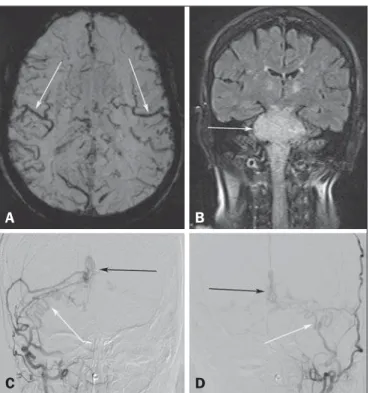
A 73-year-old woman presented with a history of at least four episodes of deep vein thrombosis. In the last five months, she had experienced severe ataxia, difficulty in swallowing, bilateral tinni-tus, and symptoms related to intracranial hypertension, such as nausea and vomiting. Magnetic resonance imaging (MRI) re-vealed a hyperintense signal on T2-weighted images and an en-larged brainstem, the swelling extending to the thalamus, cerebel-lar peduncles, and to the cervical portion of the spinal cord (Fig-ures 1A and 1B). The images could erroneously indicate a diag-nosis of brainstem tumor, glioma in particular, due to the infiltra-tive pattern of the lesion and the increased organ volume. How-ever, thorough evaluation with advanced imaging techniques, such as magnetic susceptibility-weighted sequences, demonstrated an extensive network of dilated peripheral veins, together with pro-nounced collateral circulation. Cerebral angiography showed a dural arteriovenous fistula (DAVF) with bilateral arterial supply via branches of the maxillary arteries. Venous drainage was mostly through the rectum and galenic system (Figures 1C and 1D). Involvement of the brainstem and cervical spinal cord was due to venous congestive injury. The classical surgical approach was precluded by the deep, inaccessible location, whereas endovascular therapy was precluded by the extensive involvement and bilateral nature of the fistula-sustaining arterial supply. The patient un-derwent gastrostomy and was discharged to palliative home care. Vascular lesions are often difficult to diagnose(1–8). DAVFs, which are characterized by abnormal communication between
Figure 1. A: Axial slice in a susceptibility-weighted sequence showing numerous large-caliber superficial veins, representing venous congestion. B: Coronal slice in a fluid-attenuated inversion recovery sequence showing a hyperintense signal and increased brainstem volume, mimicking a brain tumor. C,D: Digital angiography with subtraction technique, revealing the bilateral nature of the arterial supply (white arrows) and the nidus (black arrows) formed by the fistula.
A
B
Letters to the Editor
Radiol Bras. 2017 Jan/Fev;50(1):62–68
66
http://dx.doi.org/10.1590/0100-3984.2015.0186 the arterial and venous systems, without intervening capillary beds,
account for less than 10% of all cerebral vascular malformations(9). The most common place of occurrence is the transverse sinus(9), and there have been no reports of bilateral arterial supply. The two principal forms of presentation are hemorrhagic and non-hemor-rhagic, both typically occurring as a consequence of intracranial venous hypertension(9,10), which appears as the best predictor of poor prognosis(11). Cerebral angiography continues to be the gold standard for the diagnosis of DAVF, in which the nidus represents the arteriovenous shunt itself and collateral vessels develop in or-der to drain the venous congestion(12). Injury due to venous con-gestion is an example of a severe non-hemorrhagic manifesta-tion, which can be prevented through the early diagnosis of DAVF, the treatment of choice being endovascular therapy, with the objective of interrupting the arterial supply to the venous system(9).
REFERENCES
1. Cardarelli-Leite L, Velloni FG, Salvadori PS, et al. Abdominal vascular syndromes: characteristic imaging findings. Radiol Bras. 2016;49:257– 63.
2. Batista MN, Barreto MM, Cavaguti RF, et al. Pulmonary artery sarcoma mimicking chronic pulmonary thromboembolism. Radiol Bras. 2015; 48:333–4.
3. Dias DA, Afonso LHC, Abud DG. Femoral artery injury during aneu-rysm coiling. Radiol Bras. 2015;48:335–6.
4. Neves PO, Andrade J, Monção H. Coronary anomalies: what the radiolo-gist should know. Radiol Bras. 2015;48:233–41.
5. Amaral RH, Souza VVS, Nin CS, et al. Aortic lesion simulating pulmo-nary disease: a case report. Radiol Bras. 2014;47:320–2.
Bárbara Liaffa1, Fábio Noro1, Paulo Roberto Valle Bahia1, Flávia Pinto Dezonne Motta1, Edson Marchiori1
1. Universidade Federal do Rio de Janeiro (UFRJ), Rio de Janeiro, RJ, Brasil. Endereço para correspondência: Dr. Edson Marchiori. Rua Thomaz Cameron, 438, Valparaiso. Petrópolis, RJ, Brasil, 25685-120. E-mail: [email protected].
6. Ribeiro BNF, Ribeiro RN, Zanetti G, et al. Hughes-Stovin syndrome: an unusual cause of pulmonary artery aneurysms. Radiol Bras. 2016;49: 202–3.
7. Abud TG, Nguyen AD, Abud LG, et al. Anterior cerebral artery aneu-rysm rupture presenting as hemorrhage in the splenium of the corpus callosum. Radiol Bras. 2016;49:268–9.
8. Abreu Junior L, Kuniyoshi CH, Wolosker AB, et al. Vascular loops in the anterior inferior cerebellar artery, as identified by magnetic reso-nance imaging, and their relationship with otologic symptoms. Radiol Bras. 2016;49:300–4.
9. Hacein-Bey L, Konstas AA, Pile-Spellman J. Natural history, current concepts, classification, factors impacting endovascular therapy, and pathophysiology of cerebral and spinal dural arteriovenous fistulas. Clin Neurol Neurosurg. 2014;121:64–75.
10. Cognard C, Gobin YP, Pierot L, et al. Cerebral dural arteriovenous fistulas: clinical and angiographic correlation with a revised classifica-tion of venous drainage. Radiology. 1995;194:671–80.
11. Cognard C, Casasco A, Toevi M, et al. Dural arteriovenous fistulas as a cause of intracranial hypertension due to impairment of cranial venous outflow. J Neurol Neurosurg Psychiatry. 1998;65:308–16.
12. Signorelli F, Gory B, Maduri R, et al. Intracranial dural arteriovenous fistulas: a review of current management based on emerging knowl-edge. J Neurosurg Sci. 2015 Feb 13. [Epub ahead of print].
Plasmacytoma of the trachea: a surprising diagnosis
Dear Editor,
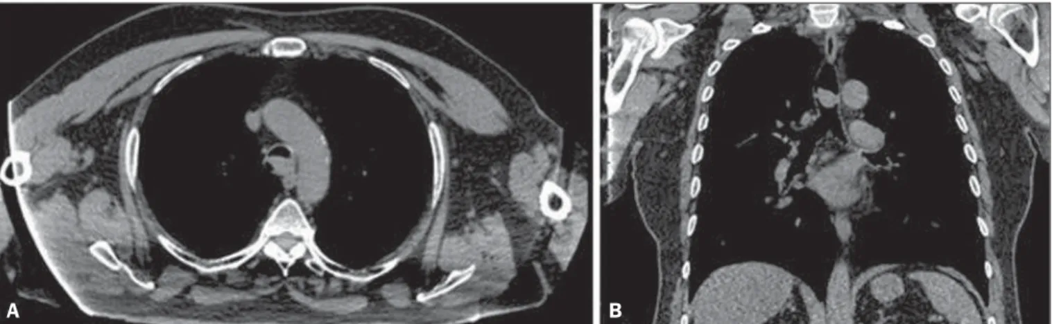
A 68-year-old man presented with a complaint of dyspnea on moderate exertion, and physical examination revealed stridor. The patient reported having previously been treated for chronic ob-structive pulmonary disease and adenocarcinoma of the prostate, the latter having been treated with 39 radiotherapy sessions. He was a former smoker with a smoking history of 150 pack-years (3 packs/day for 50 years), having quit 4 years prior. We performed contrast-enhanced computed tomography (CT) of the neck and chest, which showed an expansive, well-defined nodular mass in the distal trachea, near the carina, without enhancement or signs of invasion of the tracheal walls (Figures 1 and 2). Bronchoscopy
was requested for tumor resection, and symptom resolution was observed after the resection. The histopathological study identi-fied an outer layer with the of appearance of plasmacytoid cells, sometimes with a central eosinophilic nucleolus—“cartwheel appearance”—and hyaline intracytoplasmic inclusions suggestive of Russell bodies. The immunohistochemical profile demonstrated positivity for CD3, CD20, CD45, CD56, kappa light chain, and CD138 in plasmacytes. In the context of the clinical status and test results, the findings were consistent with solitary extramed-ullary plasmacytoma.
Diseases involving the trachea or the main bronchi are not common(1–4). Less common still are tracheal tumors, which ac-count for only 1–2% of all respiratory tract tumors(5,6), affecting mainly the lower third of the tract(7). Such tumors can be locally
Figure 1. A: Axial CT scan, without contrast, showing an extensive, well-defined nodular mass in the distal trachea, measuring 2.1 × 1.3 × 1.7 cm, without signs of tracheal wall invasion. B: Coronal CT scan, without contrast, showing an expansive, well-defined nodular mass in the distal trachea, at the level of the carina, without signs of tracheal wall invasion.