Primeira submissão em 06/02/10 Última submissão em 15/05/10 Aceito para publicação em 03/08/10 Publicado em 20/10/10
Primary sclerosing cholangitis in children and adolescents:
a clinicopathologic study with a proposal of criteria for
early diagnosis
Colangite esclerosante primária em crianças e adolescentes: uma correlação clinicopatológica com uma proposta
de critérios para primeiro diagnóstico
Wolfram F. J. Riedlinger1; Maureen M. Jonas2 ; Harpreet Pall3
Introduction: Primary sclerosing cholangitis (PSC) has been increasingly diagnosed among children
and adolescents due to better recognition of clinical, imaging and pathological features. Thus more patients are diagnosed at a younger age due to imaging and sensitivity optimization. Objective: Early liver histopathological (LH) changes are not well described and PSC is not commonly recognized before typical bile duct changes occur on cholangiography (CG). Currently, CG is considered gold standard for adults but nothing is known for early diagnosis in the pediatric age group (0- 20 years old). Methods: We reviewed clinical history, LH and CG from 47 children and adolescents with PSC (35 males, mean age 13 years old). Forty-three out of 47 patients had been through LH examination from whom 33 had also undergone CG. A clinicopathological correlation was performed. Results: LH showed active neutrophilic cholangitis in 19 patients, moderate neutrophilic pericholangitis in nine, dystrophic changes in the bile duct in eight, and concentric periductal ibrosis in 24 patients. Abnormal CG was found in 24 out of 33 patients and nine had normal results. Eleven out of these 24 patients had abnormal histology before abnormal CG and four patients had abnormal CG before histology. Data of two out of 24 patients were insuficient for correlation and 11 out of 24 had both abnormal liver histology and abnormal imaging indings. Conclusion: Our study emphasizes that even when CG is normal, PSC should be exclusively diagnosed by liver biopsy, hence cholangiography being unnecessary. Chronic portal inlammation, neutrophilic pericholangitis, periductal sclerosis and “onion skinning” are characteristic histopathological indings. Neutrophilic pericholangitis may be subtle and easily overlooked in early disease, leading to strong suspicion of PSC.
abstract
key words
Primary sclerosing cholangitis
Liver
Neutrophilic pericholangitis
Cholangiography
Inlammatory bowel disease
resumo
Introdução: Colangite esclerosante primária (CEP) é crescentemente diagnosticada em crianças e adolescentes devido ao melhor reconhecimento das apresentações clínicas por imagem e manifestações patológicas. Devido a isso, aumentaram a sensibilização e a melhora das imagens e, cada vez mais, os pacientes são diagnosticados em idade mais jovem. Objetivo: As primeiras mudanças na histopatologia do fígado (HF) não são bem descritas e a CEP não é frequentemente reconhecida antes da típica mudança do ducto biliar ocorrer na colangiograia (CG). Atualmente, a CG é considerada padrão-ouro em adultos, mas nada é conhecido para o diagnóstico precoce na faixa etária pediátrica (0-20 anos de idade). Métodos: Nós revisamos histórico clínico, HF e CG de 47 crianças e adolescentes com CEP (35 meninos, idade média de 13 anos). Desses, 43 tinham HF, sendo que 33 também possuíam CG. Uma correlação clinicopatológica foi performada. Resultados: HF mostrou colangite neutrofílica ativa em 19 pacientes, pericolangite neutrofílica moderada em nove, mudança distróica do ducto biliar em oito e ibrose periductal concêntrica em 24. Um CG anormal foi constatado em 24/33; resultados de nove pacientes eram normais. Desses 24 pacientes, 11 tiveram histologia anormal de fígado antes da CG anormal e quatro apresentaram a situação inversa. Dados disponíveis de dois pacientes eram insuicientes para propósitos de correlação e outros 11 apresentavam, ao mesmo tempo, histologia anormal de fígado e resultados anormais de imagens. Conclusão: Nosso estudo enfatiza que a CEP pode ser exclusivamente diagnosticada por biópsia de fígado, sem colangiograma necessário ou mesmo no contexto de um CG normal. Inlamação portal crônica, pericolangite neutrofílica, esclerose periductal e “cebola esfolando” são resultados característicos de achados histopatológicos. A pericolangite neutrofílica pode ser sutil e facilmente negligenciada em doença precoce, requerendo alta suspeita para CEP.
unitermos
Colangite esclerosante primária
Fígado
Pericolangite neutrofílica Colangiograia Doença inlamatória do intestino
1. Dr. med. (GER), MD (ECFMG, USA); Consultant Histopathologist, Department of Pathology, Aberdeen University Medical School, Aberdeen Royal Inirmary, Foresterhill, Aberdeen, Scotland, United Kingdom; formerly: Department of Pathology, Boston Children's Hospital, Harvard Medical School, Boston, USA.
Introduction
Primary sclerosing cholangitis (PSC) manifests itself as a rare cholestatic liver disease of unclear etiology(23). Typical histology reveals a mixed chronic inlammatory iniltrate within portal tracts, around and within bile duct walls, and a progressive, obliterative, concentric, periductal ibrosis affecting both the intra and extrahepatic bile ducts(3, 24). Especially, the periductal concentric (“onion-skinning”) fibrosis of the small intrahepatic bile ducts has long been considered the histologic hallmark of the disease(4).
No effective medical therapy has been proven to prevent disease progression. Therefore, affected patients may progress to biliary cirrhosis and liver failure consecutively making in liver transplantation the only life-extending therapeutic option for patients with end-stage disease(2, 15). However, a close to 25% recurrence rate in the allograft has been reported(12).
Most patients with PSC present with rather unspeciic signs and symptoms of chronic liver disease such as hepatosplenomegaly, abdominal pain, anorexia, fatigue, jaundice, pruritus, and cirrhosis(28). PSC has also been described in the neonatal period when patients often present with cholestasis(8).
Unfortunately, the underlying pathologic process is often not recognized on liver biopsy specimen before typical bile duct changes are evident on cholangiography because early histopathologic liver changes are not well-characterized.
Although the disease is well-known in the adult population with an estimated prevalence of about two to seven per 100,000(24, 27), its true incidence in pediatric patient population, i.e. patients in their irst two decades of life, in not accurately known(8).
Awareness of PSC in children and adolescents has only more recently increased due to better recognition of clinical and pathological manifestations as well as the more wide-spread use of invasive diagnostic techniques, i.e. endoscopic retrograde cholangio-pancreatography (ERCP) and percutaneous transhepatic cholangiography (PTC) as well as magnetic retrograde cholangiopancreatography (MRCP)(7, 12).
In the adult population, multifocal strictures of the intrahepatic and extrahepatic bile ducts are characteristic(22) and the same cholangiographic indings have been reported in pediatric patients(8, 10, 16, 28).
Again, in the adult population, cholangiography is considered the gold standard(3). However, due to the equivocal clinical and pathologic picture of very early PSC in children and adolescents, the diagnostic gold standard is currently not known for that speciic patient population.
Furthermore, systematic investigation of early histopathologic indings have never been undertaken. Additionally, the earliest time of occurrence and the distinct spatial location of the typical mixed inlammatory iniltrate have never been described, i.e. whether it irst occurs within the biliary epithelium or in the surrounding portal tracts. How the iniltrate progresses over the course of the disease, i.e. from the bile duct wall into the portal tract or vice versa, is also not known.
Therefore, the purpose of this study was not only to report over 13 years of experience with PSC at the Children’s Hospital Boston but even more to examine early pathologic manifestations and, especially, the irst spatial localization of the mixed inlammatory iniltrate and its progression within the portal tract. Also, we looked at the temporal relationship between cholangiography and liver biopsy specimens, and possible associations with previous, concomitant, or subsequent diseases, e.g. autoimmune hepatitis (AIH) and inlammatory bowel disease (IBD).
Methods
Patient population
Clinical data as well as the results of imaging and pathologic examination were taken from the pertinent medical records of the hospital archives as well as from clinical information received at time of consultation.
We reviewed age, gender, clinical history, histopathology, ERCP and/or MRCP studies, and histopathology. In cases of associated IBD, gastrointestinal biopsy results were re-examined.
Imaging studies
All ERCP and/or MRCP images were performed at our institution and interpreted by radiologists with expertise in biliary tree imaging. A clinicopathological correlation was subsequently undertaken.
Results of liver biopsy specimens and cholangiography were timely correlated to one another in an attempt to further define the temporal correlation between histopathology specimen and cholangiography.
Pathology
All liver biopsy specimens at our institution as well as all specimens seen in consultation were reviewed by a single hepatopathologist to ensure consistency in interpretation of the histopathologic indings. Liver biopsy specimens were routinely stained with Hematoxylin-Eosin, Masson’s Trichrome, Grocotts Methenamine Silver (GMS) stain, and Periodic Acid Schiff (PAS) stain with and without diastase enzymatic digestion. Iron staining was also performed. For consultation cases, we reviewed the original material from the submitting institution and in cases of equivocal indings, requested additional unstained sections to complete our routine panel of histologic sections.
The hepatic lesions were analyzed for portal and periductal inflammation, fibro-obliterative pericholangitis (“onion-skinning”), ductopenia, ductular proliferation, periportal ibrosis with possible bridging and cirrhosis, cholestasis, steatosis, interface hepatitis, necroinlammatory activity with collapse and rosetting of hepatocytes. The severity of histopathologic indings was categorized in minimal (1+), mild (2+), moderate (3+), and marked (4+).
In addition, liver specimens were evaluated for possible iron deposition to exclude hereditary hemochromatosis or secondary forms of iron deposition, idiopathic or secondary cholestasis, and any signiicant plasma cell iniltration
of portal tracts or bile ducts, indicative of concomitant autoimmune hepatitis (overlap syndrome).
In the appropriate clinical setting (i.e. no evidence of ascending cholangitis, fever, sepsis), the following histopathologic changes occurring in the background of chronic hepatitis (i.e. lymphocytic portal tract inlammation associated with mild increase in portal tract ibrosis) were interpreted as most consistent with or diagnostic of PSC:
• active mixed lymphocytic/neutrophilic cholangitis; • moderate neutrophilic pericholangitis;
• dystrophic changes of bile ducts with mild inlammation and sclerosis;
• concentric periductal ibrosis (“onion-skinning”).
Results
Our patient population consisted of a total 47 children and adolescents (35 males, 12 females) who were diagnosed with PSC at Children’s Hospital in the time frame from January 1992 to April 2005. Age at diagnosis ranged from 1.5 to 20 years (mean 13 years). Of those patients, 43 (91.5%) had undergone liver biopsy; 33/43 patients (76.7%) had received cholangiography as part of their diagnostic workup. Four of 47 patients were felt to have insuficient information for the diagnosis of PSC based on clinical grounds as well as lack of characteristic indings on imaging studies and, consecutively, did not have a liver biopsy performed.
Table 1 summarizes patients’ gender, age at time of the liver biopsy and/or cholangiogram, liver histology results, and possible concomitant IBD and/or AIH.
All 43 patients (31 males, 12 females) had diagnostic indings consistent with PSC. All patients had variably intense active mixed lymphocytic and neutrophilic portal inflammation and mild portal tract fibrosis
Table 1
Comparison of patients’ age and results of liver Bx and CG
Patient (gender)
Result Bx
Age@Bx
Result CG
Age@CG
IBD
Age@IBD
AIH
E. C. (M) Yes 11.90 N 11.95 CD 13.04 No
S. B. (M) Yes 9.76 N 9.74 UC 9.69 No
J. C. M) Yes 4.10 A 4.10 UC 3.46 No
J. W. (F) Yes 11.47 A 11.47 ID 4.92 Yes
D. W. (F) Yes 15.25 A 15.25 UC 14.62 No
S. O. (M) Yes 6.96 N 6.98 UC 7.11 No
Patient (gender)
Result Bx
Age@Bx
Result CG
Age@CG
IBD
Age@IBD
AIH
J. S. (M) Yes 11.70 A 13.11 ID 11.01 No
D. C. (M) Yes 4.49 A 4.49 UC 4.40 No
A. S. (F) Yes 7.72 N 7.77 UC 7.77 No
K. H. (M) Yes 16.08 A 16.32 UC 17.25 No
J. A. (M) Yes 10.41 N 10.35 CD 12.02 No
J. B. (M) Yes 15.48 A 15.57 UC 15.57 No
N. R. (M) Yes 16.65 A 16.46 UC 16.09 Yes
J. L. (M) Yes 14.80 A 14.70 UC 14.90 No
M. M. (M) Yes 17.1 A 16.11 No N/A No
L. S. (F) Yes 10.11 N 11.1 CD 8.70 No
J. B. (M) Yes 8.8 A 8.8 UC 6 Yes
F. T. (M) Yes 13.7 A 13.7 CD 11.11 No
M. G. (M) Yes N/E A 12 UC 12.00 No
C. F. (M) Yes 12.11 A 13.1 UC 12.11 Yes
M. T. (M) Yes 16.3 A 16.4 No N/A Yes
K. M. (F) Yes 14.4 A 14.4 N/E N/A No
J. L. (F) Yes 6.2 N/E N/E No N/A No
S. H. (F) Yes 9.9 N 10.4 UC 9.9 Yes
B. D. (F) Yes 14.2 N/E N/E No N/A Yes
G. S. (F) Yes 13.1 N/E N/E N/E NO No
R. V. (M) Yes 14.2 A N/E N/E N/A No
Z. D. (F) Yes 1.5 N/E N/E N/E N/A No
D. L. (M) Yes 12.3 N 12.6 UC 11.11 No
J. K. (M) Yes 11.7 N/E N/E UC 11.7 No
J. B. (M) Yes 8.1 A 8.1 UC 5.0 Yes
J. L. (M) Yes 11.0 N/E N/E UC 10.6 No
G. D. (M) Yes 19.8 A 19.8 CD 13.0 No
M. H. (M) Yes 11.4 N/E N/E UC <11.4 No
C. G. (M) Yes 5.3 N/E N/E No N/A No
A. S. (M) Yes 3.6 N/E N/E No N/A No
M. S. (M) Yes 14.11 N/E N/E CD 12.0 No
K. O. (M) Yes 18.0* N 18.1 No N/A No
D. R. (M) Yes 12.0 A 15.3 UC 15.3 Yes
K. T. (M) Yes 11.0 A 11.0 UC 11.0 Yes
A. A. (F) Yes 15.9 A 15.10 CD 15.1 No
M. Y. (M) Yes 15.5 A 15.5 UC 16.5 Yes
J. R. (M) N/E N/E A/N# 11.6 UC 11.0 No
D. P. (M) N/E N/E A 14.8 UC 13.8 No
G. F. (M) N/E N/E A 10.10 UC 9.4 No
J. G. (M) N/E N/E A 14.0 UC 13.7 No
Bx: biopsy; CG: cholangiogram; IBD: inlammatory bowel disease; AIH: autoimmune hepatitis; M: male; F: female; N: normal; A: abnormal; CD: Crohn disease; UC: ulcerative colitis; ID: indeterminate colitis; N/E: not examined/no information available; N/A: not applicable; *: exact time of irst diagnosis not known; #: result unclear; two magnetic retrograde cholangiopancreatography (MRCP) exams with conlicting results.
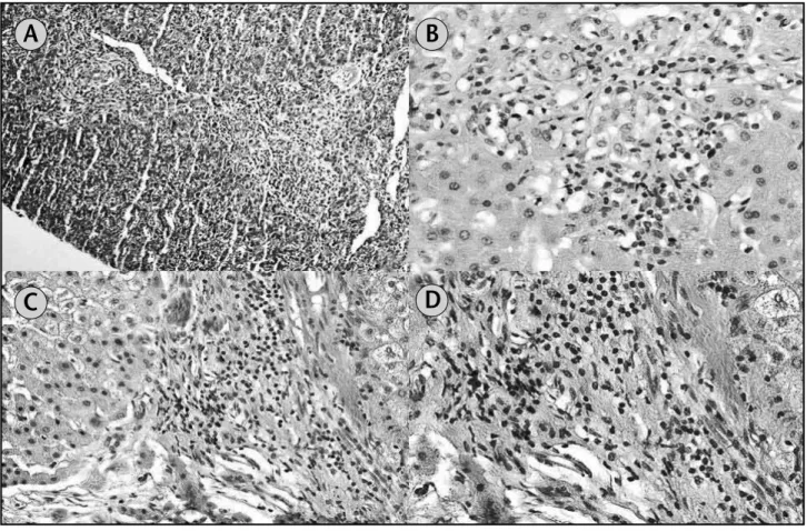
Figura 1 – Photomicrograph of different PT. (A)Medium power view of two PT with mild active mixed lymphocytic and neutrophilic inlammation surrounding and involving bile ducts (20×); (B) high power view of PT with moderate mixed inlammation (40×); (C) medium power view of PT showing marked mixed inlammatory iniltrate (20×); (D) high power view of the same PT (“C”) demonstrating marked bile duct wall involvement by mixed inlammation as well as destruction of some bile ducts (60×)
PT: portal tracts.
(Figures 1A and 3A). The inflammatory infiltrate was mainly composed of lymphocytes and monocytes with some interspersed eosinophilic and neutrophilic granulocytes.
Of those 43 patients with diagnostic liver pathology, 33 (25 males, eight females) patients also had a cholangiogram performed at some point during their evaluation; 24 of those 33 (72.7%; 19 males, ive females) showed an abnormal cholangiogram and nine patients (24.2%; six males, three females) had normal cholangiographic results. Finally, 10/43 patients (six males, four females) had no cholangiography done.
About half of the patients (11/24) (eight males, three females) who were found with abnormal liver histology presented with abnormal imaging findings at the same time. The same number (11/24) of patients (nine males, two females) had abnormal findings on liver histology and cholangiography at different points in time. Of those 11 patients, seven individuals (63.6%) (six males, one female) were noticed with abnormal liver histology before typical findings on cholangiography
were visualized. In four (36.4%) patients (three males, one female), the situation was vice versa, i.e. abnormal cholangiogram before abnormal liver histology. Available data of 2/24 patients were insufficient for correlation purposes.
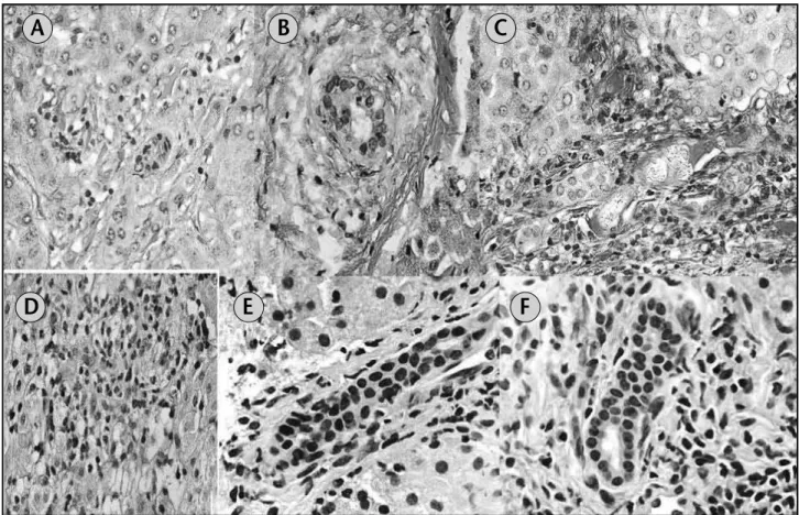
Active (> 1+) mixed lymphocytic/neutrophilic cholangitis was seen in 19 (44.2%) patients (14 males, ive females) (Figures 1B, 2C, 2E and 2F), moderate (3+) to marked (4+) neutrophilic pericholangitis in nine (20.9%) patients (six males, three females) (Figures 1C, 1D and 2D), and mild (1+) inlammation and sclerosis with dystrophic changes of bile ducts in eight (18.6%) patients (seven males, one female) (Figures 2A and 2B).
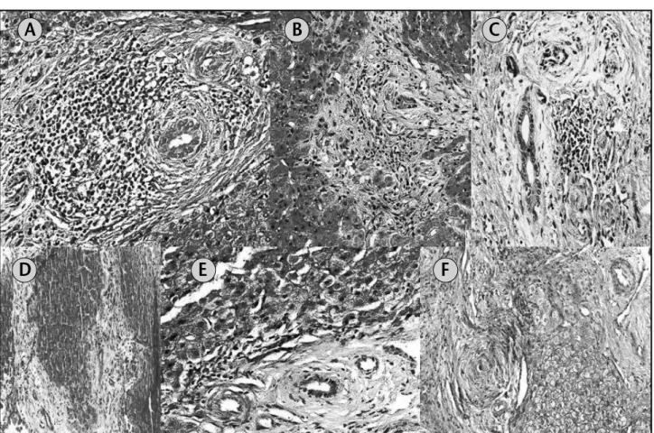
Characteristic concentric (“onion-skinning”) periductal ibrosis was noted in 24 (55.8%) patients (17 males, seven female) (Figures 3E and 3F). Of that group, one patient (J. B.) revealed an extremely severe degree of “onion-skinning”, which prevented further evaluation of bile ducts regarding the possible presence of an inlammatory component. All other 23 patients were distributed among the previously categorized groups.
A
C
B
Figura 2 – Photomicrographs of different BD. (A and B) Medium and high power views of BD showing dystrophic changes of bile ducts with mild inlammation and sclerosis (20× and 40×); (C) medium power view of PT with BD proliferation and marked mixed inlammation (20×); (D) medium power view of BD with marked neutrophilic pericholangitis (20×); (E and F) high power views of BD with minimal (early) neutrophilic pericholangitis (60×)
BD: bile ducts.
A
B
D
E
F
C
Of 43 patients with unequivocal histopathologic features of PSC, 31 (72.1%; 20 males, 11 females) had more advanced disease with compression and obliteration of bile ducts and also showed extension of portal tract ibrosis (Figures 3B and 3C) beyond the limiting plate (3+), ultimately resulting in portal to portal bridging ibrosis (4+) (Figure 3D). End-stage liver disease due to diffuse parenchymal cirrhosis was identiied in 12 patients (27.9%; ive males, seven females). Signiicant numbers of portal plasma cells, suggestive of concomitant autoimmune hepatitis as part of overlap syndrome, were found in 11 patients (25.6%; eight males, three females).
Iron deposition within the liver parenchyma was solely observed in one patient. This patient, however, only had minimal scattered traces of stainable iron which was not suggestive of a disturbance in iron metabolism. Chronic cholestasis for unclear reasons was evident in one patient and two additional patients had minimal and scattered fat droplets within hepatocytes; none of the remaining patients showed cholestasis.
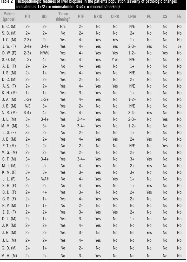
All detailed histopathologic liver indings as well as the grading regarding their severity are summarized in Table 2.
Coexistence of IBD was observed in 36 of 47 patients (76.6%; 29 males, seven females), of which 26 (55.3%; 23 males, three females) were diagnosed with ulcerative colitis, eight (17%; ive males, three females) with Crohn disease, and two (4.3%; one male, one female) with indeterminate colitis.
Liver biopsies were performed in 32 (88.9%) of 36 patients (25 males, seven females) with associated IBD. Regarding the temporal correlation of IBD and PSC, 15 (46.9%) of those 32 patients (nine males, six females) presented with IBD before PSC and six (18.8%) patients (all males) vice versa. In eight (25%) patients (seven males, one female), both diagnoses were simultaneously obtained. For three (9.4%) patients (two males, one female), insuficient clinical information were at hand to draw conclusions (Table 1).
Discussion
Figura 3 – Photomicrographs of different degrees of PT ibrosis. (A) High power view of PT with mild ibrosis surrounding bile ducts (40×); (B) medium power view of PT with moderate ibrosis and focal periportal extension (20×); (C) high power view of PT with marked ibrosis, compressing and obliterating some bile ducts (yellow arrowheads; 40×); (D) medium power view of two PT showing portal to portal bridging ibrosis and early cirrhosis (20×); (E and F) high and medium power views, respectively, of PT with several bile ducts demonstrating marked concentric periductal ibrosis (“onion skinning”) (60× [E]; 40× [F])
PT: portal tracts.
A
B
D
E
F
C
therapeutic modalities rather than attempting a systematic evaluation of early histopathologic changes to facilitate early recognition of the disease in liver biopsy specimens(12).
Another article(5) also emphasized histologic follow-up data of 20 young PSC patients rather than evaluating early histopathologic changes and most of those patients initially presented with advanced liver disease (bridging ibrosis or cirrhosis), indings that were different from those in our patient population in which about two-thirds were diagnosed in an early stage of the disease.
PSC in the pediatric age group, i.e. 0-20 years of age, has mostly been diagnosed in the second decade of life(5, 8, 10, 12, 13, 28). The age range of our patient population (n = 47) diagnosed at the Children’s Hospital matched this observation and encompassed 35 males and 12 females, ranging in age from 5 to 24 years (mean 13 years). However, earlier presentations in patients of younger age have occasionally been observed and some patients were diagnosed below 2 years of age(12). Interestingly, at the time of diagnosis, our youngest patient was 1.5 years old.
Most of our patients were males who were initially found to have unspeciic signs and symptoms of chronic liver disease, e.g. fatigue, jaundice, abdominal pain, and hepatosplenomegaly. Those features were in concordance with indings made by other observers(5, 8, 10, 12, 13, 28).
Of those patients who had received a liver biopsy, almost half of them (19/43) were diagnosed at an early stage and showed discrete minimal to mild neutrophilic pericholangitis without evidence of cholestasis. This discovery indicated that a relatively high percentage of pediatric patients were able to present with rather mild unspeciic liver histopathology, leaving a larger differential diagnosis open for further investigation. This observation consecutively highlighted the necessity of searching for possible additional clinical indings such as concomitant inlammatory bowel disease or autoimmune-hepatitis.
Table 2
Histopathologic features of liver biopsies in the patients population (severity of pathologic changes
indicated as 1+/2+ = minimal/mild; 3+/4+ = moderate/marked)
Patient
(gender)
PTI
BDI
“Onion
skinning”
PTF
BRID
CIRR
LNIA
PC
CS
FE
E. C. (M) 2+ 2+ N/E 2+ No No N/E No No No
S. B. (M) 2+ 2+ No 2+ No No 2+ No No No
J. C. (M) 2-3+ 2+ Yes 4+ Yes Yes 1+ No No No
J. W. (F) 3-4+ 3-4+ Yes 4+ Yes Yes 2-3+ Yes No 1+
D. W. (F) 2-3+ N/A% Yes 4+ Yes Yes 1-2+ No Yes No
S. O. (M) 1-2+ 4+ Yes 4+ Yes Y es N/E No No No
A. D. (F) 2+ 2+ No 4+ Yes No 1+ No No No
J. S. (M) 2+ 1+ Yes 4+ Yes No N/E No No No
D. C. (M) 2+ 2+ Yes 2+ No No 2+ No No No
A. S. (F) 2+ 2+ Yes 4+ Yes Yes N/E No No No
K. H. (M) 1+ 1+ Yes 3+ Yes No 1+ No No No
J. A. (M) 1-2+ 1-2+ Yes 4+ Yes No 1-2+ No No No
J. B. (M) N/E 3+ Yes 2+ No No N/E No No No
N. R. (M) 3-4+ 4+ Yes 4+ Yes No 3-4+ Yes No No
J. L. (M) 3+ 3-4+ Yes 3-4+ Yes No 2-3+ No Yes No
M. M. (M) 3+ 3+ No 3-4+ Yes No 1-2+ No No No
L. S. (F) 3+ 2+ No 2+ No No 1+ No No No
J. B. (M) 2+ 2+ Yes 4+ Yes Yes 2+ Yes No No
F. T. (M) 2+ 2+ No 2+ No No N/E No Yes No
M. G. (M) 2+ 2+ Yes 2+ No No 2+ No No No
C. F. (M) 3+ 3-4+ Yes 3-4+ Yes No 3+ Yes No No
M. T. (M) 2+ 2+ No 4+ Yes No 2+ Yes No No
K. M. (F) 3+ 3+ Yes 3+ Yes No 3+ No No No
J. L. (F) 3+ N/A# No 4+ Yes Yes 1+ No No No
S. H. (F) 2+ 2+ No 4+ Yes No 1+ Yes No No
B. D. (F) 2+ 4+ Yes 3+ No No 2+ Yes No No
G. S. (F) 2+ 1+ Yes 4+ Yes Yes 2+ No No No
R. V. (M) 1+ 1+ No 2+ No No No No No No
Z. D. (F) 2+ 2+ Yes 3+ Yes Yes 2+ No No No
D. L. (M) 2+ 1+ Yes 3+ Yes No 1+ No No No
J. K. (M) 2+ 2+ Yes 4+ Yes No No No No No
J. B. (M) 2+ 2+ Yes 3+ No No No Yes No No
J. L. (M) 2+ 2+ Yes 4+ Yes No No No No No
G. D. (M) 2+ 1+ No 2+ No No No No No No
that the earliest histopathologic manifestation of PSC might be seen within the bile duct wall. However, variably intense chronic inlammation of the portal tracts was observed in all patients, rendering this speciic inding not very reliable for diagnostic differentiation of PSC from other etiologies like immune-mediated or viral causes. Therefore, additional diagnostic tools, such as imaging studies and information about the clinical history, were necessary for inally sorting out the underlying disease process.
Batres et al. showed in their study that most patients (13/20) initially presented with advanced disease(5). Interestingly, most of them also demonstrated a discrete neutrophilic cholangitis without signiicant tissue cholestasis. In addition, there was ductal/ductular proliferation noted, suggesting that neutrophilic cholangitis – due to its continuous presence in later disease stages – may be a major diagnostic clue in early disease.
Ductal/ductular proliferation was not very obvious in our patients, leading to further speculation that discrete neutrophilic cholangitis may be one of the earliest – if not the earliest – histopathologic inding of PSC.
Interestingly, Feldstein et al. found that only 20% of their patients had periductal inlammation, 30% periductal ibrosis, and 30% ibro-obliterative cholangitis(12). This lower percentage might be explained by a different patient population at that institution. That medical center has been
Patient
(gender)
PTI
BDI
“Onion
skinning”
PTF
BRID
CIRR
LNIA
PC
CS
FE
C. G. (M) 2+ 2+ No 4+ Yes No 2+ No No No
A. S. (M) 1+ 1+ No 4+ Yes Yes No No No No
M. S. (M) 2+ 1+ Yes 2+ No No No No No No
K. O. (M) 2+ 2+ NO 3-4+ Yes No No No No No
D. R. (M) 2-3+ 2-3+ No 4+ Yes Yes 1+ Yes No No
K. T. (M) N/E N/E N/E N/E N/E N/E N/E Yes N/E No
A. A. (F) 2+ N/E N/E 4+ Yes Yes No No No No
M. Y. (M) N/E N/E N/E N/E N/E N/E N/E Yes N/E N/E
PTI: portal tract inlammation; BDI: bile duct inlammation (pericholangitis); PTF: portal tract ibrosis; BRID: bridging ibrosis; CIRR: cirrhosis; LNIA: lobular necroinlammatory activity; PC: plasmacells; CS: cholestasis; FE: iron deposition; N/E: not evaluated/no results available for review; N/A: not applicable; &: bile duct proliferation; %: degree of bile duct sclerosis obscured deinitive evaluation of associated pericholangitis; #: atrophy.
Grading of BDI:
= 1+ interpreted as mild cholangitis and sclerosis with dystrophic BD changes; = > 1+ interpreted as active mixed lymphocytic/neutrophilic cholangitis; = > 2+ interpreted as moderate to marked neutrophilic pericholangitis.
known to function as major referral center for PSC patients, possibly resulting in application of a different clinical threshold for performing an initial liver biopsy.
Previous studies(8, 10, 12, 13, 28) did not comment on the temporal correlation between abnormal liver biopsy and cholangiogram. Therefore, our study was the irst attempting to correlate those data. We found that of those 24 patients with an abnormal cholangiogram, i.e. beading of intrahepatic bile ducts and/or dilation of intra or extrahepatic bile ducts, 11 (eight males, three females) were found with abnormal liver histology and abnormal imaging indings at the same time.
The same number (11/24) of patients (nine males, two females) had abnormal indings on liver histology and cholangiography at different points in time. Of those 11 patients, seven (six males, one female) were noticed with abnormal liver histology before typical indings on cholangiography were visualized. In 4/11 patients (three males, one female), the situation was vice versa, i.e. abnormal cholangiogram before abnormal liver histology. Available data of 2/24 patients were insufficient for correlation purposes.
immunoglobulin serum levels, circulating autoantibodies, and interface hepatitis on liver biopsy(10, 13, 16, 28, 21).
C o n c o m i t a n t o c c u r r e n c e o f P S C a n d A I H (overlap syndrome) has been described with variable prevalence(12, 28). Rapid progression of autoimmune hepatitis in the background of PSC has been observed as well(17). Generally, percentages of overlap syndrome in children and adolescents are substantially greater than those in adults, i.e. 7.1% to 10.6%(1, 19, 26). In our study, 11 (25.6%) of our patients (eight males, three females) harbored signiicant numbers of plasma cells in their liver biopsy specimens, suggesting the possibility of concomitant AIH.
High association of concurrent PSC and IBD, mostly ulcerative colitis, in the adult population has been documented in the literature(20), ranging from 53%(28) to up to 98%(29). In the pediatric age group, this association has been much less frequently identiied(11, 25).
We observed concurrent PSC and IBD in 36 (76.6%) of our patients (29 males, seven females) and the percentage of IBD in our cohort was within the range of prior studies(12, 25, 28). Of those 36 patients, 23 males and three females (72.2%) were diagnosed with ulcerative colitis, ive males and three females (22.2%) with Crohns disease, and one male and one female (5.6%) with indeterminate colitis.
Although an autoimmune etiology for both PSC and IBD has been favored(30), deinitive evidence for this link is still lacking and much more needs to be elucidated, especially regarding the timely correlation of IBD and PSC to one another.
In our cohort, 15 (46.9%) patients (nine males, six females) had presented with IBD before clinical evidence of PSC was found. Of those, six patients (18.8%; all males) were irst diagnosed with PSC; in eight patients (25%; seven males, one female) both diagnoses were simultaneously made.
Conlicting results to our observation that a higher percentage of pediatric patients were diagnosed with IBD before PSC come from the Feldstein group who pointed out the possibility of silent IBD, which would only be detectable by endoscopy at time of clinically manifested PSC. Regrettably, these studies did not comment in detail on the speciic time course of both disease entities.
In summary, our study emphasized that in the appropriate clinical context, such as association with concurrent inlammatory bowel disease or autoimmune hepatitis, the diagnosis of primary sclerosing cholangitis can be made by liver biopsy alone, i.e. in the absence of cholangiographic examination or even when the cholangiogram is still unremarkable because of the early stage of disease.
Characteristic histopathologic features were chronic portal inlammation with neutrophilic pericholangitis and/or periductal sclerosis associated with “onion-skinning”. It should be remembered, though, that neutrophilic pericholangitis, particularly in early disease, can be subtle and, therefore, easily overlooked, especially when evaluating limited amounts of tissue. Consequently, a high level of initial suspicion for PSC by the histopathologist is recommended as well as taking into consideration other potentially associated clinical abnormalities, such as autoimmune hepatitis or inlammatory bowel disease
Acknowledgement
The authors would like to express their thanks to Antonio R. Perez-Atayde, MD, Children’s Hospital, Boston, MA, USA, for reviewing the histologic slides of the cases and for some fruitful discussions while preparing this manuscript.
Dedication
This work is dedicated to the late Tucker Collins, M.D., Ph.D., former Chairman and Pathologist-in-Chief, Children’s Hospital, Boston, MA, USA.
Dr. Collins unmatched leadership and deep commitment to clinical and basic science has constantly fertilized a highly stimulating intellectual environment at his department.
It is without any doubt that much scientiic work would not have been completed without Dr. Collins’ guidance and support. His legacy will continue to remain vividly alive in all those who were deeply inluenced by him.
References
1. ALVAREZ, F. et al. International Autoimmune Hepatitis Group
Report: review of criteria for diagnosis of autoimmune
hepatitis. J Hepatol, v. 31, n. 5, p. 929-38, 1999.
2. ANGULO, P. et al. Medical treatment for primary sclerosing
Mailing address
Wolfram F. J. Riedlinger Department of Pathology Aberdeen University Medical School Aberdeen Royal Inirmary Foresterhill Aberdeen AB25 9ZD United Kingdom Phone: +44-1224-552852 Fax: +44-1224-663002 e-mail: [email protected]
3. ANGULO, P. et al. Primary sclerosing cholangitis.
Hepatology, v. 30, n. 1, p. 325-32, 1999.
4. BARBATIS, C. et al. Histological features of sclerosing cholangitis in patients with chronic ulcerative colitis.
J Clin Pathol, v. 38, n. 7, p. 778-83, 1985.
5. BATRES, L. A. et al. Primary sclerosing cholangitis in
children: a histologic follow-up study. Pediatr Dev
Pathol, v. 8, n. 5, p. 568-76, 2005.
6. BAYER, E. M. et al. Autoimmune liver disease: diagnosis and
therapy. Z Gastroenterol, v. 42, n. 1, p. 19-30, 2004.
7. CHAVHAN, G. B. et al. Primary sclerosing cholangitis
i n c h i l d r e n : u t i l i t y o f m a g n e t i c r e s o n a n c e
cholangiopancreatography. Pediatr Radiol, v. 38, n. 8,
p. 868-73, 2008.
8. DEBRAY, D. et al. Sclerosing cholangitis in children.
J Pediatr, v. 124, n. 1, p. 49-56, 1994.
9. EBBESON, R. L. et al. Diagnosing autoimmune hepatitis
in children: is the International Autoimmune Hepatitis
Group scoring system useful? Clin Gastroenterol
Hepatol, v. 2, n. 10, p. 935-40, 2004.
10. EL-SHABRAWI, M. et al. Primary sclerosing cholangitis
in childhood. Gastroenterology, v. 92, n. 5, Pt 1,
p. 1226-35, 1987.
11. FAUBION Jr, W. A. et al. Pediatric “PSC-IBD”: a descriptive
report of associated inlammatory bowel disease among
pediatric patients with PSC. J Pediatr Gastroenterol
Nutr, v. 33, n. 3, p. 296-300, 2001.
12. FELDSTEIN, A. E. et al. Primary sclerosing cholangitis in children: a long-term follow-up study. Hepatology, v. 38, n. 1, p. 210-7, 2003.
13. FLOREANI, A. Primary sclerosing cholangitis (PSC): clinical, laboratory and survival analysis in children and
adults. Liver, v. 19, n. 3, p. 228-33, 1999.
14. GHEORGHE, L. et al. Frequency and predictive factors for
overlap syndrome between autoimmune hepatitis and primary cholestatic liver disease. Eur J Gastroenterol Hepatol, v. 16, n. 6, p. 585-92, 2004.
15. GRAZIADEI, I. W. et al. Long-term results of patients undergoing liver transplantation for primary sclerosing
cholangitis. Hepatology, v. 30, n. 5, p. 1121-7, 1999.
16. GREGORIO, G. V. et al. Autoimmune hepatitis/sclerosing
cholangitis overlap syndrome in childhood: a 16-year
prospective study. Hepatology, v. 33, n. 3, p. 544-53,
2001.
17. HONG-CURTIS, J. et al. Rapid progression of autoimmune
hepatitis in the background of primary sclerosing cholangitis. J Clin Gastroenterol, v. 38, n. 10, p. 906-9, 2004.
18. JOO, M. et al. Pathologic features of ulcerative colitis in
patients with primary sclerosing cholangitis: a
case-control study. Am J Surg Pathol, v. 33, n. 6, p. 854-62,
2009.
19. KAYA, M. et al. Overlap of autoimmune hepatitis and
primary sclerosing cholangitis: an evaluation of a
modified scoring system. J Hepatol, v. 33, n. 4,
p. 537-42, 2000.
20. KNIGHT, C. et al. Hepatobiliary associations with
inlammatory bowel disease. Expert Rev Gastroenterol
Hepatol, v. 3, n. 6, p. 681-91, 2009.
21. KOYABU, M. et al. Analysis of regulatory T cells and positive plasma cells among patients of
IgG4-related sclerosing cholangitis and autoimmune
liver diseases. J Gastroenterol, 2010. [Epub 2010
Jan 20]
22. MACCARTY, R. L. et al. Primary sclerosing cholangitis: indings on cholangiography and pancreatography.
Radiology, v. 149, n. 1, p. 39-44, 1983.
23. MAGGS, J. R. et al. An update on primary sclerosing
cholangitis. Curr Opin Gastroenterol, v. 24, n. 3,
p. 377-83, 2008. [Review]
24. OLSSON, R. G. et al. Prognostic value of cholangiography
in primary sclerosing cholangitis. Eur J Gastroenterol
Hepatol, v. 7, n. 3, p. 251-4, 1995.
25. ROBERTS, E. A. Primary sclerosing cholangitis in children.
J Gastroenterol Hepatol, v. 14, n. 6, p. 588-93, 1999. [Review]
26. VAN BUUREN, H. R. et al. High prevalence of
autoimmune hepatitis among patients with primary
sclerosing cholangitis. J Hepatol, v. 33, n. 4, p. 543-8,
2000.
27. WIESNER, R. H. Current concepts in primary sclerosing
cholangitis. Mayo Clin Proc, v. 69, n. 10, p. 969-82,
1994. [Review]
28. WILSCHANSKI, M. et al. Primary sclerosing cholangitis
in 32 children: clinical, laboratory, and radiographic features, with survival analysis. Hepatology, v. 22, n. 5, p.1415-22, 1995.
29. WOODWARD, J. et al. Autoimmune overlap syndromes.
Hepatology, v. 33, n. 4, p. 994-1002, 2001. [Review]
30. YANG, X. et al. Susceptibility to primary sclerosing
cholangitis is associated with polymorphisms of
intercellular adhesion molecule-1. J Hepatol, v. 40,