ISSN 1806-3713 © 2017 Sociedade Brasileira de Pneumologia e Tisiologia
http://dx.doi.org/10.1590/S1806-37562015000000079errata
Manuscript: Radial-probe EBUS for the diagnosis of peripheral pulmonary lesions.
Publication: J Bras Pneumol. 2016;42(4):248-53.
DOI: http://dx.doi.org/10.1590/S1806-37562015000000079
On page 251 of the original publication, left column, second paragraph, lines 14 to 18, where it is written
“Malignant nodules were found in 14 (51.8%) of the 27 cases, with a predominance of non-small cell lung cancer. The radial-probe EBUS results were positive in 10 (71.4%) of those 14 malignant nodules.”
it should read
“Tumors were found in 14 (51.8%) of the 27 nodules, with a predominance of non-small cell lung cancer. The radial-probe EBUS results were positive in 10 (71.4%) of those 14 tumoral nodules (Table 2).”
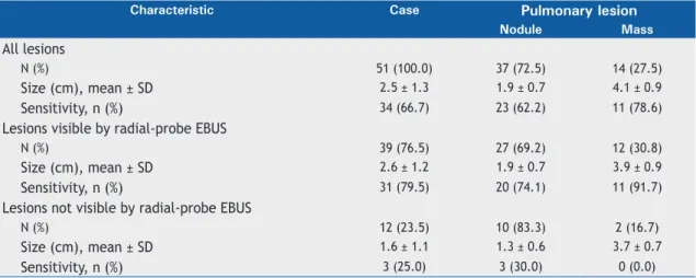
On page 251 of the original publication, Table 1 should be disregarded. The correct table should read
Table 1.Characteristics of the lesions in the patients submitted to radial-probe EBUS (N = 51).
Characteristic Case Pulmonary lesion
Nodule Mass
All lesions
N (%) 51 (100.0) 37 (72.5) 14 (27.5)
Size (cm), mean ± SD 2.5 ± 1.3 1.9 ± 0.7 4.1 ± 0.9
Sensitivity, n (%) 34 (66.7) 23 (62.2) 11 (78.6)
Lesions visible by radial-probe EBUS
N (%) 39 (76.5) 27 (69.2) 12 (30.8)
Size (cm), mean ± SD 2.6 ± 1.2 1.9 ± 0.7 3.9 ± 0.9
Sensitivity, n (%) 31 (79.5) 20 (74.1) 11 (91.7)
Lesions not visible by radial-probe EBUS
N (%) 12 (23.5) 10 (83.3) 2 (16.7)
Size (cm), mean ± SD 1.6 ± 1.1 1.3 ± 0.6 3.7 ± 0.7
Sensitivity, n (%) 3 (25.0) 3 (30.0) 0 (0.0)
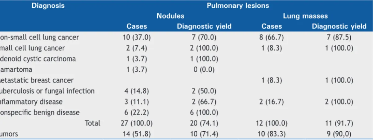
On page 252 of the original publication, Table 2 should be disregarded. The correct table should read J Bras Pneumol. 2017;43(1):78-79
Jacomelli M, Demarzo SE, Cardoso PFG, Palomino ALM, Figueiredo VR
Table 2. Final diagnoses of the lesions that were visible by radial-probe EBUS and and diagnostic yield.a
Diagnosis Pulmonary lesions
Nodules Lung masses
Cases Diagnostic yield Cases Diagnostic yield
Non-small cell lung cancer 10 (37.0) 7 (70.0) 8 (66.7) 7 (87.5) Small cell lung cancer 2 (7.4) 2 (100.0) 1 (8.3) 1 (100.0) Adenoid cystic carcinoma 1 (3.7) 1 (100.0)
Hamartoma 1 (3.7) 0 (0.0)
Metastatic breast cancer 1 (8.3) 1 (100.0)
Tuberculosis or fungal infection 4 (14.8) 2 (50.0)
Inlammatory disease 3 (11.1) 2 (66.7) 2 (16.7) 2 (100.0)
Nonspeciic benign disease 6 (22.2) 6 (100.0)
Total 27 (100.0) 20 (74.1) 12 (100.0) 11 (91.7)
Tumors 14 (51.8) 10 (71.4) 10 (83.3) 9 (90,0)
aValues expressed in n (%).
On page 252 of the original publication, left column, second paragraph, lines 16 to 18, where it is written
“The sensitivity of the procedure tripled for lesions that were visible by radial-probe EBUS compared to those that were not visible (73% vs. 25%).”
it should read
“The sensitivity of the procedure tripled for lesions that were visible by radial-probe EBUS compared to those that were not visible (79.5% vs. 25.0%).”
On page 252 of the original publication, right column, third paragraph, lines 1 to 8, where it is written
“The differential diagnosis between malignancy and infectious disease is important in Brazil. In the present
study, we identiied non-neoplastic disease in
13 (48.1%) of the 27 pulmonary nodules that were visible by radial-probe EBUS and in 2 (16.7%) of the 12
pulmonary masses that were visible by radial-probe EBUS, the inal diagnoses including fungal infections and
tuberculosis.”
it should read
“The differential diagnosis between malignancy and infectious disease is important in Brazil. In the present
study, we identiied inlammatory/infectious disease in 13 (48.1%) of the 27 pulmonary nodules that were
visible by radial-probe EBUS and in 2 (16.7%) of the 12 pulmonary masses that were visible by radial-probe
EBUS, the inal diagnoses including fungal infections and tuberculosis.”