Aortic and tricuspid endocarditis in hemodialysis
patient with systemic and pulmonary embolism
INTRODUCTION
Infective endocarditis (IE) is a severe infection that primarily involves the endocardium of the cardiac valve lealets. IE is more frequent in hemodialysis patients than it is in the general population, and S. aureus is the main infective agent.
Neurologic involvement is a frequent complication of IE. Despite the formal surgical indications for cerebral embolism, the timing of surgical intervention is controversial, particularly in cases with a coexistent intracranial hemorrhage.(1)
Here, the authors describe a case with an unusual location of IE in multiple valves in a patient on hemodialysis with severe systemic and pulmonary embolic presentation. Despite the administration of the appropriate antibiotic therapy, embolic events continued to occur with the deterioration of the patient’s clinical status. he management of this patient was a challenge due to comorbidities and concomitant intracranial hemorrhage that precluded early surgical intervention.
Silvia Aguiar Rosa1, Nuno Germano2,
Ana Santos2, Luis Bento2
1. Department of Cardiology, Centro Hospitalar de Lisboa Central, EPE - Lisboa, Portugal. 2. Critical Care Medicine, Centro Hospitalar de
Lisboa Central, EPE - Lisboa, Portugal. old Caucasian male with end-stage his is a case report of a
43-year-renal disease being treated with hemodialysis and infective endocarditis in the aortic and tricuspid valves. he clinical presentation was dominated by neurologic impairment with cerebral embolism and hemorrhagic components. A thoracoabdominal computerized tomography scan revealed septic pulmonary embolus. he patient underwent empirical antibiotherapy with ceftriaxone, gentamicin and vancomycin, and the therapy was changed to lucloxacilin and gentamicin after the
Conflicts of interest: None.
Submitted on January 13, 2015 Accepted on April 24, 2015
Corresponding author:
Silvia Aguiar Rosa Hospital de Santa Marta
Centro Hospitalar de Lisboa Central, EPE Rua de Santa Marta
1169-024 - Lisboa, Portugal E-mail: [email protected]
Responsible editor: Thiago Costa Lisboa
Endocardite aórtica e tricúspide em pacientes de hemodiálise com
embolia sistêmica e pulmonar
ABSTRACT
Keywords: Endocarditis/etiology; Renal dialysis; Embolism; Aortic valve; Tricuspid valve; Tomography, x-ray computed; Case reports
isolation of S. aureus in blood cultures. he multidisciplinary team determined that the patient should undergo valve replacement after the stabilization of the intracranial hemorrhage; however, on the 8th day of hospitalization, the patient
entered cardiac arrest due to a massive septic pulmonary embolism and died. Despite the risk of aggravation of the hemorrhagic cerebral lesion, early surgical intervention should be considered in high-risk patients.
CASE REPORT
Here, the authors report a case of a 43-year-old Caucasian man with a medical history of end-stage renal disease due to hypertensive nephropathy and a 4-year history of hemodialysis. Prior to the initiation of hemodialysis, the patient underwent renal transplantation and rejected the allograft after 5 years due to voluntary discontinuation of immunosuppressive therapy. He was admitted to the hospital with fever, malaise, altered mental state and chest pain. Physical examination revealed cognitive impairment with incoherent speech, left arm weakness, exacerbated osteotendinous relexes, the presence of the Babinski relex on the right side and three episodes of complex partial seizures. He was medicated with valproic acid and fenitoin for seizure control.
Relevant laboratory indings included creatinine 13.17mg/dL, urea 203 mg/dl, potassium 7.4mEq/L, leucocytosis 20,000/dL, C-reactive protein 411.5mg/L, troponin I 2.7ng/mL and myoglobin 317IU/L.
An electrocardiogram showed ST segment elevation in DII, DIII, aVF, V2 and V3 with upward concavity and depression of the PR segment in DII, V2 and V3.
Transthoracic echocardiography revealed normal left ventricular dimensions and systolic function with no left atrial enlargement; furthermore, the right cavities were not dilated, and the right ventricular systolic function was preserved. A mild pericardial efusion with no evidence of vegetation in the valve lealets was observed.
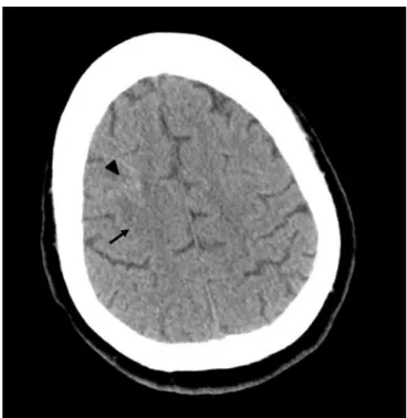
A computerized tomography scan of the brain revealed a right frontal cortical-subcortical lesion with ischemic and hemorrhagic components (Figure 1). Magnetic resonance imaging conirmed multiple hemorrhagic lesions in both cerebral hemispheres (Figure 2). A thoracoabdominal computerized tomography scan revealed septic pulmonary embolus and a splenic infarction. he diagnostic hypothesis was IE, and the patient underwent transesophageal echocardiography, which identiied vegetations in the aortic and tricuspid valves of 9.13mm and 11.73mm in size, respectively (Figures 3 and 4), with mild aortic regurgitation and moderate tricuspid regurgitation. Empirical treatment with ceftriaxone, gentamicin and vancomycin was initiated. Blood cultures were positive for methicillin-sensitive S. aureus, and the empirical treatment was changed to lucloxacilin and gentamicin. Cerebral spinal luid was unremarkable, and its culture was sterile. he pericardial efusion became bulkier and generated late diastolic inward motion of the right ventricular free wall and the right atrium, which necessitated pericardiocentesis.
Figure 1 - Cortico-subcortical right frontal hypodensity (arrow) with mild hyperdensity (arrow head) - ischemic area with intracerebral hemorrhagic component observed on brain computerized tomography.
than the incidence in the general population.(2) he risk factors in this group include recurrent bacteremia during hemodialysis, uremia, immune injury and premature degenerative heart valve disease due to abnormalities in calcium-phosphorus homeostasis and chronic inlammation.(2,3)S. aureus is the main infective agent in IE and causes 50 to 80% of IE cases (50% of which are methicillin resistant).(3) his relects the rate of infection of hemodialysis intravascular devices and the fact that 50% of all hemodialysis patients are carriers of S. aureus. Despite the relationship between hemodialysis devices and IE in these patients, infection of right-sided valves is rare. he mitral valve is the most frequently infected valve (50%), followed by the aortic valve (40%). Tricuspid IE only constitutes approximately 10% of the cases. Multiple valves, usually the mitral and aortic valves, are involved in 20% of such cases.(2,3) he degenerative disease of left-sided heart valves might explain the diference in incidence.(3)
Following cardiovascular disease, IE is the second most common cause of death in hemodialysis patients. he mortality of IE in these patients is 30 - 60%, which is considerably higher than that in the general population, probably due to the high prevalence of S. aureus and multiple comorbid conditions in hemodialysis patients. Age, diabetes, septic embolism involving cerebrovascular events, mitral valve involvement, great vegetation and S. aureus infection are factors that result in a poor prognosis in IE patients.(3)
he indications for valve replacement in the general population are deined in the European Society of Cardiology guidelines. However, there is no consensus regarding whether these guidelines are applicable to hemodialysis patients, and data regarding surgical decisions in these patients are lacking. According to the European Society of Cardiology guidelines for the management of IE, embolic events are an indication for surgical intervention. he brain and the spleen and lungs are the most frequent sites of embolism in left and right-sided IE, respectively. Embolism occurs in 22% to 50% of patients with IE. he predictors of embolism are vegetation size greater than 10mm, mitral location and infection with S. aureus.(1,4,5)
he current guidelines indicate that after a transient ischemic attack or stroke, surgery should not be delayed, but following an intracranial hemorrhage, the guidelines state that surgery must be delayed for one month.(1) However, Yoshioka et al. suggested that the risk of neurological deterioration after surgery is relatively low,
Figure 3 - Transesophageal echocardiography showing vegetation in aortic valve 9.13mm in size.
Figure 4 - Transesophageal echocardiography demonstrating vegetation in tricuspid valve11.73mm in size.
Despite the administration of efective antibiotic therapy, cerebrovascular embolic events persisted with neurologic deterioration. he multidisciplinary team decided that the patient should undergo valve replacement after the stabilization of the intracranial hemorrhage. However, on the 8th day after hospitalization, the patient entered cardiac arrest and was recovered with advanced life support. Transthoracic echocardiography after resuscitation demonstrated an enlargement of the right cavities and right ventricular hypocinesia, and the presumed diagnosis was a massive septic pulmonary embolism. he patient re-entered cardiac arrest, and although ibrinolysis was performed and advanced life support was maintained for 40 minutes, the patient died.
DISCUSSION
even in IE patients who underwent valve replacement less than 2 weeks after intracranial hemorrhage.(6) In patients who are on hemodialysis, perioperative mortality is high, probably because the patients who are selected for surgery have more severe disease and complications. It has been suggested that patients with high mortality risk should undergo earlier surgery than the general population should.(3,5) In a recent study, patients who underwent surgery had a higher survival rate than those who did not.(2)
he size and mobility of vegetations are the most important factors associated with embolism; therefore, even in the absence of an embolic event, surgical treatment should be considered in cases with vegetation with a length of 15mm or 10mm and other predictors of a complicated course.(1,7)
In addition to embolism, the other two main indications for surgery in the context of IE are heart failure and uncontrolled infection.
Heart failure in IE is caused by valve destruction and results in severe regurgitation, intracardiac istulae or valve obstruction. Heart failure is more frequent in aortic IE than it is in mitral IE. he timing of surgery depends on the patient’s clinical features. In cases of persistent pulmonary edema or cardiogenic shock, immediate intervention should
be performed. If severe valve regurgitation is well tolerated, a conservative approach with antibiotics is recommended, and surgery is indicated after resolution of IE.(1)
A perivalvular abscess, pseudoaneurysm and istulae are frequent complications of uncontrolled infection. he more common location of these complications is the aortic valve, particularly in the mitral-aortic intervalvular ibrosa. Unexplained fever or new atrioventricular block should raise the suspicion of perivalvular infection extension, and this should be conirmed by transesophageal echocardiography. In these cases, surgery is indicated as soon as possible. An intervention is also recommended in case of persistent fever or positive blood cultures and extracardiac abscesses, or if the IE is caused by a multiresistant organisms.(1,8)
CONCLUSION
his clinical report describes a case with an unusual location of infective endocarditis in multiple valves in a patient on hemodialysis with severe embolic presentation. Although there is no consensus regarding the optimal timing for surgery, it seems that early intervention could prevent disease aggravation, despite the risk of hemorrhagic lesion exacerbation.
Este artigo relata o caso de um homem caucasiano de 43 anos de idade com nefropatia terminal em tratamento com hemodiálise e apresentando endocardite infecciosa das válvulas aórtica e tricúspide. O quadro clínico foi dominado pelo comprometimento neurológico, devido à embolia cerebral e a componentes hemorrágicos. Uma tomograia computadorizada tóraco-abdominal revelou um êmbolo séptico pulmonar. O paciente foi submetido à antibioticoterapia empírica utilizando ceftriaxona, gentamicina e vancomicina, sendo o tratamento modiicado para lucloxacilina e gentamicina após o isolamento de S. aureus nas hemoculturas. A equipe
multidisciplinar determinou que o paciente deveria ser submetido à substituição de válvulas após estabilização da hemorragia intracraniana; contudo, no oitavo dia após a hospitalização, o paciente entrou em parada cardíaca causada por embolia séptica pulmonar maciça, vindo a falecer. Apesar do risco de agravamento da lesão hemorrágica cerebral, em pacientes de alto risco deveria ser considerado realizar precocemente uma intervenção cirúrgica.
RESUMO
REFERENCES
1. Habib G, Hoen B, Tornos P, Thuny F, Prendergast B, Vilacosta I, Moreillon P, de Jesus Antunes M, Thilen U, Lekakis J, Lengyel M, Müller L, Naber CK, Nihoyannopoulos P, Moritz A, Zamorano JL; ESC Committee for Practice Guidelines. Guidelines on the prevention, diagnosis, and treatment of infective endocarditis (new version 2009): the Task Force on the Prevention, Diagnosis, and Treatment of Infective Endocarditis of the European Society of Cardiology (ESC). Endorsed by the European Society of Clinical Microbiology and Infectious Diseases (ESCMID) and the International Society of Chemotherapy (ISC) for Infection and Cancer. Eur Heart J. 2009;30(19):2369-413.
2. Kamalakannan D, Pai RM, Johnson LB, Gardin JM, Saravolatz LD. Epidemiology and clinical outcomes of infective endocarditis in hemodialysis patients. Ann Thorac Surg. 2007;83(6):2081-6.
3. Nucifora G, Badano LP, Viale P, Gianfagna P, Allocca G, Montanaro D, et al. Infective endocarditis in chronic haemodialysis patients: an increasing clinical challenge. Eur Heart J. 2007;28(19):2307-12.
4. Keynan Y, Singal R, Kumar K, Arora RC, Rubinstein E. Infective endocarditis in the intensive care unit. Crit Care Clin. 2013;29(4):923-51.
5. Leither MD, Shroff GR, Ding S, Gilbertson DT, Herzog CA. Long-term survival of dialysis patients with bacterial endocarditis undergoing valvular replacement surgery in the United States. Circulation. 2013;128(4):344-51. 6. Yoshioka D, Toda K, Sakaguchi T, Okazaki S, Yamauchi T, Miyagawa S,
Nishi H, Yoshikawa Y, Fukushima S, Saito T, Sawa Y; OSCAR study group. Valve surgery in active endocarditis patients complicated by intracranial haemorrhage: the influence of the timing of surgery on neurological outcomes. Eur J Cardiothorac Surg. 2014;45(6):1082-8.
7. Thuny F, Di Salvo G, Belliard O, Avierinos JF, Pergola V, Rosenberg V, et al. Risk of embolism and death in infective endocarditis: prognostic value of echocardiography: a prospective multicenter study. Circulation. 2005;112(1):69-75. Erratum in: Circulation. 2005;112(9):e125. Disalvo, Giovanni [corrected to Di Salvo, Giovanni]; Calabro, Raffaello [corrected to Calabró, Raffaele].