OR
IGI
N
A
L
R
E
S
E
A
R
C
H
Thoracic kyphosis comparison between a patient
with chronic obstructive pulmonary disease and a
healthy individual by flexicurve method
Comparação da cifose torácica entre o paciente com doença pulmonar obstrutiva crônica e o
indivíduo saudável pelo método lexicurva
Comparación de la cifosis torácica entre el paciente con enfermedad pulmonar obstructiva
crónica y el individuo sano por el método lexicurva
Márcia Aparecida Gonçalves1, Patrícia Leite Rodovalho2, Angela Jacques Bellini2,
Ana Karla Vieira Brüggemann3, Giovana Zarpellon Mazo4, Elaine Paulin5
Study performed in the Laboratory of Respiratory Physiotherapy (LAFIR) in Santa Catarina State University (UDESC), Florianópolis (SC), Brazil.
1Physical therapist, Master in Physiotherapy, Universidade Estadual de Santa Catarina (UDESC) – Florianopolis (SC), Brazil. 2Physiotherapy undergraduate student, Universidade Estadual de Santa Catarina (UDESC) – Florianopolis (SC), Brazil. 3Physical therapist, Universidade Estadual de Santa Catarina (UDESC) – Florianopolis (SC), Brazil.
4Doctor in Sport Sciences,Universidade do Porto (U.PORTO) – Porto, Portugal. Professor of the Graduate Program in Human
Movement Sciences, Universidade Estadual de Santa Catarina (UDESC) – Florianopolis (SC), Brazil.
5Doctor in Science (Experimental Physiopathology), Universidade de São Paulo (USP) – São Paulo (SP), Brazil. Professor of the
Department of Undergraduate and Graduate Program in Physiotherapy, Universidade Estadual de Santa (UDESC) – Florianópolis (SC), Brazil.
ABSTRACT | Chronic obstructive pulmonary disease (COPD) is characterized by airlow obstruction, air entrapment and pulmonary hyperinlation. These pathophysiological factors can compromise the diaphragmatic mobility, causing deformities in the thoracic cavity and consequently increasing the angle of the thoracic curvature. We compared the angle of the thoracic curvature between COPD patients and healthy individuals by the lexicurve method. Thirty-seven patients with COPD and 37 healthy individuals participated in the study. All subjects performed the following evaluations: anthropometry, spirometry, and measurement of the thoracic curvature angle. The data were analyzed and treated with descriptive analysis such as mean and standard deviation. The Shapiro-Wilk test was used to verify the normality of the data. The Student’s t-test was used to compare the thoracic curvature angle of patients with COPD with healthy individuals. The signiicance level adopted was 5%. The mean age of the COPD group was 65.70±7.91 years, body mass index (BMI) of 26.73±5.34kg/m2, and FEV
1 (expected %) of 50.65±19.08, showing moderate obstruction degree. Healthy individuals showed an average of 7.27±62.49 years, BMI
333
of 26.97±3.55kg/m² and FEV1 (expected %) of 94.05±0 9.44. We did not observe any signiicant diference between patients with COPD and healthy individuals in the thoracic curvature angle: 56.67±11.31 and 55.42±9.61 degrees, respectively (p=0.61). The lexicurve method proved to be a useful and practical tool for assessing the thoracic kyphosis, and it also identiied no diference between the thoracic curvature of COPD patients with moderate obstruction and of healthy individuals.
Keywords | Pulmonary Disease, Chronic Obstructive; Evaluation; Spine; Kyphosis; Posture.
RESUMO | A doença pulmonar obstrutiva crônica (DPOC) é caracterizada pela obstrução do luxo aéreo, aprisionamento de ar e pela hiperinsulação pulmonar. Esses fatores isiopatológicos podem comprometer a mobilidade diafragmática, causar deformidades na caixa torácica e consequentemente aumentar o ângulo da curvatura torácica. Comparamos o ângulo da curvatura torácica entre pacientes com DPOC e indivíduos saudáveis pelo método lexicurva. Participaram do estudo 37 pacientes com DPOC e 37 indivíduos saudáveis. Todos os indivíduos realizaram as seguintes avaliações:
Mailing address: Elaine Paulin – Rua Pascoal Simone, 358 – Coqueiros – CEP 88080-350 – Florianópolis (SC), Brazil. E-mail: [email protected] – Phone: +55 48 3321 8621
antropometria, espirometria e mensuração do ângulo da curvatura torácica. Os dados foram analisados e tratados com análise descritiva como média e desvio-padrão. O teste de Shapiro-Wilk foi utilizado para veriicar a normalidade dos dados. O teste t de Student foi utilizado para comparar o ângulo da curvatura torácica dos pacientes portadores de DPOC com os indivíduos saudáveis. O nível de signiicância adotado foi de 5%. A média de idade do grupo com DPOC foi de 65,70±7,91 anos, o índice de massa corporal (IMC), 26,73±5,34kg/m2, e VEF1 (% previsto), 50,65±19,08, apresentando grau de obstrução moderada. Os indivíduos saudáveis apresentaram em média 62,49±7,27 anos, IMC de 26,97±3,55kg/m² e VEF1 (% previsto) de 94,05±9,44. Não houve diferença signiicante entre os pacientes com DPOC e os indivíduos saudáveis no ângulo da curvatura torácica: 56,67±11,31 e 55,42±9,61 graus, respectivamente (p=0,61). O método lexicurva mostrou-se uma ferramenta útil e prática para avaliar a cifose torácica, identiicando que não houve diferença entre as curvaturas torácicas dos pacientes com DPOC com obstrução moderada e dos indivíduos saudáveis. Descritores | Doença Pulmonar Obstrutiva Crônica; Avaliação; Coluna Vertebral; Cifose; Postura.
RESUMEN | La enfermedad pulmonar obstructiva crónica (EPOC) se caracteriza por la obstrucción del lujo de aire, aprisionamiento de aire e por la hiperinsulación pulmonar. Estos factores isiopatológicos pueden comprometer la movilidad diafragmática, causar deformidades en la caja torácica y
consecuentemente aumentar el ángulo de la curvatura torácica. Se comparó el ángulo de la curvatura torácica entre pacientes con EPOC y de individuos sanos mediante el método lexicurva. Participaron del estudio 37 pacientes con EPOC y 37 individuos sanos. Todos los individuos realizaron las siguientes evaluaciones: antropometría, espirometría y medición del ángulo de la curvatura torácica. Los datos fueron analizados y tratados con análisis descriptivo como media y desviación estándar. Se utilizó la prueba de Shapiro-Wilk para comprobar la normalidad de los datos, y la prueba t de Student para comparar el ángulo de la curvatura torácica de los pacientes portadores de EPOC con los individuos sanos. El nivel de signiicancia adoptado fue del 5%. La edad promedio del grupo con EPOC fue 65,70±7,91 años, el índice de masa corporal (IMC), 26,73±5,34kg/m2, y VEF
1 (% previsto), 50,65±19,08, presentando grado de obstrucción moderada. Los individuos sanos presentaron un promedio de 62,49±7,27 años, IMC de 26,97±3,55kg/m² y VEF1 (% previsto) de 94,05±9,44. No hubo diferencia signiicante entre los pacientes con EPOC y los pacientes sanos en el ángulo de la curvatura torácica: 56,67±11,31 y 55,42±9,61 grados, respectivamente (p=0,61). El método lexicurva ha mostrado ser una herramienta útil y práctica para evaluar la cifosis torácica, identiicando que no hubo diferencia entre las curvaturas torácicas de los pacientes con EPOC con obstrucción moderada y de los individuos sanos. Palabras clave | Enfermedad Pulmonar Obstructiva Crónica; Evaluación; Columna Vertebral; Cifosis; Postura.
INTRODUCTION
horacic hyperkyphosis is a condition indicated by the abnormal increase in the curvature convexity of the thoracic vertebral column1, which may be related to
increased age2, vertebral fractures3, postural alterations4,
degenerative disease in the vertebral disc5, muscle
weakness6, intervertebral ligament degeneration7, and
genetic predisposition8.
Pachioni et al.9 demonstrated that patients with
chronic obstructive pulmonary disease (COPD) have a higher angle in thoracic kyphosis when compared to healthy older adults. his increase in the thoracic kyphosis can occur both due to aging, and to pathophysiological factors related to the disease such as increase in the anteroposterior diameter of thorax10,
ribs in horizontal position11, excessive use of accessory
muscles and reduced diaphragmatic mobility12.
With the increased thoracic kyphosis, the clinical pattern of the COPD patient can be exacerbated, since thoracic hyperkyphosis may cause many adverse consequences to health13, such as harming the
pulmonary function14, increasing dyspnea14, afecting
the performance of daily activities15, reducing the quality
of life16, and increasing the risk of mortality regardless
of the subjacent vertebral osteoporosis17. As COPD
patients present all these losses as a result of the illness, the increase in the angle of the thoracic curvature can be more evident, especially in more advanced stages of the disease18, and afect even more their health.
Several instruments have been used to evaluate thoracic kyphosis19. In clinical practice, radiography
is used as the gold standard for measuring the Cobb angle2,20. Although it is a non-invasive method, easy to
he lexicurve method is equivalent to the radiographic (Cobb angle)22, 23, 24: it is not invasive,
it provides an evaluation of the thoracic curvature in the sagittal plane, it is a validated tool in Brazil, considered reliable and reproducible22-24, and it uses a
mathematical model (speciic software) to calculate the angle of thoracic curvature values from measurements obtained by the rule molded into the thoracic spine of the individual22.
here is a lack of studies that use the lexicurve method to evaluate the thoracic spine of patients with COPD. In the study by Pachioni et al.9 conducted
with COPD patients, we used the Postural Analysis Software (SAPO). However, it seems that this tool is not suitable for evaluating thoracic kyphosis, and still lacks reference values and standardization to accomplish this measure25. On the other hand,
the lexicurve rule ofers a standardized way of measurement, reference angular values and can be used in clinical practice, considering its low cost and easy use and transport.
herefore, the objective of this study was to evaluate the angle of thoracic curvature of COPD patients and healthy individuals by the lexicurve method.
METHODOLOGY
his study is an analytical cross-cutting-type research and of quantitative approach. It was approved by the Ethics Committee in Research with Human Beings at the State University of Santa Catarina, Florianópolis (CAAE: 08857612.2.0000.0118). All individuals were informed about the research, and signed a free and informed consent form, according to the resolution 466/12 of the National Health Council.
Seventy-four individuals of both genders (34 men) participated in the study. hey were divided into two groups: group 1 consisting of 37 patients with COPD, with mean age of 65.70±7.91 years, and group 2 consisting of 37 healthy subjects with a mean age of 62.49±7.27 years.
In the COPD group, patients with COPD diagnosis were included according to the classiication of the Global Initiative for Chronic Obstructive Lung Disease (GOLD)26. We also used a diagnostic record developed
by researchers linked to the Laboratory of Respiratory Physiotherapy, aiming to identify the characteristics of the individuals and verify if they met the following
inclusion criteria: 1) clinical stability during the last month and at the beginning of the evaluation protocol: absence of exacerbations characterized as persistent worsening in the stable basal condition, of acute onset (typically by the accentuation of dyspnea, with or without cough, increased expectoration volume, sputum purulence and thoracic oppression) and which may require additional treatment; 2) patients nondependent on oxygen supplementation; 3) absence of other respiratory and cardiovascular diseases; 4) patients without involvement in training programs in the 6 months prior to the beginning of this study; 5) patients who have not undergone recent surgery on the spine or in lower limbs and/or who have not had fractures in the 6 previous months.
For patients with COPD, we considered the following exclusion criteria: 1) presence of exacerbations of the disease during the research; 2) cardiorespiratory and/or musculoskeletal clinical recurrences during the evaluations; 3) incapacity to perform any of the evaluations of the study (lack of understanding or collaboration) and; 4) withdrawal of the patient during the evaluation period.
We included in the healthy group individuals with normal spirometry (FEV1/FVC ≥0.7; FEV1 ≥80% of the expected, FVC ≥80% of the expected), without associated comorbidities, and with age, weight, and BMI compatible with COPD patients. We excluded from this group individuals who were incapable to perform any of the evaluations of the study (lack of understanding or collaboration) and/or who withdrew during the process of evaluation.
Evaluated parameters
Anthropometry
For body mass measurement, we used a scale previously calibrated and a stadiometer for measuring the height, Welmy® model W200/5. After obtaining
the anthropometric values (body mass and height), we calculated the body mass index (BMI) by the equation: body mass/height2 (kg/m2). Patients were classiied,
according to the BMI, in low weight (≤18.5kg/m2),
normal weight (18.5-24.9kg/m2), overweight
(25-29.9kg/m2) and obese (≥30kg/m2)27.
Spirometry
previously calibrated in accordance with the methods and criteria recommended by the American horacic Society and the European Respiratory Society28.
Spirometric variables were expressed as absolute values and as the percentage value of the expected values of normality, according to those determined by Pereira et al.29. he criteria for normal pulmonary function test
consist of FVC and FEV1≥80% of the expected, and FEV1/FVC≥0.7.
Angle of the thoracic curvature
It was evaluated by the lexicurve method that consists of using a lexible ruler of 80cm (TRIDENT®
Precision Industry, Brazil) composed of a folding metal rod protected with lexible plastic. During the measurement of the thoracic curvature, the individual remained in an static orthostatic position, as relaxed as possible, and used a disposable apron with a posterior opening, remaining with the elbows and shoulders extended throughout the body. hen, spinous processes of C7 and T12 were located and marked with a pencil. he lexible ruler was placed initially in the spinal process of C7, being shaped with the format of the curvature of the kyphosis until the spinal process T12. hen, we performed the transcription of the dorsal column format to the graph paper, and the following straight lines were drawn: from the C7 equivalent point to the T12 point named Xtotal, from the greater angle of thoracic curve to the Xtotal straight line named H line (cm), and from the beginning of T12 to the H line named
Xmiddle (cm)22 (Figure 1).
C7
T12 H
X
total
X
middle
Xtotal – Distance between C7 and T12
Xmiddle – Distance between H and T12
H – Greater distance of the curvature with the line that unites
C7 and T12 points
Figure 1. Schematic drawing of thoracic kyphosis measurement by Flexicurve (Teixeira & Carvalho, 2007)
Finally, we performed the application of angle calculation by the third degree polynomial, in which a math formula was employed4. We considered as normal
values for the curvature of the thoracic kyphosis angles between 20° and 50° in adults22, and angles up to 56° in
the older adults30.
Statistical Analysis
Data were analyzed using the SPSS for Windows program, version 20.0, and treated with descriptive analysis as mean and standard deviation, applied in all variables. he normality of the data was veriied using the Shapiro-Wilk test. According to the data distribution, the Student’s t-test (parametric data) and the U Mann Whitney test (nonparametric data) were used for comparing the parameters between the groups. he signiicance level adopted was equal or inferior to 5%.
RESULTS
We included 44 patients diagnosed with COPD (aged from 49 to 81 years), but 07 were excluded during the study:03 for not understanding how to perform the spirometry test and 04 for not completing all evaluations. We also evaluated 48 healthy individuals (aged from 49 to 78 years), and 11 were excluded, 05 for not completing all evaluations, 05 for lack of understanding how to perform the spirometry test, and 01 due to musculoskeletal problems. herefore, 37 patients with COPD (19 men) and 37 healthy individuals (15 men) participated in the present study.
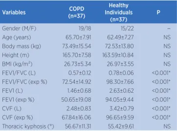
Table 1 presents the characterization of the studied groups, showing that there was no statistically signiicant diference between the groups regarding age, body mass, weight and BMI, conirming that the groups were matched in relation to the anthropometric variables.
Table 1. Basal characteristics of the studied sample
Variables COPD
(n=37)
Healthy Individuals
(n=37)
P
Gender (M/F) 19/18 15/22 --Age (years) 65.70±7.91 62.49±7.27 NS Body mass (kg) 73.49±15.54 72.53±13.80 NS Height (m) 165.70±7.58 163.59±10.84 NS BMI (kg/m²) 26.73±5.34 26.97±3.55 NS FEV1/FVC (L) 0.57±0.12 0.78±0.06 <0.001* FEV1/FVC (exp %) 72.54±14.92 98.30±7.66 <0.001* FEV1 (L) 1.46±0.68 2.63±0.62 <0.001* FEV1 (exp %) 50.65±19.08 94.05±9.44 <0.001* CVF (L) 2.48±0.83 3.42±0.79 <0.001* CVF (exp %) 67.84±16.06 96.65±9.59 <0.001* Thoracic kyphosis (°) 56.67±11.31 55.42±9.61 NS
The values are expressed as mean and ± standard deviation; COPD: Chronic obstructive pulmo-nary disease; M: male; F: female; BMI (kg/m2): body mass index in kilograms by meters2; FEV1
(L): forced expiratory volume in the irst second, in liters; FEV1 (exp %): percentage of schedule of the forced expiratory volume in the frist second; FVC (L): forced vital capacity in liters; FVC (exp %): percentage of predicted forced vital capacity; (°): degree. p: * signiicant diference (p<0.05)
he value of the angle of thoracic curvature in each group is displayed in Figure 2. We observed that there was no signiicant diference between patients with COPD and healthy individuals in the angle of the thoracic curvature (56.67±11.31; 55.42±9.61 degrees, respectively) (p=0.61), showing similarity in the curvatures.
57
56
55 56.5
55.5
54.5
=
Groups
COPD HEALTHY INDIVIDUALS
T
hor
acic k
yphosis (°)
(p=0.61). = Signiicance p>0.05
Figure 2. Comparison of the thoracic curvature angle between COPD patients and healthy individuals
DISCUSSION
In this study, the angle of the curvature of the thoracic kyphosis, evaluated by the lexicurve method of the group of COPD patients, was similar to the group of healthy individuals, probably due to the age similarity between the groups. hat is because the
thoracic hyperkyphosis is a postural change commonly observed and associated with age23, and we observed
that both groups showed aging, a characteristic that can be a determining factor for the increase in the thoracic kyphosis degree.
Few studies have investigated the thoracic kyphosis in COPD patients and compared it with healthy individuals. Our results are consistent with the study of Dias et al.18, performed with 19 CODP patients
and 19 healthy individuals, and they also found no signiicant diferences in the curvature of the cervical and thoracic column regions between the groups. However, our results contrast with those presented in the study of Pachioni et al.9, with 15 patients with
COPD and 15 healthy individuals, in which it was found a statistically signiicant diference in thoracic kyphosis between the groups.
hese diferences in results between the studies may be related to the methodology used for the analysis of thoracic kyphosis, since in the study of Pachioni et al.9, the Postural Evaluation
Software (SAPO) (with adaptation) was used to evaluate the thoracic kyphosis. Considering the complexity of the evaluation ofered by SAPO, which involves a formula of correction of values proposed by Leroux et al.25, the authors adapted
his method to other studies22,31, and based on
the angle formed between T3/T12, with apex on the most prominent vertebra, to ind the degree of thoracic kyphosis. In our study we used the lexicurve method and angles formed between C7 and T12, according to the methodology proposed by Teixeira & Carvalho22,when analyzing a larger
extension of the thoracic spine.
Although the SAPO software is used to evaluate the thoracic kyphosis, it is not considered the most suitable method because of its limitations. According to Leroux et al.25, this method of evaluation of the vertebral
column is particularly vulnerable to evaluate the thoracic kyphosis, since it depends on the placement of markers in speciic anatomical landmarks as well as the type of marker used. In addition, the small polystyrene balls used as markers are not visible in lateral view due to the scapula, and there is no standardization on the evaluation technique of the angle of curvature of the thoracic kyphosis.
in Brazil22. his method is considered as a reliable
and reproducible tool, and shows good sensitivity and speciicity for measuring the thoracic and lumbar curvature, besides having low cost, portability and being a non-invasive method19,22,23,24,32.
In this study we found that both COPD and healthy individuals do not present serious changes in the angle of the thoracic curvature. he mean of the angles in the COPD group and healthy individuals was 56.67° and 55.42°, respectively. According to Loubresse, Vialle & Wollf33, more severe angles can afect the ventilatory
function. Libby et al.34 report that more severe thoracic
curvatures feature angles above 65°.
One limitation of this study is related to lack of knowledge about the epidemiology of thoracic hyperkyphosis in patients with COPD. Another limitation was the prevalence of patients with moderate degree of obstruction, since more severe obstructions may present greater change in the thoracic cavity. hus, we need more studies to identify the causes and consequences of changes in the thoracic spine of a patient with COPD.
It is important to highlight the relevance of evaluation of thoracic kyphosis by an easily accessible and practical tool for clinical use. he early detection of changes in the angle of the thoracic curvature will provide an adequate therapeutic approach to prevent late complications caused by severe thoracic hyperkyphosis, mainly regarding losses on the pulmonary function, since patients with COPD have this feature that may be exacerbated.
CONCLUSION
he lexicurve method proved to be a useful and practical tool for assessing thoracic kyphosis, and it also identiied no diference between the thoracic curvature of COPD patients with moderate obstruction and of healthy individuals.
REFERENCES
1. Saiiari A, Khodayarib B, Bostanic M. Relation between Increasing spinal curve and anxiety. P - Soc Behav Sci. 2011;30:2246-8.
2. Nishiwaki Y, et al. Association of thoracic kyphosis with subjective poor health, functional activity and blood pressure in the community-dwelling elderly. Environ Health Prev Med. 2007;12(6):246-50.
3. Schneider DL, von Müller D, Barrtet-Connor E, Sartoris DJ . Kyphosis does not equal vertebral fractures: the Rancho Bernardo study. J Rheumatol. 2004;31:747-52.
4. Hinman MR. Comparision of thoracic kyphosis and postural stifness in younger and older women. Spine J. 2004;4(4):413-7.
5. Goh S, Price RI, Leedman PJ, Singer KP. The relative inluence of vertebral body and intervertebral disc shape on thoracic kyphosis. Clin Biomech. 1999;14:439-48.
6. Mika A, Unnithan VB, Mika P. Diferences in thoracic kyphosis and in back muscle strength in women with bone loss due to osteoporosis. Spine. 2005;30:241-6.
7. Birnbaum K, Siebert CH, Hinkelmann J, Prescher A, Niethard FU. Correction of kyphotic deformity before and after transection of the anterior longitudinal ligament: a cadaver study. Arch Orthop Trauma Surg. 2001;121:142-7.
8. Kado DM, Christianson L, Palermo L, Smith-Bidman R, Cummings SR, Greendale GA. Comparing a supine radiologic versus standing clinical measurement of kyphosis in older women: the fracture Intervention Trial. Spine. 2006;31:463-7. 9. Pachioni CAS, Ferrante JA, Panissa TSD, Ferreira DMA,
Ramos D, Moreira GL, et al. Avaliação postural em pacientes com doença pulmonar obstrutiva crônica. Fisioter Pesqui, São Paulo. 2011;18(4):341-5.
10. Oliveira, PC. Apresentações clínicas da DPOC. Pulmão RJ. 2013;22(2):15-8.
11. Soares SMTP, Carvalho CRR. Intolerância ao exercício em pacientes com doença pulmonar obstrutiva crônica. Rev Ciênc Méd. 2009;18(3):143-51.
12. Martinez FJ, Couser JI, Celli BR. Factors inluencing ventilatory muscle recruitment in patients with chronic airlow obstruction. Am Rev Respir Dis. 1990;142:276-82. 13. Kado DM, Prenovost K, Crandall C. Narrative review:
hyperkyphosis in older persons. Ann Intern Med. 2007;147(5):330-8.
14. Di Bari M, Chiarlone M, Matteuzzi D, Zacchei S, Pozzi C, Bella V, et al.. Thoracic kyphosis and ventilatory dysfunction in unselected older person: an epidemiological study in Dicomano, Italy. J Am Geriatr Soc. 2004;52(6):909-15. 15. Ryan SD, Fried LP. The impact of kyphosis on daily
functioning. J Am Geriatr Soc. 1997;45(12):1479-86.
16. Takahashi T, Ishida K, Hirose D, Nagano Y, Okumiya K, Nishinaga M. et al. Trunk deformity is associated with a reduction in outdoor activities of daily living and life satisfaction in community-dwelling older people. Osteoporos Int. 2005;16:273-9.
17. Kado DM, Huang MH, Karlamangia AS, Barrett-Connor E, Greendale GA. Hyperkyphotic posture predicts mortality in older community-dwelling men and women: a prospective study. J Am Geriatr Soc. 2004;52(10):1662-7.
18. Dias CS, Kirkwood RN, Parreira VF, Sampaio RF. Orientation and position of the scapula, head and kyphosis thoracic in male patients with COPD. Summer. Can J Resp Ther. 2009;45(2):30-4.
20. Chen Y. Vertebral centroid measurement of lumbar lordosis compared with the Cobb technique. Spine. 1999;24(17):1786-90.
21. Doody MM, Lonstein JE, Stovall M, Hacker DG, Luckyanov N, Land CEal. Breast cancer mortality after diagnostic radiography: indings from the U.S. Scoliosis Cohort Study. Spine. 2000;25(16):2052-63.
22. Teixeira FA, Carvalho GA. Coniabilidade e validade das medidas da cifose torácica através do método lexicurva. Rev Bras Fisioter. 2007;11(3):199-204.
23. Greendale GA; Nili NS, Huang MH, Seeger L, Karlamangia AS. The reliability and validity of three non-radiological measures of thoracic kyphosis and their relations to the standing radiological Cobb angle. Osteoporos Int. 2011;22:1897-905. 24. Oliveira TS, Candotti CT, la Torre M, Pelinson PPT, Furlanetto
TS, Kutchak FM, Loss JF. Validity and reproducibility of the measurements obtained using the lexicurve instrument to evaluate the angles of thoracic and lumbar curvatures of the spine in the sagittal plane. Rehab Res Practice. 2012;(2012):1-9.
25. Leroux MA, et al. A noninvasive anthropometric technique for measuring kyphosis and lordosis. Spine. 2000;25:1689-94. 26. Global initiative for chronic obstructive lung disease
(GOLD). Global Strategy for the diagnosis, management and prevention of chronic pulmonary disease. http://www. goldcopd.org/Guidelines/guidelines-resources.html, 2013.
27. World Health Organization. WHO Obesity Technical Report Series. Obesity: preventing and managing the global epidemic. Report of a World Health Organization Consultation. Geneva: World Health Organization. 2000;284:256.
28. Miller MR. Series “ATS/ERS task force: Standardisation of lung function testing”. Standardisations of spirometry. Eur Respir J. 2005;26:319-38.
29. Pereira CA, Sato T, Rodrigues SC. New reference values for forced spirometry in white adults in Brazil. J Bras Pneumol. 2007;33:397-406.
30. Bandeira FM, Delíno FC, Carvalho GA, Valduga R. Comparação entre a cifose torácica de idosos sedentários e praticantes de atividade física pelo método lexicurva. Rev Bras Cineantropom Desempenho Hum. 2010;12(5):381-6. 31. Baraúna M.A., et al. Validade e coniabilidade intra-indivíduo
do cifolordômetro na avaliação da convexidade torácica. Rev Bras Fisiot. 2005;9 (3):319-25.
32. Tillotson KM, Burton AK. Noninvasive measurement of lumbar sagital mobility: an assessment of the lexicurve technique. Spine. 1991;16:29-33.
33. Loubresse CG, Vialle R, Wolf S. Cyphoses pathologiques Pathological kyphosis. EMC-Rhumatologie Orthopédie. 2005;294-334.