J Bras Pneumol. 2007;33(3):347-350 347
Case Report
Application of the anthropometric index for the assessment of
Pectus
excavatum
in patients submitted to the Nuss technique: two cases*
Rodrigo Ribeiro Brigato1, José Ribas Milanez de Campos2, Fabio Biscegli Jatene3
Abstract
Pectus excavatum (PEX) is the most frequent congenital deformity of the anterior chest wall and is defined as the dislocation of the medial or inferior portion of the sternal region toward the spinal column. There are various ways to measure the deformity. In this study, we present an objective method of assessing such deformity, the anthropometric index for PEX (AI-PEX). The AI-PEX was developed in the Thoracic Surgery Department of the Heart Institute - University of São Paulo School of Medicine Hospital das Clínicas. The anthropometric measurements are taken during the physical examination. We herein report two cases involving patients with PEX assessed using the AI-PEX and treated with the minimally invasive Nuss technique. The measurements were always taken at the point of greatest deformity. The patients were assessed on the day of the operation and again at 60 days after the surgery. The AI-PEX allowed us to obtain a satisfactory assessment of the defect. In both patients, the post-operative evolution was favorable.
Keywords: Thoracic wall; Thoracic surgery, Video-assisted; Funnel chest.
* Study carried out in the Thoracic Surgery Department of the University of São Paulo School of Medicine Hospital das Clínicas, São Paulo (SP) Brazil. 1. PhD in Plastic Surgery from the Universidade de São Paulo – USP, University of São Paulo – São Paulo (SP) Brazil.
2. PhD in Thoracic Surgery from the Universidade de São Paulo – USP, University of São Paulo – São Paulo (SP) Brazil.
3. Full Professor in the Department of Cardiopulmonology of the Hospital das Clínicas da Faculdade de Medicina da Universidade de São Paulo – HCFMUSP, University of São Paulo School of Medicine Hospital das Clínicas – São Paulo (SP) Brazil.
Correspondence to: Rodrigo Ribeiro Brigato. Rua Onze de Agosto, 798 apto. 81, Campos Elíseos, CEP 14085-030, Ribeirão Preto, SP, Brasil. Phone 55 16 3979-6749. Fax 55 16 3916-5208. E-mail: rrbrigato@incor.usp.br
Submitted: 18 April 2006. Accepted, after revision: 1 June 2006.
Introduction
Pectus excavatum (PEX) is the most common deformity of the anterior chest wall and is characterized by a decrease in the anteroposterior diameter of the chest cavity, princi-pally in the most caudal portion of the sternum. It is three times more common in males. It can be symmetrical (the most prevalent form) or asymmetrical. When it is asymmet-rical, the greatest depression is typically on the right side.(1)
The etiology of PEX is unknown, and the most widely accepted idea is that its pathogenesis is attributable to the disorderly, abnormal growth of the costal cartilages.(2)
Although there have been descriptions of clinical or
conservative treatment suggesting that skeletal remodeling can be achieved through the use of orthopedic braces, the results obtained have been poor or questionable, principally in cases of deformities involving chest retraction.
348 Brigato RR, Campos JRM, Jatene FB
J Bras Pneumol. 2007;33(3):347-350
PEX is accepted by the majority of authors, and the decision of whether to administer such treatment is based on esthetic and functional findings as well as on patient quality of life.(2)
There is as yet no consensus regarding the defin-itive or ideal surgical technique, and it is difficult to draw comparisons among the results obtained in studies applying a variety of techniques, espe-cially among those employing subjective methods of assessing deformity. Since it is common for PEX patients to present psychological problems or social withdrawal, subjective assessment of the results is often complex and questionable.(3)
With the aim of creating a simple and objec-tive method of assessing the deformity, the Thoracic Surgery Department of the Heart Institute (a branch of the University of São Paulo School of Medicine Hospital das Clínicas) developed the PEX anthro-pometric index (PEX-AI).(4) To calculate the PEX-AI,
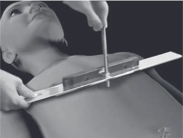
two measurements suffice: measurement A, defined as the greatest anteroposterior distance (Figure 1); and measurement B, which is the greatest depth of the deformity (Figure 2). Patients with an PEX-AI under 0.12 are considered normal, whereas those who present PEX-AI values equal to or greater than 0.12 are considered to have PEX. This index is easily obtained through physical examination of a patient in a medical office, i.e. the PEX-AI does not depend on radiologic or imaging exams, which implies a reduction in costs and morbidity. In addition to evaluating the deformity, the PEX-AI can assess the
surgical results irrespective of the operatory tech-nique used.
We herein report two cases of patients who were assessed using the PEX-AI and submitted to the minimally invasive Nuss technique.(5,6)
Case reports
The first patient, case 1, was a 14-year-old white male. The second patient, case 2, was a 20-year-old white male. Both presented normal cardiac and pulmonary function without any symptoms or prior clinical complaints. Therefore, the surgical procedure was recommended with the objective of minimizing the structural alterations of the anterior chest wall and facilitating the social inclusion of the patients. In case 1, the parents considered their son incapable of carrying out normal physical and social activi-ties for his age, and the patient therefore presented psychological alterations.
In both cases, the deformity was the most pronounced in the caudal portion of the sternum. There were no evident alterations of the pectoral musculature or sternal notch. The deformed position of the costal arches was considered compensatory and was responsible for the smaller anteroposte-rior diameter of the chest cavity. The patients were examined on two occasions: on the day of the oper-ation; and on post-operative day 60. The clinical measurements for obtaining the PEX-AI were taken with the patient in the horizontal dorsal decubitus position on a flat table that was parallel to the floor. Each measurement was taken during a deep
Figure 1 - Measurement A = distance between the
horizontal surface supporting the patient and the highest plane of the rib cage.
Figure 2 - Measurement B = distance from the point of
Application of the anthropometric index for the assessment of Pectus excavatum in patients submitted to the Nuss technique: two cases
J Bras Pneumol. 2007;33(3):347-350 349
inspiration. The numeric value of the ratio of the second measurement to the first provides the index: PEX–AI = B/A. Therefore, the index varies between 0 and 1. The lower the value, the lower the degree of deformity.
The surgical technique developed by Nuss(6) for the
treatment of PEX is described as minimally invasive. Using videothoracoscopy under direct vision, a curved metal bar (molded in accordance to the deformity of each patient) is inserted into the lateral chest wall from right to left and fixed in retrosternal position at the point of greatest deformity. After its introduc-tion, the bar is rotated 180° so that the curvature of the bar and that of the anterior chest wall coincide. Subcutaneous metal stabilizers at the ends of the bar maintain it in the correct position until its removal, which typically occurs after 36 months.
General anesthesia should always be performed using a peridural catheter for continuous infusion. The catheter should be left in place at least until post-operative day 3 in order to avoid intense pain and facilitate the respiratory therapy of the patients. The mean duration of surgery was 90 min. The patients remained hospitalized for five days. They spent the first 24 h after the surgery in an intensive care unit to guarantee appropriate monitoring of vital signs and analgesia.
Table 1 shows the pre- and post-opera-tive measurements of the patients, together with the respective indices. The proposed treatment corrected the deformity of the anterior chest wall in both cases.
Discussion
The pre- and post-operative PEX-AI values show the reduction of the sternal deformity in both patients after surgical treatment with the Nuss technique, and the thoracic contours assumed an appearance esthetically closer to normality. It is of note that the measurements that served as the basis
for calculating the PEX-AI were obtained from the external surface of the chest wall, i.e. they correlate more closely with the appearance of the patients, which makes the deformity data more reliable and consistent with the observations of doctors, family and the patient himself. Therefore, the esthetic deformity is not underestimated or maximized as occurs when we take into consideration measure-ments derived from X-rays or computed tomography scans of the chest which take into consideration the bone surfaces (internal) represented in the exams. Therefore, the PEX-AI is essentially an objective method. However, it also takes into consideration the external appearance of the chest, which is quite important in a deformity affecting a region of the body in which appearance is highly valued and directly related to patient quality of life.
By being an objective and numeric index obtained only through physical examination, it is possible for doctors in the same specialty or in corre-lated specialties to use it and easily comprehend the results. Therefore, it can be used to evaluate the results of a therapeutic intervention in a single patient, as well as to compare different groups oper-ated on in distinct departments, or even to make comparisons among patients submitted to different surgical techniques, in terms of outcomes.
This minimally invasive Nuss technique, performed with the aid of videothoracoscopy, was developed based on the fact that the chest wall (of children, adults and even the elderly) is malleable. The use of rigid metal orthopedic braces on long bones provides excellent esthetic and functional results. The principal advantages of this technique over the conventional (open) technique, which was developed by Robicsek,(1) are as follows: less surgical
trauma on the structures of the anterior chest wall, shorter duration of surgery, minimal blood loss and immediate esthetic results.
The principal criticism of the Nuss technique relates to the cost of the surgical material, the provisory (30-day) limitation of the movements of the chest cavity to avoid early dislocation of the bar, and the rigorous control of analgesia over the first few days after surgery.(7)
In order to identify acute complications, meas-urements were taken on postoperative day 60. The literature recommends maintaining the metal bars for at least 36 months in order to avoid recurrence of the deformity. Our patients were evaluated with
Table 1 - Measurements for the calculation of the
PEX-AI in both patients, taken on the day of surgery and on post-operative day 60.
B (cm) A (cm) IA = B/A Patient 1 Pre-operative 3.3 18.7 0.18
Post-operative 0 20.8 0
350 Brigato RR, Campos JRM, Jatene FB
J Bras Pneumol. 2007;33(3):347-350
the bars still in place, and our results are therefore preliminary. However, the presence of the bars does not prevent us from concluding that the outcomes of these two cases constitute a demonstration of the practical applicability of the PEX-AI.
Since surgical times and hospitalization stays are relatively shorter when the Nuss technique is employed, the total cost of the surgery can be equal to or even less than that of other techniques, such as the Ravitch and Robicsek techniques. Our two patients did not present any significant complaints or complications and expressed satisfaction with the immediate surgical results, as did the families.
References
1. Robicsek F, Daugherty HK, Mullen DC, Harbold NB Jr, Hall DG, Jackson RD, et al. Technical considerations in the surgical management of pectus excavatum and carinatum. Ann Thorac Surg. 1974;18(6):549-64.
2. Wu PC, Knauer EM, McGowan GE, Hight DW. Repair of pectus excavatum deformities in children: a new perspective of treatment using minimal access surgical technique. Arch Surg. 2001;136(4):419-24.
3. Fonkalsrud EW, Beanes S, Hebra A, Adamson W, Tagge E. Comparison of minimally invasive and modified Ravitch pectus excavatum repair. J Pediatr Surg. 2002;37(3):413-7. 4. Rebeis EB, Samano MN, Dias CTS, Fernandez A, Campos JRM,
Jatene FB, et al. Índice antropométrico para classificação quantitativa do pectus excavatum. J Bras Pneumol. 2004;30(6):501-7.
5. Molik KA, Engum SA, Rescorla FJ, West KW, Scherer LR, Grosfeld JL. Pectus excavatum repair: experience with standard and minimal invasive techniques. J Pediatr Surg. 2001;36(2):324-8.
6. Nuss D, Kelly RE Jr, Croitoru DP, Katz ME. A 10-year review of a minimally invasive technique for the correction of pectus excavatum. J Pediatr Surg. 1998;33(4):545-52.