*Correspondence: E.L. Souza. Departamento de Nutrição, Centro de Ciências da Saúde, Universidade Federal da Paraíba, Campus I, Cidade Universitária, 58051-900 - João Pessoa - PB, Brasil. E-mail: [email protected]
A
vol. 48, n. 4, oct./dec., 2012
Adhesion and biofilm formation by
Staphylococcus aureus
from
food processing plants as affected by growth medium, surface type
and incubation temperature
Heloísa Maria Ângelo Jerônimo, Rita de Cássia Ramos do Egypto Queiroga, Ana Caroliny Vieira
da Costa, Isabella de Medeiros Barbosa, Maria Lúcia da Conceição, Evandro Leite de Souza
*Laboratory of Food Microbiology, Department of Nutrition, Health Sciences Center, Federal University of Paraíba
This study assessed the effect of different growth media [BHI broth, BHI broth plus glucose (10 g/100 mL) and BHI broth plus NaCl (5 g/100 mL)] and incubation temperatures (28 or 37 ºC) on the adherence,
detachment and bioilm formation on polypropylene and stainless steel surfaces (2 x 2 cm coupons) for
a prolonged period (24-72 h) by some strains of Staphylococcus aureus (S3, S28 and S54) from food
processing plants. The eficacy of the sanitizers sodium hypochlorite (250 mg/mL) and peracetic acid (30 mg/mL) in reducing the number of viable bacterial cells in a preformed bioilm was also evaluated.
S. aureus strains adhered in highest numbers in BHI broth, regardless of the type of surface or incubation temperature. Cell detachment from surfaces revealed high persistence over the incubation period. The
number of cells needed for bioilm formation was noted in all experimental systems after 3 days. Peracetic acid and sodium hypochlorite were not eficient in completely removing the cells of S. aureus adhered onto polypropylene and stainless steel surfaces. From these results, the assayed strains revealed high
capacities to adhere and form bioilms on polypropylene and stainless steel surfaces under the different growth conditions, and the cells in bioilm matrixes were resistant to total removal when exposed to the
sanitizers sodium hypochlorite and peracetic acid.
Uniterms: Staphylococcus aureus/food processing. Bioilm. Surfaces. Sanitizers/eficacy.
Este estudo teve como objetivo avaliar o efeito de diferentes meios de crescimento [caldo BHI, caldo BHI adicionado de glucose (10 g/100 mL) e caldo BHI adicionado de NaCl (5 g/100 mL)] e temperaturas
de incubação (28 e 37 °C) sobre a adesão, separação e formação de bioilme sobre superfícies (2 x 2 cm) de polipropileno e aço inoxidável durante longo tempo de incubação (24-72 h) por parte de cepas
de Staphylococcus aureus (S3, S58 e S54) isoladas de plantas de processamento de alimentos. Também
foi avaliada a eicácia dos sanitizantes hipoclorito de sódio (250 mg/mL) e ácido peracético (30 mg/mL) na redução do número de células bacterianas viáveis presentes em um bioilme pré-formado. As cepas
de S. aureus aderiram em número mais elevado quando incubadas em caldo BHI em ambos os tipos de
superfícies e temperaturas de incubação testadas. A separação das células das superfícies revelou alta persistência ao longo do período de incubação. Número de células necessário para a formação do bioilme foi detectado depois de três dias de incubação em todos os sistemas experimentais. O ácido peracético e o hipoclorito de sódio não foram eicientes em remover completamente a células de S. aureus aderidas
sobre as superfícies de polipropileno e aço inoxidável. Os resultados obtidos revelaram alta capacidade das cepas ensaiadas em aderir e formar bioilme sobre superfícies de polipropileno e aço inoxidável sobre diferentes condições de crescimento e que as células na matriz do bioilme apresentaram-se resistentes à total remoção quando expostas aos sanitizantes hipoclorito de sódio e ácido peracético.
Unitermos: Staphylococcus aureus/ processamento de alimentos. Adesão. Superfícies. Bioilme.
INTRODUCTION
The relevance of contaminated surfaces in spreading pathogenic microorganisms to foods is already well estab-lished for food processing plants (Kusumaningru et al., 2003; Fuster-Valls et al., 2008). Some pathogenic bacteria are able to adhere to food-contact surfaces and remain viable even
after cleaning and disinfection (Ammor et al., 2004). One of
the most common ways for bacteria to live is adhering to
sur-faces and forming bioilms in which they are embedded in an organic, extracellular polymeric matrix (Chae, Schraft, 2000).
Bioilm-adhered bacteria can detach during process -ing and contaminate food as it passes the surfaces. This cross-contamination is a high-risk source of pathogenic bacteria and may seriously affect the safety and quality of foods (Bagge-Ravn et al., 2003).
Staphylococcus aureus is among the most common pathogenic bacteria isolated from surfaces in food process-ing plants (Pastoriza et al., 2002), where it can adhere and
form bioilms (Kunig, Almeida, 2001). Foodborne disease
caused by S. aureus is typically intoxication due to the
ingestion of enterotoxins preformed in food by enterotoxi -genic strains (Normanno et al., 2007). Most investigations regarding biofilm formation by staphylococci using S. epidermidis as the model microorganism have focused on clinical aspects in association with implants and medi-cal devices (Rode et al., 2007). Whereas the capacity for adhesion and biofilm formation of some food-related pathogenic bacteria has already been elucidated (Conlon
et al., 2002), studies focusing on the adhesion and bioilm
formation of S. aureus are still rare. Regarding these as-pects, it is important to identify the conditions under which
S. aureus is able to survive and multiply on food process-ing surfaces. Early studies found that bacterial adhesion
and bioilm formation depended upon the bacterial species,
the nature of the surface, the growth medium and other en-vironmental conditions (Pompermayer, Gaylarde, 2007). The present study aimed to evaluate the capability of strains of S. aureus from surfaces of food processing plants
to adhere and form bioilms on polypropylene and stain
-less steel surfaces when exposed to different growth media
and incubation temperatures. The study also assessed the effects of the sanitizers peracetic acid and sodium hypo-chlorite in reducing the number of viable bacterial cells
in a preformed bioilm.
MATERIAL AND METHODS
Test strains
S. aureus S3, S. aureus S28 and S. aureus S54
ob-tained from the Microorganism Collection, Laboratory of Food Microbiology, Health Sciences Center, Federal University of Paraíba (João Pessoa, Brazil), were used as test microorganisms. These strains were isolated from dif-ferent surfaces of Food Processing Plants using standard
procedures. Stock cultures were kept on Nutrient Agar (NA) (Difco, Brazil) slants under refrigeration (7 ± 1 ºC).
Unless otherwise stated, inocula (10 log cfu/mL) used in assays were obtained from suspensions of the strains in the stationaryphase of growth and prepared fol-lowing a previously described procedure (Carson et al., 2002). The suspensions obtained were serially diluted in PBS (10-1 - 10-2) to provide a viable cell count of approxi -mately 8.0 log cfu/mL.
Surfaces
AISI 304 stainless steel (2 x 2 x 0.2 cm) and polypro
-pylene coupons (2 x 2 x 0.4 cm) were used as test surfaces.
The coupons were individually cleaned, sanitized and sterilized according to a previously described procedure (Marques et al., 2007).
Experimental conditions
The adherence, detachment and biofilm
forma-tion of the test strains were assessed under six different experimental conditions: i) in the presence of a
nutrient-rich medium using Brain Heart Infusion Broth (BHI) at 28 ºC; ii) in the presence of a nutrient-rich medium (BHI) at 37 ºC; iii) in the presence of a nutrient-rich and adhesion-promoting medium, BHI supplemented with 10 g/L of glucose (BHI-Glucose) at 28 ºC; iv) in the presence of a nutrient-rich and adhesion-promoting medium (BHI-Glucose) at 37 ºC; v) in the presence of a selective medium, BHI supplemented with 5 g/Lof NaCl (BHI-NaCl) at 28 ºC; and vi) in the presence of a selective medium (BHI-NaCl) at 37 ºC.
Adhesion to surfaces and quantification of adhered cells
An aliquot of 100 µL of the growth media was mixed with 50 µL of the bacterial inoculum, plated onto the center
of each coupon and incubated under the pre-established
temperatures. After 24, 48 and 72 h of incubation, coupons
SPW with vigorous vortexing for 30 s. The mixture was
serially diluted (10-1-10-5) in SPW, and aliquots of 100 µL
were spread plated onto sterile NA plates. The plates were
incubated for 24 h at 37 ºC (Herrera et al., 2007; Rode et al., 2007). After the incubation period, the number of vi
-able cells was counted, and the results were expressed as
Log cfu/cm2.
Detachment of adhered cells
An aliquot of 100 µL of the growth media was mixed with 50 µL of the bacterial inoculum and plated
onto the center of each coupon, followed by incubation
under the pre-established temperatures. After 24, 48 and
72 h of incubation, coupons (two for each treatment) were withdrawn and immersed in SPW for 15 s to release
non-adhered cells. Each coupon was placed onto a sterile NA
plate, and after 2 min, removed and placed onto a second
sterile NA plate. This procedure was repeated through 7 sterile NA plates. The number of detached cells on the NA plates with order number of 1, 2, 3, 4, 5, 6 and 7 was
found by transferring the agar blotting from each plate to 10 mL of SPW followed by blending using a Stomacher.
The mixture was serially diluted (10-1-10-5) in SPW, and
aliquots of 100 µL were spread plated onto NA plates.
The plates were incubated for 24 h at 37 ºC (Herrera et al.,
2007). After the incubation period, the number of viable cells was counted and the results expressed in Log cfu/cm2.
Biofilm development and quantification
The level of bioilm formation by S. aureus S3 on polypropylene and stainless steel surfaces incubated in vegetable broth at 7 ºC and 28 ºC over 15 days was assessed.
For this measurement, ive stainless and ive polypropylene
coupons were immersed in sterile Petri dishes containing 20 mL of the growth media and 2 mL of the bacterial inoculum. The Petri dishes were sealed and incubated statically at the
pre-established temperatures. After 3, 6, 9, 12 and 15 days
of incubation, the coupons were withdrawn and washed
with SPW to remove the non-adherent cells. Once again,
the coupons were immersed in a fresh medium containing the same amount of inoculum, and the process was repeated four times over a 15-day period.
At each incubation interval, two coupons from each treatment were submitted to bioilm matrix bacterial counting. For this counting, each bioilm was scraped with
two moistened sterile swabs and resuspended in 9 mL of
SPW with vortexing for 30 s. Serial dilutions were pre
-pared in SPW, and aliquots of 100 µL were spread plated onto sterile NA plates followed by incubation at 37 ºC for
24 h (Marques et al., 2007). After the incubation period,
the number of viable cells was counted and the results
expressed as Log cfu/cm2.
Sanitizer application
The eficacies of the sanitizers sodium hypochlorite
(250 mg/L) and peracetic acid (30 mg/L) (Meira et al., 2012) in removing the cells of S. aureus S3 from the
bio-ilm matrix grown in the vegetable-based broth at 7 and 28 ºC were assessed. For this assessment, ive coupons were allowed to develop bioilms according to the experimen
-tal conditions cited above. After 15 days of incubation,
the coupons were washed in SPW and immersed for 30 s in sterile Petri dishes containing 20 mL of the sanitizer
solution. Afterwards, the coupons were withdrawn from
the sanitizer solution and immersed for 3 s in a neutral-izing solution (0.1 M Na2S2O3). The remaining cells were counted after scraping with two sterile moistened swabs
and resuspended by vigorously vortexing in 9 mL of SPW.
Serial dilutions were prepared in SPW, and aliquots of 100
µL were spread plated onto NA plates and incubated at 37 ºC for 24 h (Ammor et al., 2004). After the incubation pe -riod, the number of viable cells was counted and the results
expressed as Log cfu/cm2. In control assays, the sanitizer solutions were replaced by sterile distilled water. The
ef-iciency of each sanitizer was calculated as the difference
between the counts obtained for the control surfaces and
for the surfaces exposed to the sanitizers.
Reproducibility and statistical analysis
All analyses were carried out in triplicate, and the results are expressed as the average of the assays. Counts
were converted to decimal Logarithmic values (Log cfu/ cm2) to nearly match the assumption of a normal distri-bution. Counts obtained for adhesion, detachment and
bioilm formation were submitted to Analysis of Variance (ANOVA) followed by the Duncan test to determine the signiicance of the inluences of the incubation tempera -tures and contact surfaces. Counts obtained for tests of the effects of the sanitizers (before and after the application)
on the biofilm matrix were compared using the paired
Student´s t-test. Data were analyzed using the software
Statistica 7. A probability value P <0.05 was accepted
as indicating signiicant differences (Meira et al., 2012).
RESULTS AND DISCUSSION
growth media (nutrient rich-media, with or without added glucose and NaCl) and incubation temperatures (28 and
37 ºC) were tested to determine their possible inluences on cell adherence and bioilm formation by isolates of S. aureus on polypropylene and stainless steel surfaces. The temperatures 37 ºC and 28 ºC were chosen as the ideal temperature for the growth of S. aureus and the common environmental temperature found in Brazilian Food and Nutrition Services, respectively.
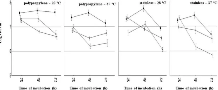
The numbers of S. aureus cells adhered to poly-propylene and stainless steel surfaces under different
experimental conditions over 72 h of incubation are shown
in Figures 1-3. The highest numbers of adhered cells
(6 – 8 log cfu/cm2) on polypropylene and stainless steel surfaces were found when the strains were cultivated in BHI. There was no indication that adherence increased in BHI-NaCl or BHI-Glucose media. It appears surprising that the presence of glucose and NaCl in the growth media caused no increase in the adhesion capacity of the assayed strains. Moretro et al. (2003) found that the presence of NaCl (2 g/100 mL) in tryptic soy broth (TSB) resulted
in increased adhesion and bioilm formation by standard
cultures of staphylococci from food and food processing environments. Herrera et al. (2007) also noted the same behavior for a standard cultureof S. aureus cultured in TSB supplemented with glucose (1 g/100 mL).
S. aureus S3 and S54 cultured in BHI at both tested temperatures revealed a clear two-phase adhesion pat-tern regardless of the surface type: an initial phase with a progressively increasing numbers of adhered cells, with highest counts after 48 h of incubation, followed by a
second phase (72 h) with a decreasing number of adhered cells. These results suggest that under static conditions, the adhered cells may be present in high numbers, but the number of adhered cells do not constantly increase over the incubation time.
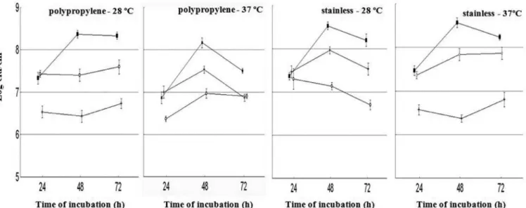
S. aureus S54 presented a different pattern of
adhe-sion in most experimental systems, with a decrease in the
numbers of adhered cells over the evaluated time intervals at 28 and 37 ºC. Data for the number of adhered cells on stainless steel and polypropylene surfaces did not differ (P > 0.05) for strains S3 and S28 in all cultivation media at 37 ºC. For strain S54, there were greater numbers (P < 0.05) of adhered cells on stainless steel surfaces than on polypropylene in BHI and BHI-glucose.
Regarding the effect of the incubation temperature on the adherence capability of the tested strains, it was surprising to note that no difference (P > 0.05) was found between 28 ºC and 37 ºC. Some previous studies have reported a positive effect of lower temperature on the adhesion pattern of S. aureus. Herald and Zottola (1988) noted that Listeria monocytogenes and Yersinia enteroco-litica cultivated in laboratorial media adhered to stainless surfaces in greater numbers at 21 ºC than at 30 ºC. Rode
et al. (2007) found higher attachment capacity for S. au-reus on polystyrene when cultivated in tryptic soy broth at sub-optimal temperatures (20, 25 and 30 ºC). Morton et al. (1998) reported that regardless of the species or surface
assayed, the adhesion process occurs at maximum inten -sity when microorganisms are allowed to grow at their optimum temperatures.
Among the tested strains, S. aureus S3 was used for
FIGURE 1 - Adhesion of S. aureus S3 to polypropylene and stainless steel surfaces as affected by different experimental conditions
FIGURE 2 - Adhesion of S. aureus S28 to polypropylene and stainless steel surfaces as affected by different experimental conditions
(■: BHI; ◊: BHI-Glucose; +: BHI-NaCl) over 72 h of incubation.
FIGURE 3 - Adhesion of S. aureus S54 to polypropylene and stainless steel surfaces as affected by different experimental conditions
(■: BHI; ◊: BHI-Glucose; +: BHI-NaCl) over 72 h of incubation.
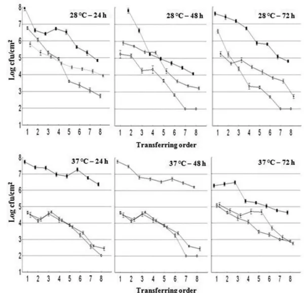
further assays of detachment and bioilm formation. Data
for cell detachment from polypropylene and stainless steel surfaces for S. aureus S3 under different experimental
conditions are shown in Figures 4 and 5, respectively. Bacterial counts revealed a linear decrease in the
detach-ment rate over the contact sequence for all experidetach-mental systems. Regarding the inluence of the growth media,
the results obtained showed higher detachment values (P
< 0.05) when the strain was grown in BHI compared to
other media. In all experimental systems, the cell detach -ment was at least 3 Log cfu/cm2 during the irst 5 contacts (blots), suggesting high persistence of cells on the surfaces
over 72 h. No signiicant inluence (P > 0.05) of the surface
type or time of incubation was noted on the detachment rate. The highest numbers of detached cells were observed when S. aureus S3 was incubated in BHI, which could be related to increased bacterial growth on this substrate relative to the other growth media. These data about the detachment of cells over a large number of contacts with blot agar reveal a high risk of dissemination for S. aureus
in food processing plants.
Levels of biofilm formation by S. aureus S3 on polypropylene and stainless surfaces over 15 days under
different experimental conditions were also evaluated. For most systems, the number of cells in the bioilm matrix
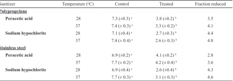
TABLE I - Effect of peracetic acid (30 mg/L) and sodium hypochlorite (250 mg/L) on counts (Log cfu/cm2) of S. aureus S3 (grown
in BHI broth at 28 and 37 °C) adhered to polypropylene and stainless surfaces
Sanitizer Temperature (oC) Control Treated Fraction reduced
Polypropylene
Peracetic acid 28 7.3 (±0.3) a 3.8 (±0.2) b 3.5
37 7.4 (± 0.3) a 3.3 (± 0.2) b 4.1
Sodium hypochlorite 28 7.1 (±0.4) a 2.7 (±0.3) b 4.4
37 7.4 (± 0.4) a 2.6 (± 0.3) b 4.8
Stainless steel
Peracetic acid 28 6.9 (±0.2) a 4.1 (±0.2) b 2.8
37 7.7 (± 0.2) a 4.2 (± 0.4) b 3.6
Sodium hypochlorite 28 6.9 (±0.4) a 2.6 (±0.4) b 4.3
37 7.7 (± 0.3) a 3.1 (± 0.3) b 4.6
Values followed by the same letters in each line differ signiicantly (p<0.05) according to the Student t test.
FIGURE 4 - Detachment of S. aureus S3 of polypropylene surfaces as affected by different experimental conditions (■: BHI;
FIGURE 5 - Detachment of S. aureus S3 of stainless steel surfaces as affected by different experimental conditions (■: BHI;
◊: BHI-Glucose; +: BHI-NaCl).
periods (data not showed ). For most experimental condi
-tions, the maximum number of cells (approximately 6 log
cfu/cm2) was found after 6 days of incubation. The bacte
-rial counts indicated bioilm formation on both surfaces under all experimental systems after only 3 days. Greater than 6 – 7 log cfu/cm2 of viable cells are needed for bioilm formation, and lower counts could indicate an adhesion process (Planchon et al., 2006).
Counts of S. aureus S3 cells adhered to polypro-pylene and stainless surfaces before and after application of peracetic acid (30 mg/L) and sodium hypochlorite are shown in Table I. The decrease in cell counts caused by
sodium hypochlorite (250 mg/L) was 2.6–3.1 Log cfu/
cm2, while for peracetic acid, the decrease was 3.3 – 4.2 Log cfu/cm2. However, in all experimental systems, both sanitizers greatly decreased (P < 0.05) the counts of cells
adhered to the assayed surfaces. According to the results of these experiments, the sanitizers peracetic acid and
sodium hypochlorite, in the concentrations assayed, were
not eficient in completely removing the cells of S. aureus
that had formed bioilms on polypropylene and stainless
steel surfaces. Residual cells adhered to the surfaces after the application of sanitizers reinforce the concept of
bio-ilm as a substantial source of cross contamination in food
processing plants.
CONCLUSIONS
The results of this study have clear implications for designing strategies to control cross-contamination in food processing environments because the strains used as test organisms were isolated directly from food-contact
surfaces and the assays for adherence and bioilm forma -tion were carried out using surfaces commonly found in food processing plants. The strains assayed here revealed
-sayed surfaces when exposed to different environmental
conditions, suggesting that the clumping phenotype (ag-gregation) of the tested strains did not show any clear
in-luence of the surrounding environment with regard to the
surface type, growth media or temperature of incubation.
REFERENCES
AMMOR, S.; CHEVALLIER, I.; LAGUE, A.; LABADILE, J.; TALON, R.; DUFOUR, E. Investigation of the selective
bactericidal effect of several decontaminating solutions on bacterial biofilms including useful, spoilage and/or
pathogenic bacteria. Food Microbiol., v.21, n.1, p.11-17,
2004.
BAGGE-RAVN, D.; GARDSHODN, K.; GRAM, L.; VOGEL,
B.F. Comparison of sodium dypochlorite-based foam and
peroxyacetic acid-based fog sanitizing procedures in a salmon smokehouse: survival of the general micolora and
Listeria monocytogenes. J. Food Protect., v.66, n.4, p.592-598, 2003.
CARSON, C.F.; MEE, B.J.; RILEY, T.V. Mechanism of action
of Melaleuca alternifolia (Tea tree) oil on Staphylococcus aureus determined by time-kill, lysis, leakage, and salt
tolerance assay and electron microscopy. Antimicrob.
Agents Chemother., v.46, n.6, p.1914-1920, 2002.
CHAE, M.S.; SCHRAFT, H. Comparative evaluation of
adhesion and biofilm formation of different Listeria
monocytogenes strains. Int. J. Food Microbiol., v.62, n.1-2, p.103-111, 2000.
CONLON, K.M.; HUMPREYS, H.; O`GARA, J.P. içaR encodes a transcription repressor involved in environmental
regulation of ica operon expression and bioilm formation
in Staphylococcus epidermidis. J. Bacteriol., v.184, n.16, p.4400-4408, 2002.
FUSTER-VALLS, N.; HERNÁNDEZ-HERRERO, M.; MARÍN-DE-MATEO, M.; RODRÍGUEZ-JEREZ, J.J.
Effect of different environmental conditions on the bacterial
survival on stainless steel surfaces. Food Control., v.19, n.3,
p.308-314, 2008.
HERALD, P.J.; ZOTTOLA, E.A. Scanning electron microscopic examination of Yersinia enterocolitica attached to stainless
steel at selected temperature and pH values. J. Food Prot.,
v.51, n.6, p.445-448, 1988.
HERRERA, J.J.R.; CABO, M.L.; GONZÁLEZ, A.; PAZOS, I.; PASTORIZA, L. Adhesion and detachment kinectics
of several strains of Staphylococcus aureus subsp. aureus
under three different experimental conditions. Food Microbiol., v.24, n.6, p.585-591, 2007.
KUNIGK, L.; ALMEIDA, M.C.B. Action of peracetic acid on
Escherichia coli and Staphylococcus aureus in suspension
or settled on stainless steel surfaces. Braz. J. Microbiol.,
v.32, n.1, p.38-41, 2001.
KUSUMANINGRU, H.D.; RIBOLDI, G.; HAZELEGER,
W.C.; BEUMMER, R.R. Survival of food-borne pathogens on stainless steel surfaces and cross-contamination to foods. Int. J. Food Microbiol., v.85, n.3, p.227-236, 2003.
MARQUES, S.C.; REZENDE, J.G.O.S.; ALVES, L.A.F.; SILVA, B.C.; ALVES, E.; ABREU, L.R.; PICCOLI, R.H. Formation of bioilm by Staphylococcus aureus on stainless steel and glass surfaces and its resistance to some selected
chemical sanitizers. Braz. J. Microbiol., v.38, n.3,
p.538-543, 2007.
MEIRA, Q.G.M.; BARBOSA, I.M.; ATAHAYDE, A.J.A.A.; SIQUEIRA-JÚNIOR, J.P.; SOUZA, E.L. Influence of
temperature and surface kind on biofilm formation by Staphylococcus aureus from food-contact surfaces and
sensitivity to sanitizers. Food Control., v.25, n.1,
p.469-475, 2012.
MORETRO, T.; HERMANSEN, L.; HOLCK, A.L.; SIDHU, M.S.; RUDI, K.; LANGSRUD, S. Bioilm formation and
the presence of the intercellular adhesion locus ica among Staphylococci from food and food processing environments. Appl. Environ. Microbiol., v.69, n.9, p.5648-5655, 2003.
MORTON, L.H.G.; GREENWAY, D.I.A.; GAYLARDE, C.C.; SURMAN, S.B. Consideration of some implications of the resistance of bioilms to biocides. Int. Biodeter. Biodegrad., v.41, n.3-4, p.247-259, 1998.
NORMANNO, G.; LA SALANDRA, G.; DAMBROSIO, A.; QUAGLIA, N.C.; CORRENTE, M.; PARISI, A.; SANTAGADA, G.; FIRINU, A.; CIRSETTI, E.; CELANO, G.V. Occurrence, characterization and antimicrobial resistance of enterotoxigenic Staphylococcus aureus
isolated from meat and dairy products. Int. J. Food
PASTORIZA, L.; CABO, M.L.; BERNÁRDEZ, M.; SAMPEDRO, G.; HERRERA, J.R. Combined effects of
modified atmosphere packaging and lauric acid on the
stability of pre-cooked ish products during refrigerated
storage. Eur. Food Res. Technol., v.215, n.3, p.189-193,
2002.
PLANCHON, S.; GAILLARDE-MARTINIE, B.; DORDET-FRISONI, E.; BELLON-FONTAINE, M.N.; LEROY, S.; LABADIE, J.; HEBRAUD, M.; TALON, R. Formation
of biofilm by Staphylococcus xylosus. Int. J. Food
Microbiology, v.109, n.1-2, p.88-96, 2006.
POMPERMAYER, D.M.C.; GAYLARDE, C. The inluence of temperature on the adhesion of mixed cultures
of Staphylococcus aureus and Escherichia coli to
polypropylene. Food Microbiol., v.17, n.4, p.361-165, 2007.
RODE, T.M.; LANGSRUD, S.; HOLCK, A.; MORETRO, T. Different patterns of bioilm formation in Staphylococcus aureus under food-related stress conditions. Int. J. Food Microbiology, v.116, n.3, p.372-383, 2007.
Received for publication on 20th March 2012