Biofilm formation by
Staphylococcus aureus
from food contact surfaces in a
meat-based broth and sensitivity to sanitizers
Evandro Leite de Souza
1, Quênia Gramile Silva Meira
1, Isabella de Medeiros Barbosa
1,
Ana Júlia Alves Aguiar Athayde
1, Maria Lúcia da Conceição
1, José Pinto de Siqueira
Júnior
21Laboratório de Microbiologia de Alimentos, Departamento de Nutrição, Centro de Ciências da Saúde,
Universidade Federal da Paraíba, João Pessoa, PB, Brazil.
2Laboratório de Genética de Microrganismos, Departamento de Biologia Molecular, Centro de Ciências
Exatas e da Natureza, Universidade Federal da Paraíba, João Pessoa, PB, Brazil.
Submitted: July 26, 2012; Approved: September 9, 2013.
Abstract
This study assessed the capacity of adhesion, the detachment kinetic and the biofilm formation by Staphylococcus aureusisolated from food services on stainless steel and polypropylene surfaces (2 x 2 cm) when cultivated in a meat-based broth at 28 and 7 °C. It was also to study the efficacy of the sanitizers sodium hypochlorite (250 mg/L) and peracetic acid (30 mg/L) in inactivating the bacterial cells in the preformed biofilm.S. aureusstrains adhered in high numbers regardless the assayed sur-face kind and incubation temperature over 72 h. Cells detachment of sursur-faces revealed high persis-tence over the incubation period. Number of cells needed for biofilm formation was noted at all experimental systems already after 3 days. Peracetic acid and sodium hypochlorite were not efficient in completely removing the cells ofS. aureusadhered on polypropylene and stainless steel surfaces. From these results, the assayed strains revealed high capacity to adhere and form biofilm on poly-propylene and stainless steel surfaces under different growth conditions. Moreover, the cells in biofilm matrix were resistant for total removal when submitted to the exposure to sanitizers.
Key words: Staphylococcus aureus, food processing environment, cross-contamination, biofilm,
sanitizers.
Introduction
The surfaces that come into contact with foods are important sources for the transmission of microorganisms in food processing plants (BARNESet al., 1999). Many pathogenic or spoilage bacteria can be found attached to surfaces in the form of planktonic cells or sessile cells forming a biofilm (Bragaet al., 2005). Biofilms are aggre-gates of microbial cells surrounded by a matrix of exopoly-mers, which confers resistance to these microorganisms (Costerton et al., 1999). Bacteria that aggregate to form biofilms are known to possess greater resistances to stress conditions than their planktonic counterparts, which are dispersed in the environment, including the susceptibility to sanitizers and other antimicrobials (Fux et al., 2004).
Early studies have found that bacterial adhesion and bio-film formation depend on the bacterial species, the nature of the surface, the growth medium and other environmental conditions (Malheiroset al., 2010; Oulahalet al., 2008).
To remove biofilm organisms, the solution of saniti-zation must penetrate the matrix of exopolymers and gain access to the microbial cells causing biofilm inactivation and removal. The oxidizing substances like chlorine or peracetic acid are frequently used for sanitizations of food processing plants. Peracetic acid may be used as a disinfec-tant because this agent decomposes into safe and environ-mental friendly residues (acetic acid and hydrogen perox-ide), hence it can be applied without rinsing risks for food contamination by toxic residues and its efficacy is not af-fected by protein residues. Sodium hypochlorite (NaClO) is
Send correspondence to E.L. Souza. Departamento de Nutrição, Centro de Ciências da Saúde, Universidade Federal da Paraíba, Campus I, 58051-900 Cidade Universitária, João Pessoa, PB, Brasil. E-mail: [email protected].
a chemical compound used for sanitization of food-contact surfaces (Meyer, 2003; Sreyet al., 2012).
The formation of biofilms on the surfaces of food pro-cessing plants by pathogenic bacteria is a high risk source of cross-contamination because biofilm-adhering bacteria can detach during production and contaminate food as it passes over these surfaces.Staphylococcus aureushas been frequently found in food processing plants and is recog-nized as being responsible for outbreaks related to the con-sumption of fresh and processed foods worldwide (Rodeet al., 2007).
The pathogenesis of the intoxication caused by S. aureusdepends on the ability of the strain to survive, multi-ply under a variety of conditions and produce various extra-cellular compounds (Carsonet al., 2002). The capability of S. aureusto adhere to and form biofilms enhances its sur-vival and growth in food processing plants, providing a physiological advantage to S. aureus as the etiological agent of foodborne diseases (Herrera et al., 2007). Al-though some researchers have studied the ability of mem-bers of theStaphylococcus genera to adhere to and form biofilms (Kuzmanet al., 2007; Møretroet al., 2003), most studies have addressed the clinical aspects related to bio-film formation byS. intermediuson medical implants and materials. Moreover, the few studies regarding the ability ofS. aureusto form biofilms have employed standard type strains inoculated into synthetic media, and little is known about the ability of wild strains ofS. aureusto adhere to and form biofilms when they are exposed to conditions simulat-ing those environments found in food processsimulat-ing plants.
With regard to these aspects, this study assessed the capability ofS. aureuscells from food processing plants to adhere to and form biofilms on polypropylene and stainless steel surfaces when cultivated in a meat-based broth under different temperatures. In addition, this study aimed to ob-serve the effect of sanitizers on the reduction of viable cells in a biofilm matrix.
Materials and Methods
Test strains
S. aureusS3,S. aureusS28 andS. aureusS54 were obtained from the Microorganism Collection, Laboratory of Food Microbiology, Health Sciences Center, Federal University of Paraíba (João Pessoa, Brazil) and were used as the test microorganisms. These strains were isolated us-ing standard procedures (Vanderzant and Splittsstoesser, 1992; Downes and Ito, 2001) from different surfaces of food processing plants of Food and Nutrition Services in the city of João Pessoa (Paraíba, Brazil). Stock cultures were maintained on nutrient agar (NA; Difco, Brazil) slants under refrigeration (7±1 °C).
Unless otherwise stated, the inocula used in the as-says were obtained from cell suspensions grown to station-ary phase and prepared by inoculating two colonies of each
bacterium from overnight cultures, which were grown on Brain-heart infusion agar (Himedia, India), into 400 mL of Braheart infusion broth (Himedia, India), which was in-cubated at 37 °C for 18 h. After incubation, the bacteria were separated from the growth medium by centrifugation (10,000 xg) for 12 min at 4 °C, washed twice with phos-phate-buffered saline(PBS; pH 7.4), and resuspended in PBS. Suspensions were adjusted so that the optical density at 620 nm (OD620) of a 1-to-100 dilution was approximately 0.3, which was approximately 10 log colony forming units per milliliter (10 log cfu/mL) (Carsonet al., 2002). The sus-pensions were serially diluted in PBS (10-1-10-2) to provide a viable cell count of approximately 8.0 log cfu/mL.
Test surfaces and experimental conditions
AISI 304 stainless steel (2 x 2 x 0.2 cm) and poly-propylene coupons (2 x 2 x 0.4 cm) were used as the test surfaces. The coupons were individually cleaned, sanitized and sterilized according to a previously described proce-dure (Marqueset al., 2007).
The adherence, detachment and biofilm formation of the test strains on the polypropylene and stainless steel sur-faces, when inoculated into a meat-based broth, were as-sessed at two different incubation temperatures, 7 and 28 °C.
Preparation of the meat-based broth
Bovine meat steaks were trimmed of all external fat and cut into pieces of a uniform size (3 x 3 x 3 cm). The meat pieces were boiled in distilled water for 20 min. Ap-proximately 500 mL of meat broth was obtained and vac-uum filtered using Whatman nº 1 filter paper, and the filtrate was sterilized by autoclavation for 15 min (1.21 atm). The obtained broth was stored at -20 °C in aliquots of 50 mL, and when required, one aliquot was thawed under refrigeration (7±1 °C) and used for the
ex-perimental analysis.
The meat broth was characterized with regard to its physico-chemical characteristics (acidity, moisture, pro-tein, fat, carbohydrate, mineral content and pH value) ac-cording to standard procedures. The physico-chemical characteristics for the meat broth used in the assays are shown in Table 1.
Adhesion to surfaces and quantification of adhered cells
An aliquot of 100mL of the growth media was mixed
into 50mL of the bacterial inoculum (initial amount of
with two moistened swabs, which were resuspended in SPW by vigorously vortexing for 30 s. The mixture was se-rially diluted (10-1-10-5) in SPW, and aliquots of 100
mL
were spread plated onto sterile NA plates. The plates were incubated for 24 h at 37 °C (Herreraet al., 2007; Rodeet al., 2007). After the incubation period, the number of viable cells was counted, and the results were expressed in log cfu/cm2.
Detachment of adhered cells
An aliquot of 100mL of the growth media was mixed
into 50mL of the bacterial inoculum (initial amount of
bac-terial cells in the system, 106cfu) and plated onto the center of each coupon, followed by incubation under the pre-established temperatures. After 24, 48 and 72 h of incuba-tion, the coupons (four for each treatment) were withdrawn and immersed in SPW for 15 s to release the non-adherent cells. Each coupon was placed on a sterile NA plate and, af-ter 2 min, was removed and placed onto a second saf-terile NA plate. This procedure was repeated through 7 sterile NA plates. The numbers of detached cells on the NA plates, us-ing an order number of 1, 2, 3, 4, 5, 6, and 7, were found by transferring the agar blotted from each plate to 10 mL of SPW followed by blending using a stomacher. The mixture was serially diluted (10-1-10-5) in SPW, and aliquots of 100mL were spread plated onto NA plates. The plates were
incubated for 24 h at 37 °C (Herreraet al., 2007). After the incubation period, the number of viable cells was counted, and the results were expressed in log cfu/cm2.
Biofilm development and quantification
The level of biofilm formation by S. aureusS3 on polypropylene and stainless steel surfaces incubated in meat broth at 7 °C and 28 °C over 15 days was assessed. For this experiment, eight stainless coupons and eight poly-propylene coupons were immersed in sterile petri dishes containing 20 mL of the growth media and 2 mL of the bac-terial inoculum (initial inoculum, approximately 107 cfu/mL of meat broth). The petri dishes were sealed and statically incubated at the pre-established temperatures. Af-ter 3, 6, 9, 12 and 15 days of incubation, the coupons were withdrawn and washed with SPW to remove the non-adherent cells. The coupons were then again immersed in
fresh media containing the same amount of inoculum. Be-cause the process was repeated four times, the experiment required a 15-day period.
At each incubation interval, four coupons for each treatment were submitted for bacterial counts from their biofilm matrices. For these counts, the biofilm was scraped with two moistened sterile swabs, which were resuspended in 9 mL of SPW by vortexing for 30 s. Serial dilutions were prepared in SPW, and aliquots of 100 mL were spread
plated onto sterile NA plates and then incubated at 37 °C for 24 h (Marqueset al., 2007; Meiraet al., 2012). After the in-cubation period, the number of viable cells was counted, and the results were expressed in log cfu/cm2.
Sanitizer application
The efficacy of the sanitizers sodium hypochlorite (25 mL/L) and peracetic acid (30 g/L), which are the con-centrations these sanitizers commonly applied for disin-fecting surfaces in food processing plants, were assessed for their ability to removeS. aureusS3 cells from a biofilm matrix grown in the meat-based broth at 7 and 28 °C. For this experiment, five coupons were allowed to develop biofilms under the experimental conditions cited above. After 15 days of incubation, the coupons were washed in SPW and immersed for 30 s in sterile petri dishes contain-ing 20 mL of the sanitizer solution (18). The coupons were then withdrawn from the sanitizer solution and immersed for 3 s in a neutralizing solution (0.1 M Na2S2O3). The re-maining cells were counted after scraping, which employed two sterile moistened swabs. The swabs were then vigor-ously resuspended by vortexing them in 9 mL of SPW. Se-rial dilutions were prepared in SPW, and aliquots of 100mL
were spread plated onto NA plates and incubated at 37 °C for 24 h (Ammoret al., 2004). After the incubation period, the number of viable cells was counted, and the results ex-pressed in cfu/cm2. In control assays, the sanitizer solutions were replaced with sterile distilled water. The efficiency of each sanitizer was calculated with regard to the difference between the counts obtained for the control surfaces and for the surfaces exposed to the sanitizers.
Procedures for scanning electron microscopy
Samples were submitted to biofilm formation on the polypropylene and stainless steel surfaces (28 °C), accord-ing to the already cited procedure, and after exposure to the sanitizers or distilled water, the samples were observed for remaining viable cells in the biofilm matrix using scanning electron microscopy. The coupons were pre-fixed with glutaraldehyde (2 mL/100 mL) for 2 h at 4 °C and post-fixed with osmium tetroxide (2 g/100 mL) for 30 min at 30 °C. Afterward, the cells were washed twice with PBS, dried at a critical point in liquid CO2under 95 bar of pres-sure, gold covered (fine coat ion sputter JFC-1100, JEOL Ltd., Tokyo, Japan) and examined using a scanning
elec-Table 1- Physico-chemical characteristics of meat-based broth.
Physico-chemical parameters Amounts
Protein 0.96 g/100 g
Fat 0.43 g/100 g
Moisture 97.75 g/100 g
Carbohydrates 0.65 g/100 g
Ashes 0.21 g/100 g
tron microscope (FEI QUANTA 200F) as previously de-scribed (Kumar and Anand, 1998; Zoltaiet al., 1981).
Reproducibility and statistics
All assays were performed in triplicate on three sepa-rate occasions, and the results are expressed as averages for each of the assays. Counts were converted to decimal loga-rithmic values (log cfu/cm2) to match the assumption of a normal distribution. Counts obtained for adhesion, detach-ment and biofilm formation were submitted to an analysis of variance (ANOVA) followed by Duncan’s test to deter-mine whether the incubation temperature and contact sur-face had a significant influence. Counts obtained for the effect of the sanitizers (before and after the application) on the biofilm matrix were compared using a paired Students t-test. Data were analyzed using the software Statistica 7. A probability value of p < 0.05 was accepted to indicate a sig-nificant difference.
Results and Discussion
A meat-based broth and two incubation temperatures (7 and 28 °C) were tested with regard to their possible influ-ence on cell adherinflu-ence and biofilm formation byS. aureus to polypropylene and stainless steel surfaces. The tempera-tures 7 and 28 °C were chosen because these are the typical temperature used to store pre-prepared meat products and the common temperature found in rooms used by Brazilian food service agencies, respectively. With regard to the nu-trient availability in foods and bacterial interactions with their surrounding environment, some researchers have pro-posed the use of a liquid food-based media as a substrate for assessing microbial growth (in a planktonic or sessile form) because this medium may be useful for obtaining more re-alistic results, as compared with the use of a laboratory broth (Herreraet al., 2007).
The number ofS. aureusS3, S28 and S54 cells ad-hered to polypropylene and stainless steel surfaces over 72 h of incubation at 7 and 28 °C is shown in Figure 1. The highest numbers of adhered cells (5-6 log cfu/cm2) on the
polypropylene and stainless steel surfaces over 72 h were found forS. aureusS54 in all experimental systems. The counts of adherent S. aureus S3 cells were about 5 log cfu/cm2over the evaluated time intervals. However, for
S. aureusS28, the counts were in the range of 4-5 log cfu/cm2. S. aureusS3 and S54 presented a similar pattern of adhesion, with similar bacterial counts over the three as-sessed time intervals. At both tested temperatures, S. aureusS28 displayed a clear two phase adhesion pattern on the stainless steel coupons, with an initial increase in the number of adherent cells up to 5 and 5.9 log cfu/cm2at 7 and 28 °C, respectively. This increase was followed by a decrease at 72 h to 4.8 and 4.9 log cfu/cm2at 7 and 28 °C, respectively. Herreraet al.(2007) noted a similar behavior with S. aureus growing in a phosphate buffer solution (mimicking a nutrient-lacking media) over 25 h at 25 °C. These findings suggest that under static conditions, adher-ent cells may be presadher-ent in high numbers but do not always increase over time.
No significant influence (p > 0.05) of surface type or incubation temperature on the adherence ofS. aureus S3 and S54 was noted. For S. aureus S28, a significantly greater number (p < 0.05) of adherent cells was found on the stainless steel surface at 28 °C, with respect to 7 °C. Otherwise, the number of adherentS. aureusS28 cells on polypropylene did not differ (p > 0.05) at 28 and 7 °C. In cultivating cells in tryptic soy broth at sub-optimal temper-atures (20, 25 and 30 °C), Rodeet al.(2007) found the high-est attachment capacity withS. aureuson polystyrene. To the contrary, Mortonet al.(1998) found that, regardless of the species or assayed surface, bacterial adhesion occurs at a maximal intensity when the bacteria are allowed to grow at their optimal temperature.
Cell detachment (kinetics of the detachment) from the polypropylene and stainless steel surfaces forS. aureusS3, S28 and S54 at 7 and 28 °C are displayed in Figure 2. The bacterial counts revealed a linear decrease in the detach-ment rate over the contact sequence in all the experidetach-mental systems. Cell detachment was at least 3 log cfu/cm2during
the first 6 contacts (blot order) for most systems, suggesting a cell high persistence on the surfaces over 72 h.
No significant influence (p > 0.05) for surface type or incubation temperature was noted for the detachment rate
of S. aureus S28 and S54 over the evaluated incubation times. After 24 h of incubation, the highest numbers (p < 0.05) of detachedS. aureusS3 cells were found for stainless steel at 7 °C, while after 48 and 72 h, the highest
counts (p < 0.05) were noted for polypropylene at 28 °C. Rossoni and Gaylarde (2000) states that stainless steel is more easily disinfected compared to polyethylene; how-ever studies performed with different types of stainless steel that have undergone a corrosion process indicate an increase in bacterial adhesion. Stainless steels although more resistant than others surfaces used for processing food (due to their chemical and mechanical/physical stability at various food-processing temperatures, cleanability and high resistance to corrosion), has been noted to suffer pit-ting and formation of local deep pits on these free surfaces, because for example the liberation of free chlorine from the use of chlorine compounds used in sanitization processes, thereby facilitating bacterial adhesion and biofilm forma-tion (Zottola and Sasahara, 1994; Van Houdt and Michiels, 2010).
These data, regarding the detachment of S. aureus cells over a significant number of contacts to the blot agar, suggest a high-risk source for the cross-contamination of foods as they come into contact with contaminated sur-faces. The cells released from adhered-cells aggregates or biofilms retain its high level of resistance to antimicrobial agents, and may contain sufficient numbers of bacteria to represent a potential infectious dose. Thus, it is assumed that adhered-cells aggregates or biofilm formation can mean a potential risk for food cross-contamination, espe-cially if contamination of the food occurs after an operation or bactericidal treatment (Spoering and Lewis, 2001; Fuxet al., 2004; Meiraet al., 2012).
The maturation of a bacterial biofilm occurs between three to six days after the initial adhesion, and only after 10 days is an increased population density with a pronounced production and deposition of exopolysaccharide reached (a mature biofilm) (Chenget al., 2007; Heydornet al., 2000).
Levels of biofilm formation byS. aureusS3 on polypropy-lene and stainless surfaces over 15 days and under different experimental conditions are shown in Figure 3. These re-sults indicated a similar capability (p > 0.05) for biofilm formation when the strain was submitted to the different combinations of surface types and growth temperatures. For most systems, the maximum number of cells (6-7 log cfu/cm2) was found after six days of incubation, followed by a linear decrease in the remainder of the assessed time intervals.
The bacterial counts indicated biofilm formation on both surfaces at 7 and 28 °C after 3 days. A number of via-ble cells, from 5-7 log cfu/cm2(105to 107cfu/cm2), is re-quired for biofilm formation, and lower values could indi-cate an adhesion process (Ronner and Wong, 1993; Wirtanenet al., 1995). The highest intensity of biofilm for-mation on the stainless steel and polypropylene surfaces was found at 28 °C. Malheiroset al.(2010) studying the ad-herence ofS. aureuson polyethylene and stainless steel sur-faces noted increase in counts of adhered cells at 20 °C in comparison to lower incubation temperatures (7, 10, 12 and 15 °C). Previous studies also showed that regardless the mi-crobial specie or assayed surface, the adhesion process oc-curs at maximum intensity when bacteria are kept next to or at their optimum growth temperature (Mortonet al., 1998; Kusumaningrumet al., 2003; Malheiroset al., 2010).
Marqueset al.(2007) studied biofilm formation byS. aureuson stainless steel and glass surfaces immersed in BHI and determined bacterial counts of 7 and 8 log cfu/cm2, respectively, after 15 days of incubation. Oulahal et al. (2008) assessed the survival ofS. aureuson artificially con-taminated stainless steel and polypropylene surfaces im-mersed in dairy products (pasteurized skim milk, raw milk and cheese curd) at 12 °C and 25 °C and observed that the
cells remained viable on these surfaces for 8 days. These authors suggested that bacterial survival might be related to the possible formation of a biofilm during the incubation period.
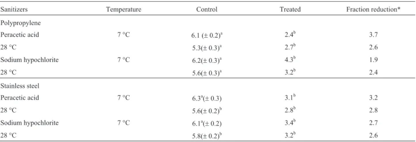
Counts ofS. aureusS3 adhered to polypropylene and stainless surfaces after the application of peracetic acid and sodium hypochlorite are shown in Table 2. The log reduc-tion caused by peracetic acid ranged from 2.6 to 3.7 log cfu/cm2, while for sodium hypochlorite, the reduc-tion was 1.9-2.7 log cfu/cm2. However, in all the experi-mental systems, both tested sanitizers largely decreased (p < 0.05) the counts of cells adhered to the assayed sur-faces.
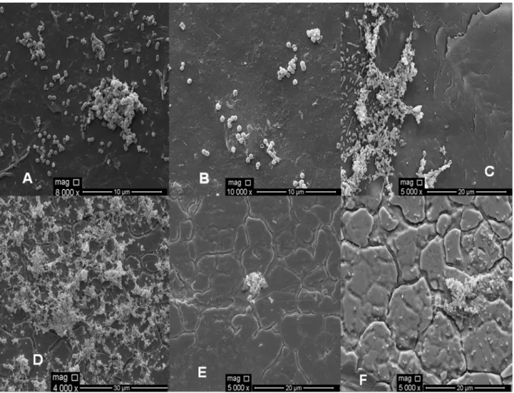
The electron microphotography of the polypropylene and stainless steel surfaces after the application of the sanitizers, sodium hypochlorite and peracetic acid, is shown in Figure 4. The findings for the electron micros-copy are in accord with the results obtained from the analy-sis of the viable cell counts on the surfaces when some remaining cells were found after applying peracetic acid (Figure 4 - B and E) and sodium hypochlorite (Figure 4 - C and F). The obtained results indicate that the sanitizers, peracetic acid (30 mg/L) and sodium hypochlorite (250 mg/L), were not efficient in completely removing the S. aureuscells adhered to the polypropylene and stainless steel surfaces. Residual cells adhered to the surfaces after the application of these sanitizers reinforce the biofilm as a likely source of cross-contamination in food processing plants. Meira et al. (2012) also found results similar to those found in this study when studied biofilm formation on stainless steel and polypropylene surfaces, where they found that peracetic acid and sodium hypochlorite were not efficient in completely removing the cells of biofilm ofS. aureusunder the same conditions used in this study.
The real quantification of the microorganisms ad-hered to the surfaces, both before and after the disinfection
procedure, is still not a completely solved problem, making it difficult to choose a reliable and effective method. There are publications on methods to quantify cells attached to in-ert materials, including swabbing (Herrera et al., 2007; RODEet al., 2007), impedance measurements (Asseré et al., 2008; Revol-Junelleset al., 2005) and real-time PCR (Bruhnet al., 2006). However, impedance measurements and real-time PCR are still only considered for usage inin vitro studies (Asseré et al., 2008). Swabbing, the oldest method, is still the main method employed in field studies; however, this method is known to not detach all microor-ganisms (Midelet and Carpentier, 2002). The removal effi-ciency of the swabbing procedure depends on several aspects. For instance, regarding the swab material, cotton swabs are better at removing adhered cells than polyester and rayon swabs. Moreover, using a moistened swab is more efficient than a dry one, and multiple swabbings may be performed at the same location (Asseré et al., 2008; Roseet al., 2004). All these concerns were considered in our study to increase the efficiency of the swabbing proce-dure, providing a real estimation of the surface-adhered cell population.
With regard to the limitations of this study in assess-ing the adhesion, detachment and biofilm formation of a monospecies inoculum under static conditions, there are clear implications for our results in designing strategies to control cross-contamination in the food service industry because the strains utilized as the test microorganisms were directly isolated from food contact surfaces and because the assays were performed with materials (surfaces) that are commonly used in food processing when the materials are immersed in a food-based media. These results are encour-aging for further studies focusing on the capacity of these strains to adhere to, detach and form biofilms on the sur-faces of equipment and utensils used in the food service in-dustry under continuous flux and for the efficacy of
differ-Table 2- Effects of peracetic acid (30 mg/L) and sodium hypochlorite (250 mg/L) on the count (log cfu/cm2) ofS. aureusS3 cells (grown in meat-based
broth at 7 and 28 °C) forming a 15-days biofilm onto polypropylene and stainless steel surfaces.
Sanitizers Temperature Control Treated Fraction reduction*
Polypropylene
Peracetic acid 7 °C 6.1 (±0.2)a 2.4b 3.7
28 °C 5.3(±0.3)a 2.7b 2.6
Sodium hypochlorite 7 °C 6.2(±0.3)a 4.3b 1.9
28 °C 5.6(±0.3)a 3.2b 2.4
Stainless steel
Peracetic acid 7 °C 6.3a(±0.3) 3.1b 3.2
28 °C 5.6(±0.2)b 2.8b 2.8
Sodium hypochlorite 7 °C 6.1a(±0.2) 3.4b 2.7
28 °C 5.8(±0.2)b 3.2b 2.6
* in relation to the counts found in control assay.
ent sanitization procedures designed to decrease cells in a biofilm matrix.
Acknowledgments
The authors are grateful to CNPq (Conselho Nacional de Desenvolvimento Científico e Tecnológico, Brazil) for the scholarship of the first author, and to CETENE (Centro de Tecnologia do Nordeste, Recife, Brazil) for the technical support in electronic microscopy analysis.
References
Ammor S, Chevallier I; Laguet A; Labadie J; Tallon R; Dufour E (2004) Investigating of the selective bactericidal effect of several decontaminating solutions on bacterial biofilms in-cluding useful, spoilage and/or pathogenic bacteria. Food Microbiol 21:11-17.
Asséré A, Oulahal N, Carpentier B (2008) Comparative evalua-tion of methods for counting surviving biofilm cells adher-ing to a polyvinyl chloride surface exposed to chlorine or drying. J Appl Microbiol 104:1692-1702.
Barnes LM, Lo MF, Adams MR, Chamberlain AHL (1999) Effect of milk proteins on adhesion of bacteria to stainless steel sur-faces. Appl Environ Microbiol 65:4543-4548.
Braga LC, Shupp JW, Cummings C, Jett M, Takahashi JÁ, Carmo LS, Chartone-Souza E, Nascimento AMA (2005) Pome-granate extract inhibitsStaphylococcus aureusgrowth and
subsequent enterotoxin production. J Ethnopharmacol 96:335-339.
Bruhn JB, Hagensen JAJ, Bagge-Ravn D, Gram I (2006) Culture conditions of roseobacter strain 27-4 affect its attachment and biofilm formation as quantified by real-time PCR. Appl Environ Microbiol 72:3011-3015.
Carson CF, Mee BJ, Riley TV (2002) Mechanism of action of
Melaleuca alternifolia (Tea tree) oil on Staphylococcus aureusdetermined by time-kill, lysis, leakage, and salt toler-ance assay and electron microscopy. Antimicrob Agents Chemother 46:1914-1920.
Cheng G, Zhang Z, Chen S, Bryers JD, Jiang S (2007) Inhibition of bacterial adhesion and biofilm formation on zwitterionic surfaces. Biomaterials 28:4192-4199.
Costerton JW, Steward PS, Greenberg EP (1999) Bacterial bio-films: a common cause of persistent infections. Science 284:1318-1322.
Downes FP, Ito K (2001) Compendium of methods for the micro-biological examination of foods. American Public Health Association (APHA), Washington.
Fux CA, Wilson S, Stoodley P (2004) Detachment characteristics and oxacillin resistance ofStaphylococcus aureusbiofilm
emboli in an in vitro catheter infection model. J Bacteriol 186:4486-4491.
Heydorn A, Nielsen AT, Hentzer M, Stenberg C, Givskov M, Esbol BK, Molin S (2000) Quantification of biofilm struc-tures by the novel computer program COMSTAT. Micro-biol 146:2395-2407.
Herrera JJR, Cabo ML, González A, Pazos I, Pastoriza L (2007) Adhesion and detachment kinetics of several strains of
Staphylococcus aureussubsp.aureusunder three different experimental conditions. Food Microbiol 24:585-591. Kumar C, Anand SK (1998) Significance of microbial biofilms in
food industry: a review. Int J Food Microbiol 42:9-27. Kusumaningrum HD, Riboldi G, Hazeleger WC, Beumer RR
(2003) Survival of foodborne pathogens on stainless steel surfaces and cross-contamination to foods. Int J Food Microbiol.85:227-236.
Kuzman L, Rózalski M, Walenka E, Rózalska B, Wysokinska H (2007) Antimicrobial activity of diterpenoids from hairy roots ofSalvia sclarea L.: salvipisone as a potential
anti-biofilm agent active against antibiotic resistant Staphylococ-ci. Phytomed 14:31-35.
Malheiros OS, Passos CT, Casarin LS, Serraglio L, Tondo EC (2010) Evaluation of growth and transfer ofStaphylococcus aureusfrom poultry meat to surfaces of stainless steel and
polyethylene and ther disinfection. Food Cont 21:298-301. Marques SC, Rezende JGOS, Alves LAF, Silva BC, Alves E,
Abreu LR; Piccoli RH (2007) Formation of biofilm by
Staphylococcus aureuson stainless steel and glass surfaces and its resistance to some selected chemical sanitizers. Braz J Microbiol 38:538-543.
Meira QGS, Barbosa IM, Athayde AJAA, Siqueira-Júnior JP, Souza EL (2012) Influence of temperature and surface kind on biofilm formation byStaphylococcus aureusfrom food-contact surfaces and sensitivity to sanitizers. Food Cont 25:469-475.
Meyer B (2003) Approaches to prevention, removal and killing of biofilms. Int Biodeter Biodegrad 51:249-253.
Midelet G, Carpentier B (2002) Transfer of microorganisms, in-cludingListeria monocytogenes, from various materials to beef. Appl Environ Microbiol 68:4015-4024.
Møretro T, Hermansen L, Holck AL, Sidhu MS, Rudi K, Langsrud S (2003) Biofilm formation and the presence of the intercellular adhesion locus ica among Staphylococci from food and food processing environments. Appl Environ Microbiol 69:5648-5655.
Morton LHG, Greenway DIA, Gaylarde CC, Surman SB (1998) Consideration of some implications of the resistance of biofilms to biocides. Int Biodeter Biodegrad 41:247-259. Oulahal N, Brice W, Martial A, Degraeve P (2008) Quantitative
analysis of survival ofStaphylococcus aureus orListeria innocuaon two types of surfaces: Polypropylene and
stain-less steel in contact with three different dairy products. Food Cont 19:78-185.
Revol-Junelles AM, Miguindou-Mabiala R, Roger-Maigne D, Milliere JB (2005) Behavior of Escherichia coli cells and Bacillus cereus spores on poplar wood crates by impedance measurements. J Food Prot 68:80-84.
Rode TM, Langsrud S, Holck A, Moretro T (2007) Different pat-terns of biofilm formation inStaphylococcus aureusunder food-related stress conditions. Int J Food Microbiol 116:372-383.
Ronner AB, Wong ACL (1993) Biofilm development and sani-tizer inactivation ofListeria monocytogenesandSalmonella typhimuriumon stainless steel and buna-n rubber. J Food Prot 56:750-758.
Rose L, Jensen B, Peterson A, Banerjee SN, Arduino MJ (2004) Swab materials andBacillus anthracisspore recovery from nonporous surfaces. Emerging Infect Dis 10:1023-1029. Rossoni EMM, Gaylarde CC (2000) Comparison of sodium
hypo-chlorite and peracetic acid as sanitizing agents for stainless steel food processing surfaces using epifluorescence micros-copy. Int J Food Microbiol 61:81-85.
Spoering AL, Lewis K (2001) Biofilms and planktonic cells of Pseudomonas aeruginosa have similar resistance to killing by antimicrobials. J Bacteriol 183:6746-6751.
Srey S, Jahid IK, Ha SD (2012) Biofilm formation in food indus-tries: A food safety concern. Food Cont 31:572-585. Van Houdt R, Michiels CW (2010) Biofilm formation and the
food industry, a focus on the bacterial outer surface. J Appl Microbiol 109:1117-1131.
Vanderzant C, Splittsstoesser DF (1992) Compendium of meth-ods for the microbiological examination of fometh-ods. American Public Health Association (APHA), Washington.
Wirtanen G, Ahola W, Mattila-Sandholm T (1995) Evaluation of cleaning procedures in elimination of biofilm from stainless steel surface in process equipment. Food Bioprod Process 73:9-16.
Zoltai PT, Zottola EA, Mckay LL (1981) Scanning electron mi-croscopy of microbial attachment to milk contact surfaces. J Food Prot 44:204-208.
Zottola EA, Sasahara KC (1994) Microbial biofilms in the food processing industry - Should they be a concern? Int J Food Microbiol 23:125-148.