DEVELOPMENTAL CHARACTERISTICS OF TEMPORAL
SHARP TRANSIENTS IN THE EEG OF NORMAL
PRETERM AND TERM NEWBORNS
Magda Lahorgue Nunes
1,
Fernando T. Gameleira
2,
Andréa J. Oliveira
3, Jaderson Costa da Costa
4ABSTRACT - Objective: To describe developmental characteristics, morphological aspects and incidence of temporal sharp transients (TST) in normal preterm and term newborns at matched conceptional ages (CA). Method: Neonatal EEGs from two groups of normal newborns were evaluated in order to identify and characterize TST. Group I (n=40) consisted of newborns from 34 to 40 weeks of gestational age (GA) that were submitted to a single EEG between 24 and 48 hours of life. Group II consisted of 10 preterm newborns with GA between 30-32 weeks, followed with a weekly EEG until they reached term. Morphology of TST was divided in 3 groups (temporal sawtooth, isolated transients or repetitive transients). TST index, density and total number were calculated in each polysomnography and related to sleep stages and CA. Laterality (right/ left) was also evaluated. The groups were compared at 34, 36, 38 and 40 weeks of CA. Results: TST index and density decreased with the increase of CA in both groups (p<0.0001). The temporal sawtooth feature was registered in both groups only at 34 weeks. Although rare, repetitive and isolated TST were the most prevalent morphology between 36 - 40 weeks CA. Significant intragroup difference was observed in the comparison of TST density in REM and transitional sleep in GI. Moreover, isolated TST morphology was significant higher in GI at 34 weeks when compared to the others CA. No intragroup differences were observed on GII. No significant differences between the groups were observed considering TST number, index, density, morphology or laterality, at the matched CA. Conclusion: TST are normal features of neonatal EEG, as they are registered in normal newborns. Its incidence varies accordingly to morphology and they tend to disappear following the increase of CA. Temporal sawtooth appears more often in preterm newborns. Our results suggest that TST index, density and morphology variability may be a function of CA.
KEY WORDS: neonatal EEG, temporal sharp waves, preterm newborn, sleep ontogenesis.
Características das ondas agudas temporais no EEG de recém-nascidos normais pré-termo e a termo
RESUMO - Objetivo: Descrever as características, aspectos morfológicos e incidência das ondas agudas temporais (OAT) em recém - nascido pré-termo e a termo pareados por idade concepcional (IC). Método: OAT foram identificadas e analisadas nos EEGs de dois grupos de recém – nascidos normais. O GI (n=40) foi constituído de recém - nascidos com idade gestacional (IG) de 34 a 40 semanas, submetidos a EEG com 24-48 horas de vida. O GII consistia de 10 recém – nascidos pré - termo com IG entre 30 - 32 semanas, seguidos com EEG semanal até atingirem IC de 42 semanas. As OAT foram divididas de acordo com sua morfologia em serrilhada, isolada e repetitivas. O número total, índice e densidade de OAT foram calculados em cada EEG neonatal e relacionados ao estágio do sono e IC. A lateralidade (esquerda/direita) também foi analisada. Os grupos foram comparados nas seguintes semanas de IC: 34, 36,38 e 40 semanas. Resultados: O índice e a densidade de OAT diminuíram com o aumento da IC (p<0.001). A morfologia serrilhada foi registrada em ambos os grupos somente até a IC de 34 semanas. As OAT com morfologia isolada e repetitiva foram visualizadas mais freqüentemente entre 36-40 semanas. No GI foram observadas diferenças significativas em relação à morfologia e densidade de OAT em sono REM, em associação ao aumento da IC. A densidade das OAT isoladas foi significativamente superior no GI na 34ª semana de IC. Não foram observadas diferenças no GII, assim como também não foi observada diferença significativa entre os recém - nascidos dos grupos I e II quando pareados por IC. Conclusão: OAT são elementos normais do EEG neonatal já que são registradas em recém - nascidos
Serviço de Neurologia do Hospital São Lucas (HSL) e Faculdade de Medicina (FAMED) da Pontifícia Universidade Católica do Rio Grande do Sul Porto Alegre RS, Brasil (PUCRS): 1Professora Adjunta de Neurologia e Pediatria da FAMED – PUCRS; 2Neurologista e Neurofisiologista
Clínico, Ex - aluno do Curso de Especialização em Neurociências/ EEG Neonatal do Serviço de Neurologia do HSL – PUCRS; 3Neurofisiologista
Clínica, Ex - aluna do Curso de Especialização em Neurociências/ EEG Neonatal do Serviço de Neurologia do HSL – PUCRS; 4Professor
Titular de Neurologia, FAMED - PUCRS
Received 11 October 2002, received in final form 30 January 2003. Accepted 19 February 2003.
normais. Sua incidência varia de acordo com a morfologia e tende a desaparecer com o aumento da idade concepcional. A morfologia serrilhada é mais freqüente em prematuros. Nossos resultados sugerem que o índice, a densidade e a morfologia das OAT variam de acordo com a idade concepcional.
PALAVRAS–CHAVE: EEG neonatal, ondas agudas temporais, prematuro, ontogênese do sono.
Sharp fast transients are generally normal features
in newborn EEG and tend to occur more over
fron-tal and temporal derivations. Temporal sharp
tran-sients (TST) usually occur in young preterm neonates
and they become more prominent and frequent
bet-ween 30 – 32 weeks of conceptional age (CA)
1,2. Since
the first systematic descriptions of neonatal EEG, the
record of TST has been considered a representative
pattern of prematurity
3. These transients have been
previously called temporal sawtooth, temporal theta
bursts and premature temporal theta
4,5,6. It has been
reported that the number and duration of TST
in-creases until 31 weeks of CA and they tend to
disap-pear until 34 weeks. They usually are recorded
bilate-rally, but asynchronously, and are more evident
du-ring REM sleep
7.
Different types of TST have been reported in
pre-term and pre-term newborns, with variable morphology,
besides temporal sawtooth
8. In a previous study we
have evaluated the presence of TST and a possible
correlation to CA, and we concluded that the
pre-sence of TST suggests a preterm newborn EEG
9.
Al-though related to preterm, TST were also observed
in EEGs from close to term newborns. Hughes
6re-ported TST until 39 weeks of CA, with a peak of
unila-teral TST at 36 weeks of CA.
In previous studies in the literature the
morpho-logy of TST, besides the temporal sawtooth pattern,
has not been systematically analyzed. Based on the
hypothesis that morphological features and
inciden-ce of TST varies according to CA, this study was
desig-ned to evaluate the morphological variability of TST
and if its rate of occurrence can be influenced by
the extrauterine development in preterm newborns.
METHOD
Patient selection
We report the data of 50 neurologically normal new-borns from the São Lucas Hospital – PUCRS School of Me-dicine, Porto Alegre, Brazil. The newborns were divided into two groups. Group I consisted of 40 preterm and term newborns with gestational age (GA) of 34 weeks (n=4), 36 weeks (n=8), 38 weeks (n=14), 40 weeks (n=14), submitted to one EEG between 24-48 hours after birth. Group II consisted of 10 preterm newborns with GA between 30 and 32 weeks, who were followed up with a weekly EEG, until they reached 42 weeks CA. Newborns that met the following criteria were sequentially included
in the study: admitted to NICU because of preterm or born in our hospital; 5 minutes Apgar score greater than 7; GA calculated by the neonatologist using the Capurro me-thod10; adequate weight for GA; normal clinical
examina-tion; no evidence of CNS disorder; adequate prenatal ma-ternal care during gestation. The preterm newborns were submitted to at least one cranial ultrasound in the first two weeks of life, and after hospital discharge were follo-wed in the Neonatal Outpatient Follow – Up Clinic at the same Institution. Conceptional age was calculated as adding the number of days since birth to gestational age. All parents gave informed consent to have their new-borns included in the study.
Polysomnographic recordings
The recordings were performed on a 16 channel EEG and consisted of 10 channels of EEG, electro-oculogram, submental electromyogram, nasal and abdominal respiratory monitoring and electrocardiogram9,11. Bipolar
montage was used, with double distance electrodes, placed based on the 10-20 system as modified for newborns12,13. The state of the newborn and all the
movements during the exam were recorded by the technician. All studies were performed in the morning, after the babies were fed, cleaned and dried. The babies were in the supine position and each exam lasted at least 50 minutes or until a complete sleep cycle ( REM - NREM – REM) was recorded1.
Other aspects of the EEG data from some of these newborns have been reported elsewhere9,11.
Morphology and quantification of TST
TST were identified in the EEG and divided according to morphology in : temporal sawtooth, isolated transients or repetitive transients.
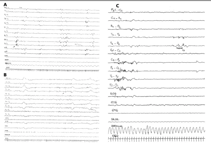
Temporal sawtooth - brief bursts of four to six sharp or sharp contoured, rhythmic waves at 4-7 Hz, with amplitude between 150 and 250µV, in the temporal regions8 (Fig 1a).
Isolated transients - single monophasic or diphasic transients, with maximal expression at temporal region, amplitude 50-250 µV (Fig 1b).
Repetitive transients - burst of 2 or more sharp contoured or comb-like rhythmic transients, separated by an interval inferior to 2 seconds, amplitude <100 µV (Fig 1c).
Positive temporal sharp transients were not included in this analyses.
The total number of TST during each exam was calcu-lated regarding morphology, laterality and synchrony, and was related to CA.
Fig 1. Examples of the different morphologycal features of TST, all EEGs were recorded with speed 15mm/s. In a) example of temporal sawtooth in transitional sleep (NREM – REM) registered at T5 in a newborn with CA = 34 weeks, b) an isolated TST registered in NREM sleep at T4-O2 in a 38 weeks CA newborn, c) example of the repetitive morphological feature registered at T6, in NREM sleep in a 34 weeks CA newborn.
eventual differences related to different total sleep time in each EEG. TST density was calculated and related to sleep stages (TST number / minutes in each sleep stage).
To determine laterality, TST were counted as they were registered either in left or right side.
The age-related changes in TST index, laterality and morphology were evaluated, and groups were matched at ages 34,36,38 and 40 weeks. The results were compared considering inter and intra group variability using paired
and unpaired t Test. We have established as statistically significant, p values under 0.05.
RESULTS
Group I consisted of 5 female and 5 male
newborns. Group II consisted of 25 female and 15
male newborns. We have not observed sex
differen-Fig 2. The total number of TST decreased as a function of CA in both groups (p< 0,001).
B
A
Fig 3. Among the different morphological features of TST suggested in this study the sawtooth pattern was registered only at 34 weeks CA. The isolated morphology predominated on GI at all ages.
ces in any of the comparisons performed considering
TST number, density, morphology or laterality. We
have analyzed 40 EEGs from group I newborns (4
for 34 weeks , 8 for 36 weeks, 14 for 38 and 40
weeks CA) and 74 from group II. However, for
sta-tistics we have included only 29 EEGs from group II
that corresponded to CA 34,36,38,40 weeks.
In Group I (one single EEG per newborn), we have
found TST in 14/40 newborns (35%). Regarding CA,
in the preterm newborns , all EEGs for 34 weeks had
TST and 25% of the EEGs from 36 weeks had TST. In
the term newborns, 28.5% of the EEGs from 38 and
40 weeks CA had TST.
In Group II we have found TST in 8/10 preterm
newborns (80%), and in 55.5% of the EEGs from 34
weeks, 37.5% of the EEGs from 36 weeks, 16.7% of
the EEGs from 38 weeks and in 33.4% of the EEGs
from 40 weeks.
Number of TST
- The total number of TST and TST
index have a tendency to decrease in both groups
ac-cording to CA (p<0.001), (Fig 2), (Table 1).
Compa-ring both groups, TST index was higher in Group I at
34 and 38 weeks CA. However, differences between
the groups were not significant considering the
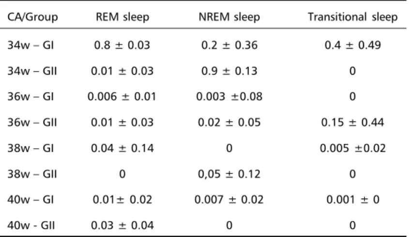
matched CA. TST density calculated in relation to sleep
phases, decreased in all sleep stages , in both groups,
with the increase of CA. Significant intragroup
difference was observed comparing REM and
transitional sleep in GI (Table 2). No significant
dif-ferences were observed between the groups at the
matched CA, regarding sleep phases and TST density.
Morphology -
The temporal sawtooth pattern
was registered only at 34 weeks CA in both groups.
Although rare the unique transients registered
beyond 34 weeks were repetitive or isolated TST (Fig
3). Isolated TST was significant higher in group I at
Table 1. TST index.
CA/Group Minimum Maximum Median Mean ± sd
Index Index
34w - GI 1.3 20.4 5.39 11.23 ± 8.65
34w - GII 0 9.0 1.08 2.33 ± 3.20
36w - GI 0 3.42 0 0.57 ± 1.21
36w - GII 0 5.5 0 1.53 ± 2.28
38w - GI 1.6 8.8 0 1.02 ± 2.37
38w - GII 0 4.8 0 0.81 ± 1.98
40w - GI 0 3.4 0 0.54 ± 1.00
40w - GII 0 4.2 0 1.05 ± 1.77
OBS: TST index (total number of TST / total sleep time X 60 ).
Table 2. TST density related to sleep stage (mean ± sd).
CA/Group REM sleep NREM sleep Transitional sleep
34w – GI 0.8 ± 0.03 0.2 ± 0.36 0.4 ± 0.49
34w – GII 0.01 ± 0.03 0.9 ± 0.13 0
36w – GI 0.006 ± 0.01 0.003 ±0.08 0
36w – GII 0.01 ± 0.03 0.02 ± 0.05 0.15 ± 0.44
38w – GI 0.04 ± 0.14 0 0.005 ±0.02
38w – GII 0 0,05 ± 0.12 0
40w – GI 0.01± 0.02 0.007 ± 0.02 0.001 ± 0
40w - GII 0.03 ± 0.04 0 0
34 weeks when compared to higher CA (p=0.04).
However, there was no significant difference
between the groups concerning each morphological
feature at the matched CA.
Laterality
- No specific pattern or predominance
was identified in the groups regarding laterality.
DISCUSSION
TST have been studied by many authors either in
normal or high risk newborns
6,9,14-17.There is a
consensus that these transients are normal features
of neonatal EEG and are related to preterm
newborns, although some authors have described
their appearance in term or near term newborns with
different morphologies
7,8.
In our study we observed that the total number
of TST have a tendency to decrease in both groups
in all sleep stages corresponding with the increase
of CA, suggesting that these transients varies as a
function of CA. This finding corroborates to what
has been previously reported by other authors
1,6. TST
peak has been described at lower ages than 31
weeks
6,16. However as we have not studied newborns
below 32 weeks CA we cannot compare our data. In
spite of that we have observed a descendent slope
on TST index and density after 34 weeks.
Many studies have attempted to correlate EEG
maturation patterns to extrauterine and intrauterine
development; some authors believe that EEG
maturation is a function of CA, and is independent
of extrauterine life development
18-20. However, other
authors have reported differences in EEG
matura-tion
21or behavior, when matching term newborns
to preterm newborns at same CA
9,22.
We have not found significant differences
bet-ween the groups at the matched CA’s studied, when
considering TST index, density, morphology or
laterality; these findings differed from a previous
study from Scher
17, who had observed different
peaks and greater number of TST when comparing
preterm infants at term post - conceptional ages.
Differences observed in the results could be due to
methodological aspects, since we did not included
positive transients, as did the authors from the
previous study. We decided to exclude positive
transients in order to clearly differentiate TST from
rolandic or temporal positive waves, that could have
a pathological significance
1,2,23.
Regarding laterality, Hughes
6,14has described a
predominance of TST on the right side after 30 weeks
of CA. Although we have observed, also, a
predomi-nance of right sided transients in GI at 38 and 40
weeks, this finding was not significant, probably due
to the reduced number of transients registered on
term newborns.
In our study, TST were occasionally registered in
term newborns, and the predominant morphological
patterns were isolated or repetitive TST. We have not
found temporal sawtooth after 34 weeks CA,
ho-wever, Scher
16has described a peak of TST at 39-40
weeks when evaluating physiological sharp transients
on healthy fullterm newborns.
Finally, Biagioni
15described a relationship between
incidence of TST and neurological outcome; this author
suggested that a higher incidence of TST over 33-34
weeks CA was associated to mild abnormalities on
follow-up. Our study was not designed to follow these
infants. Nevertheless, all preterm newborns from
Group II, and some of the newborns from Group I
were followed in our Neonatal Outpatient Follow Up
Clinic, from a period varying from 3 months to 4 years
and their neuropsychological development, assessed
by the Denver screening test and neurological
examination was considered normal
24.
Our results allow to conclude that TST are
nor-mal transients of the neonatal EEG, as they are
re-gistered in asymptomatic normal newborns. They are
a predominant feature of the preterm EEG and they
tend to disappear following increase of CA. The
mor-phological feature temporal sawtooth is more related
to preterm, while isolated and repetitive unilateral
transients could be registered on preterm and term
newborns. TST characteristics in term newborns
when matched to preterm newborns at the same
CA did not significantly differ. These findings allows
us to conclude that TST index, density and
morpho-logy varies as a function of CA.
REFERENCES
1. Lombroso CT. Neonatal electroencephalography. In Niedermeyer E, Lopez Da Silva F. (eds) Electroencephalography: basic principles, clinical applications and related fields, 3. Ed. Baltimore: Urban Schwarzenberg 1993;803-875.
2. Lombroso CT. Neonatal polygraphy in full-term and premature infants: a review of normal and abnormal findings. J Clin Neurophysiol 1985;2:105-155.
3. Dreyfus-Brisac C. The electroencephalogram of the premature infant. Wld Neurol 1962;3:5-15.
4. Werner SS, Stockard JE, Bickford RG. Atlas of neonatal electroence-phalography. New York: Raven Press, 1977.
5. Torres F, and Anderson C.The normal EEG of the human newborn. J Clin Neurophysiol 1985;2:89-103.
6. Hughes JR, Fino JJ, Hart LA. Premature temporal theta. Electroen-cephalogr Clin Neurophysiol 1987;67:7-15.
7. Lamblin MD, Andre M, Challamel MJ, et al. Electroencephalographie du nouveau-ne premature et a terme. Aspects maturatifs et glossaire. Neurophysiol Clin 1999;29:123-219.
9. Nunes ML, Da Costa JC, Moura-Ribeiro MVL. Polysomnographic quantification of bioelectrical maturation in preterm and fullterm newborns at matched conceptional ages. Electroencephalogr Clin Neurophysiol 1997;102:186-191.
10. Capurro H, Konicheki S, Fonseca D, Caldeyro-Barcia R. A simplified method for diagnosis of gestational age in the newborn infant. J Pediatr 1978;93:120-122.
11. Nunes ML, Penela MM, Da Costa JC. Differences in the dynamics of frontal sharp transients in normal and hypoglycemic newborns. Clin Neurophysiol 2000;111:305-310.
12. Anders T, Emde K, Parmelee AH. A manual of standardized terminology, techniques and criteria for scoring of states of sleep and wakefulness in newborn infants. UCLA Brain Information Service, Los Angeles, 1971.
13. Curzi - Dascalova LC, Mirmiran M Manual of methods for recording and analyzing sleep-wakefulness states in preterm and full term infants. INSERM, Paris, 1996.
14. Hughes JR, Kohrman MH. Topographic mapping of the EEG in premature infants and neonates. Clin Electroencephalogr 1989;20:228-234.
15. Biagioni E, Bartalena L, Boldrini A, Cioni G, Giancola S, Ipata AE. Background EEG activity in preterm infants: correlation of outcome with selected maturational features. Electroencephalogr Clin Neurophysiol 1994;91:154-162.
16. Scher MS, Bova JM, Dokianakis SG, Steppe DA. Physiological significance of sharp wave transients on EEG recordings of healthy
pre-term and full-term neonates. Electroencephalogr Clin Neurophysiol 1994;90:179-185.
17. Scher MS, Bova JM, Dokianakis SG, Steppe DA. Positive temporal sharp waves on EEG recordings of healthy neonates: a benign pattern of dysmaturity in pre-term infants at post-conceptional term ages. Electroencephalogr Clin Neurophysiol 1994; 90:173-178.
18. Dreyfus-Brisac C, Flescher J, Plassart E. L’electroencéphalogramme: critére d ‘age conceptionel du nouveau né à terme et prematuré. Biol Neonat 1962;4:154-173.
19. Borsini W, Lambruschini P, Marcacci G. Lo studio del sonno nel neonato a termine e pretermine com L’EEG –poligrafia. Riv Neurol 1990;60:234-235. 20. Ferrari F, Toricelli A, Giustardi A, et al. Bioelectric brain maturation in
fullterm infants and in healthy and pathological preterm infants at term post-menstrual age. Early Hum Dev 1992;28:37-63.
21. Scher MS, Steppe D, Dahl RE, Astana S, Guthrie RD. Comparison of EEG sleep measures in healthy full-term and preterm infants at matched conceptional ages. Sleep 1992;15:442-448.
22. Duffy FH, Als H, Mc Anulty GB. Behavioral and electrophysiological evidence for gestational age effects in healthy preterm and fullterm infants studied two weeks after expected due date. Child Dev 1990;61:1271-1286. 23. Da Costa JC, Lombroso CT. Neurphysiological correlates of neonatal