MORPHOQUANTITATIVE EFFECTS OF
ACUTE DIABETES ON THE MYENTERIC NEURONS
OF THE PROXIMAL COLON OF ADULT RATS
Maria Montserrat D.P. Furlan
1, Sônia L. Molinari
2, Marcílio H. de Miranda Neto
3ABSTRACT - The effects of acute diabetes on the density and size of the myenteric neurons of the proximal colon of adult rats were investigated. The injection of streptozotocin was followed by a period of observation of seven days, during which the diabetic animals showed weight loss, excessive food and water intake, large urinary debt and hyperglicemia. The whole-mounts from the proximal colon were stained with the techniques of Giemsa and of the NADH-diaphorase, and the employment of these techniques made it possible to verify a decrease on the neuronal density and on the cell body size of the myenteric neurons in the colon of the diabetic rats. These observations were discussed in terms of the pathophysiology of the diabetes and the experimental protocol. KEY WORDS: acute diabetes, myenteric neurons, proximal colon.
Efeitos morfoquantitativos do diabetes agudo sobre os neurônios mioentéricos do colo proximal de ratos adultos Efeitos morfoquantitativos do diabetes agudo sobre os neurônios mioentéricos do colo proximal de ratos adultos Efeitos morfoquantitativos do diabetes agudo sobre os neurônios mioentéricos do colo proximal de ratos adultos Efeitos morfoquantitativos do diabetes agudo sobre os neurônios mioentéricos do colo proximal de ratos adultos Efeitos morfoquantitativos do diabetes agudo sobre os neurônios mioentéricos do colo proximal de ratos adultos RESUMO - Foram investigados os efeitos do diabetes agudo sobre a densidade e o tamanho dos neurônios mioentéricos do colo proximal de ratos adultos. À injeção de estreptozootocina seguiu-se um período de observação de sete dias, durante os quais os animais diabéticos apresentaram perda de peso, ingestão excessiva de alimento e água, grande débito urinário e hiperglicemia. Os preparados de membrana do colo proximal foram corados pelas técnicas de Giemsa e da NADH-diaforase. A aplicação dessas técnicas permitiu constatar uma redução da densidade neuronal e do tamanho do corpo celular dos neurônios mioentéricos no colo dos ratos diabéticos. Essas observações foram discutidas em termos da patofisiologia do diabetes e do protocolo experimental. PALAVRAS-CHAVE: diabetes agudo, neurônios mioentéricos, colo proximal.
Pesquisa realizada no Departamento de Ciências Morfofisiológicas da Universidade Estadual de Maringá, Maringá PR, Brasil: 1Professora
Assistente; 2Professora Associada; 3Professor Titular.
Received 22 January 2002. Accepted 28 March 2002.
Dra. Maria Montserrat Diaz Pedrosa Furlan Universidade Estadual de Maringá Avenida Colombo 5690 – 87020900 Maringá PR -Brasil. E-mail:[email protected]
The research on the changes induced by experi-mental diabetes on the several tissues and organs of laboratory animals is quite large. Among those systems under investigation are the gastrointestinal tract and its intrinsic enteric nervous system, the neu-ronal network responsible for the control of the activ-ities of the bowel. It is reported, for instance, that the myenteric neurons of the stomach, duodenum and cecum are numerically reduced in diabetes1-3, and that
specific neurochemical groups show response pat-terns to diabetes which depend on the intestinal seg-ment and the duration of the diabetic state4-7. These
neuronal changes, as well as those associated to the autonomic innervation of the gut8-10, stand among
the responsible by the clinical gastrointestinal symp-toms of diabetes11-13.
Recently, we described an increase in the NADH-diaphorase positive myenteric neuronal population
in the duodenum of rats subjected to acute diabe-tes, although the total number of neurons was not affected14. Aiming at evaluating the response of the
neurons of the proximal colon to this same condition, we subjected adult rats to streptozotocin-induced diabetes for a period of seven days. The number of neurons stained with Giemsa and NADH-diaphorase (NADH-diaphorase) was assessed, and the neuronal sizes in the proximal colon were measured as well.
METHOD
indi-vidual metabolic cages, where they were kept during seven days with daily supply of ration (NUVILAB®) and water ad
libitum. The cages were maintained under controlled con-ditions of temperature (22oC) and light/dark cycles (12/12
hr) for the whole experimental period. Food and water intake and urinary debt of each animal were recorded daily. On the day before killing, the rats were weighted and subjected to overnight fast. The purpose of this fasting was to reduce the volume of the intestinal material so as to ease cleaning of the collected segments and return the colon to its resting dimensions.
The animals were killed by neck dislocation15,16. Blood
samples were collected for the evaluation of blood glu-cose. The colon was removed, washed, measured and weighted. Washing and distension were made with the appropriate solution and ligature of the extremities. Those segments destined to Giemsa staining were washed and filled with acetic formaldehyde and then processed ac-cording to the description17. The segments selected to the
technique of the NADH-d were washed and filled with Krebs solution, pH 7.3, and treated as described18. Times
were set as follows: the samples were washed twice (10 min each) in Krebs, kept in 0.3% Triton X-100 for 5 min, again washed twice in Krebs and transferred to incuba-tion medium, were they remained for 45 min, as estab-lished in the protocol of our laboratory. Reaction was in-terrupted by immersion in 10% buffered formalin.
The whole-mounts were made under stereomicros-cope. Only the mucosa and the submucosa were comple-tely dissected out so as to avoid removal of myenteric neu-rons, adhered to the circular smooth muscle. Next the who-le-mounts were mounted in slide according to standard histological procedures.
Neurons were counted under a BX40 microscope with 40X objective. Eighty microscopic fields (17.68 mm2),
equally distributed on the intermediate and antimesocolic regions of the proximal colon circumference, had their neurons counted. Half-seen neurons were counted in alter-nate fields. The counts were carried out in the whole-mounts stained with Giemsa and those stained with the NADH-d.
The Giemsa-stained myenteric neurons were measured under microscope with 40X objective coupled to a com-puterized image analyzer (ImagePro Plus). Due to the prox-imity of the cell bodies in the myenteric ganglia, the cell body and nucleus profiles were drawn manually. The ar-eas of 65 neurons and their nuclei of each circumferential region in each whole-mount were measured, yielding 520 neurons per group.
The results were statistically analyzed by the test t of Student with significance level of 5%. The values presented are the mean ± SEM of each set of data and the number of data considered is indicated in each case.
RESULTS
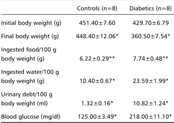
During the days the rats were in the cages, the diabetic animals exhibited more irritability than the
Table 1. Physiological features of the control and diabetic rats. Controls (n=8) Diabetics (n=8) Initial body weight (g) 451.40±7.60 429.70±6.79 Final body weight (g) 448.40±12.06* 360.50±7.54* Ingested food/100 g
body weight (g) 6.22±0.29** 7.74±0.48** Ingested water/100 g
body weight (g) 10.40±0.67* 23.59±1.99* Urinary debt/100 g
body weight (ml) 1.32±0.16* 10.82±1.24* Blood glucose (mg/dl) 125.00±3.49* 218.00±11.10*
*Values differ significantly between controls and diabetics (p<0.01) **Values differ significantly between controls and diabetics (p<0.05)
Table 2. Measures of length, weight, circumference and area of the colon of control and diabetic rats.
Controls (n=8) Diabetics (n=8) Length (cm) 16.33±1.57 14.49±0.61
Weight (g) 2.90±0.42 2.51±0.11
Circumference (cm) 2.60±0.15* 2.05±0.10* Area (cm2) 42.58±5.04* 29.91±2.27*
*Values differ significantly between controls and diabetics (p<0.05)
controls, making their handling difficult. None of the animals of any group died during this period or was discarded because of any alterations. Also, control animals injected with vehicle had no changes, when compared to non-injected rats (data not shown). Table 1 presents the parameters recorded during the one-week period; the means of food and water intake and urinary debt are presented by 100 g of mean body weight to make comparisons easier. Table 2 shows the measures of the colon obtained in both groups.
At the microscope, most of the myenteric neurons were found clustered in ganglia, with isolated neurons being scarce. Although the fibers of the plexus were not stained by either technique, the course of the thick primary connectives could be followed with relative ease in the whole-mounts stained with NADH-d. Most of the myenteric ganglia were polygonal and often fused with their neighbours or extended towards them. Their predominant orientation was parallel to the circular smooth muscle layer, but some had exten-sions in other directions, forming a patchy pattern.
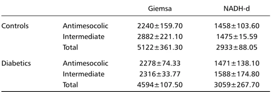
pre-sented in Table 3. The number of neurons stained with Giemsa was not different between the circumferential regions in either group. The same applied to the NADH-d positive myenteric neurons. However, with both tech-niques, there was a trend towards more neurons in the intermediate regions of the colon. This larger neu-ronal density could be visually observed in some whole-mounts.
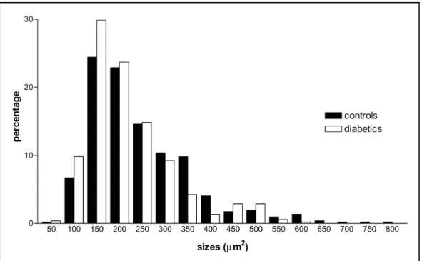
There was no significant change on the neuronal density of the diabetic group relative to the control. Figure 1 shows the percent distribution of the areas of the neuronal profiles in the studied groups. The general pattern for both groups was very similar, with the largest percentages between 100 and 250 µm2.
But it was observed that neurons with cell body profile above 600 µm2 were found only in the control animals.
Accordingly, the size range of the neurons of the diabetic animals was narrower than that of the controls (from 49.43 to 756.63 µm2 in the control group and
from 38.61 to 589.14 µm2 in the diabetic group).
On average, the areas of the neuronal profiles of the diabetic rats were smaller than those of the con-trols (Table 4). The decrease was more pronounced in the total area of the cell body than on the area of the nucleus, so that the percentage of the total profile occupied by the nucleus had a significant increase.
DISCUSSION
The experimental diabetes induced by streptozo-tocin is currently the most used tool to evaluate the
effects of diabetes mellitus under controlled con-ditions. Streptozotocin is a diabetogenic agent of quite stable action and dose-response relationship19;
it allows the investigator to follow organic alterations for predetermined periods of time. In experiments carried out by our research group, the dose of 35 mg/kg body weight has had efficacy in the diabetes induction. It is also the minimal single dose capable of triggering a consistent diabetic state in the rat19.
The diabetic rats of the present work showed fea-tures which are typical of the diabetic state: weight loss, excessive food and water intake, large urinary debt and hyperglicemia (Table 1). The impaired in-sulin secretion in these animals, caused by strepto-zotocin, increases lipolysis, proteolysis, glicogenolysis and neoglicogenesis, leading to above-normal blood glucose levels and reduced body mass20. Despite this
abundant blood supply of nutrients, there is parado-xically a state of “cellular fast”, once glucose uptake by most of the body cells is insulin-dependent. As a compensatory response, the diabetic animals eat more. However, there is a reduced food conversion, as increased food ingestion is not translated into in-creased body weight.
The water alterations are also related to the me-tabolic imbalances caused by the partial or total lack of insulin20. The hyperglicemia is accompanied by
urinary loss of glucose, once the renal tubular me-chanisms of glucose reabsorption get saturated with
the large glucose load filtered by the renal glomerulli. Being an osmotically active compound, the glucose which is filtered and not reabsorbed causes excre-tion of an increased amount of water, building up the urinary debt. To compensate for this loss of volu-me, neural mechanisms are put into action, which increase water intake. In this way, the most charac-teristic clinical and behavioural signs of diabetes mellitus were observed early in this work.
Although significantly high, the blood glucose level was inferior to that recorded in other investi-gations of our laboratory2, and this deserves some
consideration. Streptozotocin has as one of its distin-ctive features the fact that the dose has a good corlation with the severity of the resulting diabetes, re-gardless of this being assessed by blood glucose lev-els, plasma insulin content or pancreatic insulin con-tent19. However, as our standard dose is always 35
mg/kg body weight, the possibility that different do-ses could have caused different degrees of severity (assessed by the blood glucose level) can be discar-ded. Other factors then could account for this varia-tion. In those early studies, diabetes induction was made in rats aging 2.5 months and the animals were killed after several weeks, while the present work employed seven-month old rats that were kept dia-betic for only a week. These differences could mean,
for instance, differences in the sensitivity of the pan-creatic β cells to the cytotoxic effects of streptozo-tocin, then less destruction of these cells and less pronounced decrease in insulin secretion. As far as we were able to investigate, there are no systematic data on the literature which can throw some light on this issue.
The seven-days period could also have been too short to allow a wide destruction of the β cells, so that a residual release of insulin could be present. Progressive morphological alterations were observed in the pancreatic islets of mice from 5 to 18 days after streptozotocin injection21, and the plasma
glu-cose levels also changed during this time, with the highest levels being observed after 10 days.
The rats of the control group did not gain weight during the experimental period. This can be an inher-ent feature of the animals at this age, but the physical restrain imposed by the size of the metabolic cages should be considered as a preclusion to the weight gain. The dimensions of the colon of the diabetics were inferior to those of the controls, with circumference and total area reaching statistical significance (Table 2). This decrease can probably be explained by the smaller body weight of the diabetic animals, which was accompanied, although not proportionately, by
Table 4. Areas of cell body and nucleus profiles of the myenteric neurons of the proximal colon of control and diabetic rats.
Controls Diabetics (n=520) (n=520) Total profile of the cell body (µm2) 219.20±4.99* 193.60±4.32*
Nucleus profile (µm2) 81.88±1.57** 76.77±1.31**
% of the profile occupied by the nucleus 39.89±0.46* 42.28±0.42*
*Values differ significantly between controls and diabetics (p<0.001). **Values differ significantly between controls and diabetics (p<0.05).
Table 3. Number of neurons stained by the techniques of Giemsa and NADH-d in the antimesocolic and intermediate regions of the proximal colon of control and diabetic rats (n=4 for each technique per group).
Giemsa NADH-d
Controls Antimesocolic 2240±159.70 1458±103.60 Intermediate 2882±221.10 1475±15.59
Total 5122±361.30 2933±88.05
Diabetics Antimesocolic 2278±74.33 1471±138.10 Intermediate 2316±33.77 1588±174.80
a diminishment of the internal organs: in percentual terms, the diabetics had a body weight loss of 16.10% relative to their initial weight, while the colon showed a weight decrease of 13.45% as compared to that of the controls. Smaller intestinal segments were fre-quently observed in our laboratory, specially in ani-mals losing weight due to protein desnutrition22,23.
Insulin deficiency is also an explanation for the reduced dimensions observed here, once insulin is an anabolic hormone and, as such, a promoter of cellular devel-opment. On the other hand, there are reports that rats subjected to experimental diabetes have dimen-sional and weight increases of the small and large intestines2,4,6,7,12. The absence of hypertrophy of the
proximal colon in this study could be related to fac-tors such as the duration of the experimental pe-riod, the age of the animals and the moderate diabe-tic state. Also, there must be myenteric neurons still functionally capable of maintaining the tonus (and hence the dimensions) of this intestinal segment.
The staining techniques employed here evidence larger numbers of neurons in the intermediate re-gion of the proximal colon, in both groups. Although not attaining significance, these circumferential diffe-rences were observed in other instances24, correlated
to the fact that the intermediate region of the colon has a thicker layer of longitudinal smooth muscle, analogous to the human, which must require a larger neuronal population for its innervation and control24.
This kind of regional variation was also observed in other segments of the bowel25-29.
The Giemsa staining is easy to use and allows the visualization of the whole myenteric neuronal po-pulation in a given intestinal segment. The neuronal counts made with Giemsa yielded a neuronal den-sity corresponding to 29,000 neurons/cm2 of
proxi-mal colon, a value much similar to that found in other age-and weight-matched rats28. In younger rats, is
was found a number of neurons/cm2 larger than
this30, which can be related to the smaller intestinal
area and correspondingly lesser neuronal dispersal. On the other hand, the technique of the NADH-d stains only a fraction of the myenteric neuronal popu-lation. Although there are reports that it stains al-most all the myenteric neurons27,31, this has not been
the case in our research group14,28. In this study, the
number of NADH-d positive neurons in the proximal colon was of 57-66% that found with Giemsa. As the dissection could have caused neuronal loss, all care was taken to avoid the removal of the muscu-lar tunicas, to which the myenteric neurons are fixed. The incubation times adopted in these investigations
are probably the most satisfactory explanation for the discrepancies; especially important are the peri-ods during which the intestine is kept in the media containing the emulsifying agent and the substrates for the NADH-d activity, respectively. The longer are these periods – and they vary a lot from one study to another – the greater the possibility of staining cells of low enzymatic activity. To minimize these effects, the periods adopted in our laboratory in these me-dia are those described in the section Methods. The neuronal counts with the NADH-d revealed a den-sity of about 16,600 neurons/cm2 of intestinal area.
The density of neurons in the proximal colon of the diabetics did not have significant changes. Ne-vertheless, when considering that the colon area was 30% smaller in this group, it would be expected that the neuronal density was 30% greater, because the smaller growth makes neuronal density larger. The Giemsa technique displayed 10.3% less neurons in the diabetics than in the controls, suggesting that there was a mean loss of 40.3% of the neurons. With the NADH-d, the neuronal density was 4.3% greater in the diabetics, and thus this neuronal subpopula-tion had a percentual loss of 25.7% relative to the controls. Alternatively, neuronal activity could only be reduced to the point that the neurons did not stain after 45 minutes. In the duodenum, on the other hand, there was an increase in the NADH-d positive neuronal population in conditions of acute diabe-tes14. This shows that the NADH-d positive neurons
react differently from the general neuronal popula-tion to the diabetic condipopula-tion and that the locapopula-tion of the myenteric neurons in the bowel length influ-ences how they react to modifications in their envi-ronment4,7.
Large neurons (above 600 µm2) were found only in
the control animals. The histogram of Figure 1 resem-bles those of other investigations31,32, although neurons
having areas above 800 µm2 had not been recorded.
The comparisons of the neuronal areas between the two groups showed that the mean area of the neurons in the diabetics was significantly smaller than that in the controls. In other words, the myenteric neurons of the proximal colon of the diabetic rats reduced their size, despite the short duration of the experimental period. The smaller neuronal area was caused primarily by a reduction in the cytoplasmic area of the cell body. There are reports of changes in the nuclear and/or cytoplasmic areas in other regions of the nervous system in diabetics9,33,34, which were
translation of biomolecules and of electrical activity. The role of cellular dehydration to the reduced size also has to be taken into account.
In summary, it was verified that the acute diabe-tes induced by streptozotocin caused increased blood glucose levels, body weight loss, excessive food and water ingestion, and large urinary debt; it reduced the area of the colon and the sizes of the myenteric neurons and their nuclei and decreased the overall neuronal population.
Aknowledgements: Aknowledgements: Aknowledgements: Aknowledgements:
Aknowledgements: The authors wish to thank the help of the technicians Maria Aparecida Pantoja Agostinho and José Antônio de Souza.
REFERENCES
1. Buttow NC, Miranda-Neto MH, Bazotte RB. Morphological and quan-titative study of the myenteric plexus of the duodenum of streptozo-tocin-induced diabetic rats. Arq Gastroenterol 1997;34:34-42. 2. Zanoni JN, Miranda-Neto MH, Bazotte RB, Souza RR. Morphological
and quantitative analysis of the neurons of the myenteric plexus of the cecum of streptozotocin-induced diabetic rats. Arq Neuropsiquiatr 1997;55:696-702.
3. Fregonesi CEPT, Miranda-Neto MH, Molinari SL, Zanoni JN. Quantitative study of the myenteric plexus of the stomach of rats with streptozotocin-induced diabetes. Arq Neuropsiquiatr 2001;59:50-53. 4. Lincoln J, Bokor JT, Crowe R, Griffith SG, Haven AJ, Burnstock G.
Myenteric plexus in streptozotocin-treated rats: neurochemical and histochemical evidence for diabetic neuropathy in the gut. Gastro-enterology 1984;86:654-661.
5. Ballmann M, Conlon JM. Changes in the somatostatin, substance P and vasoactive intestinal polypeptide content of the gastrointestinal tract following streptozotocin-induced diabetes in the rat. Diabetologia 1985;28:355-358.
6. Belai A, Lincoln J, Milner P, Burnstock G. Progressive changes in adrenergic, serotonergic, and peptidergic nerves in proximal colon of streptozotocin-diabetic rats. Gastroenterology 1988;95:1234-1241. 7. Belai A, Lincoln J, Milner P, Burnstock G. Differential effect of
strep-tozotocin-induced diabetes on the innervation of the ileum and distal colon. Gastroenterology 1991;100:1024-1032.
8. Monckton G, Pehowich E. Autonomic neuropathy in the streptozotocin diabetic rat. J Can Scien Neurol 1980;7:135-142.
9. Kniel PC, Junker U, Perrin IV, Bestetti GE, Rossi GL. Varied effects of experimental diabetes on the autonomic nervous system of the rat. Lab Invest 1986;54:523-530.
10. Yagihashi S, Sima AAF. Neuroaxonal and dendritic dystrophy in diabetic autonomic neuropathy: classification and topographic distribution in the BB-rat. J Neuropathol Exp Neurol 1986;45:545-565. 11. Abrahamsson H. Gastrointestinal motility disorders in patients with
diabetes mellitus. J Intern Med 1995;237:403-309.
12. Karakida T, Ito S, Homma S. In vitro motor activity of intestinal seg-ments of streptozotocin diabetic rats. J Auton Nerv Sys 1989;26:43-50.
13. Öztürk Y, Aydin S, Özçelikay AT, Altan VM, Yildizoglu-Ari N. Calmodulin content and in vitro contractility of duodenum from streptozotocin-induced diabetic rats: effects of insulin therapy and calmodulin antagonism. Eur J Pharmacol 1997;321:59-65.
14. Furlan MMDP, Miranda-Neto MH, Sant’Ana DMG, Molinari SL. Num-ber and size of myenteric neurons of the duodenum of adult rats with acute diabetes. Arq Neuropsiquiatr 1999;57:740-745.
15. Belai A, Cooper S, Burnstock G. Effect of age on NADPH-diaphorase-containing myenteric neurons of rat ileum and proximal colon. Cell Tissue Res 1995;279:379-383.
16. Michel K, Sann H, Schaaf C, Schemann M. Subpopulations of gastric myenteric neurons are differentially activated via distinct serotonin receptors: projection, neurochemical coding and functional impli-cations. J Neurosci 1997;17:8009-8017.
17. Barbosa AJA. Técnica histológica para gânglios nervosos intramurais em preparados espessos. Rev Bras Pesq Med Biol 1978;11:95-97. 18. Gabella G. Detection of nerve cells by a histochemical technique.
Experientia 1969;25:218-219.
19. Junod A, Lambert AE, Stauffacher W, Renold AE. Diabetogenic action of streptozotocin: relationship of dose to metabolic response. J Clin Invest 1969;48:2129-2139.
20. Guyton AC, Hall JE. Textbook of medical physiology. 9.Ed. Philadel-phia: W.B. Saunders, 1996.
21. Papaccio G, Esposito V. Ultrastructural observations on cytotoxic ef-fector cells infiltrating pancreatic islets of low-dose streptozocin treated mice. Virchows Archiv Pathol Anat 1992;420:5-10.
22. Meilus M, Natali MRM, Miranda-Neto MH. Study of the myenteric plexus of the ileum of rats subjected to proteic undernutrition. Rev Chil Anat 1998;16:9-14.
23. Sant’Ana DMG, Molinari SL, Miranda-Neto MH. Effects of protein and vitamin B deficiency on blood parameters and myenteric neurons of the colon of rats. Arq Neuropsiquiatr 2001;59:493-498.
24. Gabella G. Innervation of the gastrointestinal tract. Int Rev Cytol 1979;59:129-193.
25. Irwin DA. The anatomy of Auerbach’s plexus. Am J Anat 1931;49:141-166. 26. Leaming DB, Cauna N. A qualitative and quantitative study of the
myen-teric plexus of the small intestine of the cat. J Anat 1961;95:160-169. 27. Gabella G. Neuron size and number in the myenteric plexus of the
newborn and adult rat. J Anat 1971;109:81-95.
28. Sant’Ana DMG, Miranda-Neto MH, Molinari SL, Sant’Ana MA. Neu-ron number in the myenteric plexus of the ascending colon of rats. Arq Neuropsiquiatr 1997;55:460-466.
29. Fregonesi CEPT, Miranda-Neto MH, Molinari SL. Estudo morfológico e quantitativo dos neurônios do plexo mioentérico do corpo do estômago de Rattus norvegicus. Acta Scientiarum 1998;20;221-224. 30. Romano EB, Miranda-Neto MH, Cardoso RC. Preliminary
investiga-tion about the effects of streptozotocin-induced chronic diabetes on the nerve cell number and size of myenteric ganglia in rat colon. Rev Chil Anat 1996;14:139-145.
31. Santer RM, Baker DM. Enteric neuron numbers and sizes in Auerbach’s plexus in the small and large intestine of adult and aged rats. J Auton Nerv Sys 1988;28:59-67.
32. Barbosa AJA. Auerbach’s plexus of the albino rat: I. Quantitative study of the ganglia and nerve cells in the caecum and colon. Rev Bras Pesq Med Biol 1973;6:253-262.
33. Dheen ST, Tay SSW, Wong WC. Ultrastructural changes in the hypo-thalamic supraoptic nucleus of the streptozotocin-induced diabetic rat. J Anat 1994a;184:615-623.