Arq Neuropsiquiatr 2002;60(3-A):624-627
POLYMYOSITIS
Clinical investigation in two sisters
Margô Gomes de Oliveira Karnikowski
1, Bruno Rodrigues Veloso Costa
2,
Oscar Francisco Sanchez Osella
3, Otávio de Tolêdo Nóbrega
1ABSTRACT - We present an investigation of a case of polymyositis affecting two sisters of one same parenthood. Their cases have been documented for almost two decades, being investigated by means of a diagnostic protocol which combined clinical findings as well as laboratorial, histopathological and image tests. In both cases, clinical manifestations started in childhood, without signs of involvement of the central and peripheral nervous system. Both patients proved to respond to a therapeutics based on corticosteroids. The degree of relatedness between their parents corroborate the notion that genetic factors may contribute to the development of the disease.
KEY WORDS: polymyositis, inflammatory myopathy.
P PP
PPolimiosite: investigação clínica em duas irmãsolimiosite: investigação clínica em duas irmãsolimiosite: investigação clínica em duas irmãsolimiosite: investigação clínica em duas irmãsolimiosite: investigação clínica em duas irmãs
RESUMO - Apresentamos a investigação de dois casos de polimiosite, ocorridos entre irmãs de uma mesma filiação. Seus casos foram documentados ao longo de quase duas décadas, tendo sido diagnosticados utilizando-se de protocolo diagnóstico que combinou achados clínicos, exames laboratoriais, histopatológicos e por imagem. Em ambos os casos, as manifestações clínicas se iniciaram ainda na infância, sendo constatada ausência de acometimento do sistema nervoso central ou periférico. Ambas as pacientes responderam satisfatoriamente a terapia baseada em corticosteróide. O grau de parentesco entre os genitores das pacientes sugere que fatores genéticos podem predispor ao desenvolvimento da doença.
PALAVRAS-CHAVE: polimiosite, miopatia inflamatória.
Department of Physiotherapy, Catholic University of Brasilia, Brasilia DF, Brasil; 1Ph.D. in Molecular Pathology and Professor of
Immunol-ogy and Human Genetics; 2Medical student; 3Ph.D. in Medicine and Professor of Cardiology.
Received 2 January 2002. Accepted 18 March 2002.
Dra. Margô Gomes de Oliveira Karnikowski - Curso de Fisioterapia Prédio São João Bosco (bloco G) – Universidade Católica de Brasília, EPCT Q.S. 7, Lote 1 – 72030-170 Brasília DF - Brasil. E-mail: [email protected]
Idiopathic myopathies are a heterogeneous group
of acquired muscle diseases which affect 1 in
100,000 individuals
1. Polymyositis (PM) comprises an
inflammatory myopathy that results from an
abnor-mal immune response to skeletal muscle fibers and
associated tissues. Activation of auto-reactive
T-cyto-lytic lymphocytes and molecular mimicry are
proba-ble mechanisms underlining this pathology
2.
More-over, the majority of patients diagnosed with PM
display high levels of antibodies to self-antigens, such
as cell membrane components and nuclear
pro-teins
3,4. Current diagnosis of PM is assessed by means
of compatible clinical findings (proximal muscle
weak-ness) and measurements of serum muscle enzymes,
electromyography and muscle biopsy
5. Even though
causes for PM remain undetermined, an increasing
awareness that genetic factors are implicated has
evolved from different studies
6,7. In spite of that, the
occurrence of more than one case within the same
family still consists in a rare event
6,8,9.
This study has the purpose to describe
anamne-sis and testing procedures which resulted in the
diag-nosis of two patients with PM within the same
paren-thood, and to report treatment and evolution of both
patients documented over almost two decades.
CASES
Arq Neuropsiquiatr 2002;60(3-A) 625
were performed: complete hemogram (normal); summary of urine (normal); oxalacetic transaminase 145 UI/l max RV 30 UI/l; pyruvate transaminase 186 UI/l (max RV 37 UI/ l); lactate dehydrogenase 419 UI/l (max RV 240 UI/l); cre-atine kinase (CK) 2,600 UI/l (max RV 70 UI/l); aldolase 26.3 UI/l (max RV 7.6 UI/l). X-ray analysis of the lumbar and the sacrum-coccyx regions of the vertebral column yielded normal results.
Studies on nerve-motor conduction revealed no altera-tion on either latency or amplitude of the acaltera-tion potentials of the nerves studied (median, ulnar and fibular), being the speed of conduction preserved as well. Studies on sensitive conduction of the same nerves have also failed to demon-strate alterations. Nonetheless, quantitative electromyogra-phy yielded motor units of greatly reduced length and am-plitude, being polyphasic in their majority, mainly on the biceps, deltoid and quadriceps muscles in spite of no signs of denervation. Tomographic analysis failed to demonstrate any intracranial expansive processes either above or below the tentorium as well as absence of any pathological calci-fications, attesting a cranium-encephalic status within the parameters of normality. Additional tomographic analysis revealed a similarly healthy vertebral condition, character-ized by canal with dimensions and configuration compat-ible with normality, lacking pathological compression upon the dural sac. All findings above are consistent with a myo-pathic condition of idiomyo-pathic etiology.
Biopsy of biceps and quadriceps were performed under local anesthesia, being samples fixed before histological processing. Both tissues revealed a degenerative process characterized by disorganization of striated muscle fibers with various degrees of atrophy, displaying multiple endo-mysial infiltrates by polymorphonuclear cells. In some ar-eas, intensive fagocytosis of muscle fibers by inflammatory mononuclear cells could be observed, with signs of bundle regeneration and endomysial fibrosis. Such findings are compatible with a myopathic condition of acute and chronic polymyositis.
Patient 2. M.R.V.C., 23 years old, female, Caucasian, sister of G.R.V.C., was born presenting Apgar 8. The first symptom of mobility disorder arose at the age of nine, by walking with her left foot in valgus. Having in mind her sister’s condition, myopathy was taken under considera-tion. At that time, the following laboratorial tests were performed: complete hemogram with eosinophilia at 9% (max RV 3%); oxalacetic transaminase 24.5 UI/l; pyruvate transaminase 29 UI/l; lactate dehydrogenase 273 UI/l; cre-atine kinase 1,580 UI/l; aldolase 19.7 UI/l. Max RV were as cited previously. X-ray analysis of hands and feet revealed osseous age within the normality. Similarly to her sister’s case, studies of nerve-motor conduction have not shown major alterations of either latency or amplitude of the ac-tion potentials of the nerves investigated. Accordingly, elec-tromyographic examination confirmed motor units of duced length and amplitude, largely polyphasic and re-stricted to the proximal muscles. Biopsies of these muscle
revealed an interstitial microscopic aspect similar to the previous case, with several degenerative inflammatory ar-eas and bundles being regenerated. On what concerned its intensity, the degenerative process that was observed in these samples exceeded the severity of the preceding case, in spite of the lower levels of muscular enzymes. The accentuated disappearance of the striated aspect and the formation of large vacuoles in the muscle cells could trans-late this increased myopathy. Both biochemical evidence and histopathological findings attest an intense myopathic process, greater in severity than the preceding case, and consistent with acute polymyositis.
In both cases, initial therapeutics consisted on oral ad-ministration of 60 mg of the corticosteroid prednisone per day, which resulted in marked decrease of the serum enzyme levels within the first month (Fig 1). Dosage was gradually reduced according to the therapeutic response. Nowadays, both patients present stable clinic conditions which allow regular daily activities: walking without assis-tance, absence of dysphasia and minor difficulty in shift-ing from the seated to the ortostatic position. Both pa-tients attend to physiotherapy sessions twice a week in order to strengthen the pelvic and scapular musculature. M.R.V.C employs a maintenance dose of 5 mg of corticos-teroids in alternate days. G.R.V.C. sustains her medication suspended since October/2000, following a 16 years period of stable condition under administration of a similar main-tenance scheme.
DISCUSSION
The first reports of polymyositis were based on
findings of anamnesis
10,11. These studies used to
ge-nerate inaccurate diagnosis since inflammatory
myo-pathies share clinical symptoms such as mialgia,
pro-ximal muscle weakness and electromyographic
al-terations with several other idiopathic forms of
myo-pathy
1,12. The development of diagnostic protocols
that couple laboratorial, histopathological and
im-age procedures has greatly facilitated the precise
identification of myopathies with inflammatory
etio-logy
13,14, what in turn allows hiring an effective
thera-peutics. In this study, an investigation based on
clini-cal, biochemical and images findings was
success-fully hired to diagnose polymyositis in two non-twin
sisters from the same parenthood.
626 Arq Neuropsiquiatr 2002;60(3-A)
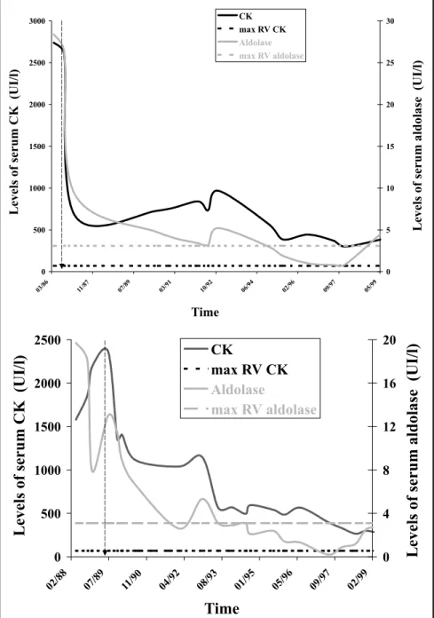
Fig 1. Levels of the muscular enzymes CK (left axis) and aldolase (right axis) in the serum of both patients, G.R.V.C. (A) and M.R.V.C. (B), throughout the treatment with corticosteroid. Each solid line represents the union between independent dosages of CK (black line) and aldolase (gray line) determined over the time. Horizontal dotted lines represent maximum referential values (max RV) for each enzyme. A vertical dotted arrow indicates the beginning of the treatment of each patient.
0 500 1000 1500 2000 2500 3000
03/86 11/87 07/89 03/91 10/92 06/94 02/96 09/97 05/99
Time
Levels
of
serum
CK
(UI/l)
0 5 10 15 20 25 30
CK max RV CK Aldolase max RV aldolase
Levels
of
serum
aldolase
(UI/l)
0
500
1000
1500
2000
2500
02/88 07/89 11/90 04/92 08/93 01/95 05/96 09/97 02/99
Time
Levels
of
serum
CK
(UI/l)
0
4
8
12
16
20
CK
max RV CK
Aldolase
max RV aldolase
Levels
of
serum
aldolase
(UI/l)
In addition, histopathological analysis of proximal
muscle biopsies of each patient revealed an intense
degenerative process characterized by marked
in-flammatory infiltrates, showing a clinical condition
compatible with PM. Nonetheless, symptoms usually
associated to inflammatory myopathies such as fever,
dysphasia and arthralgia
12,13have not been observed.
Exclusion of dermatomyositis (DM) was possible
ba-sed on the absence of cutaneous involvement in
ei-ther patients. Both cases proved to respond to a
the-rapeutics of corticosteroids, noticed the great
reduc-tion of the serum muscle enzymes within the first
month of treatment (Fig 1). In spite of the long-term
administration of steroids, periodic osseous
densi-tometry revealed no major pathological alterations
of their bone constitution.
Arq Neuropsiquiatr 2002;60(3-A) 627
with the origin of inflammatory myopathies. Some
studies implicate viral and bacterial infections as
possible triggering factor of the pathogenic
pro-cess
9,15,16. Others enroll immune disturbances and
genetic profiles as causative determinants for the
onset of PM and DM. In the latter, disruption of
im-munological tolerance is considered a factor that
probably accounts to the development of muscular
lesions, since such lesions may consist in a
patho-logical outcome of the autoantibodies found in a
significant number of patients with PM
6,17. In
addi-tion, the augmented frequency of the HLA-B8 and
DR3 haplotype among patients diagnosed with this
pathology suggests that a genetic profile may
pre-dispose to the its onset
18. In the present study, both
patients are sisters from one same parenthood,
whe-re the pawhe-rents awhe-re cousins in first degwhe-ree. It is
note-worthy that related individual share sets of
haplo-type and that, for this reason, consanguine unions
tend to predispose their descendants to the onset
of recessive and multifactorial phenotypes. Thus,
even though a definitive relationship between
im-munogenetic factors and polymyositis has not been
established, we consider that the cases reported in
this study corroborate such an association.
The comprehension that the immune system
works as an agent that ensures the equilibrium within
the organism implies that its disturbance allows or
generates unbalance. In autoimmune diseases, it is
desirable to apply medicinal treatments that
contri-bute to the recovery from the unbalanced state. In
the cases above, treatment with corticosteroid
pro-ved to be appropriate for such purpose, considering
that patient G.R.V.C. remains with a stable
condi-tion despite of having her medicacondi-tion suspended for
one year. Nonetheless, it is important to emphasize
the need for an early and accurate diagnosis of
in-flammatory myopathies, so that an appropriate
therapeutics may take place around the dawn of the
pathological process to allow satisfying outcomes,
similar to those described in this study.
Acknowledgments - Acknowledgments - Acknowledgments -
Acknowledgments - Acknowledgments - The authors gratefully thank both patients and their family for the consent on this study, and the Catholic University of Brasília for providing the environment for its development.
REFERENCES
1. Dalakas MC. Polymyositis, dermatomyositis and inclusion-body myo-sitis. N Engl J Med 1991;325:1487-1498.
2. Miller FW, Love LA, Barbieri SA, Balow JW, Plotz PH. Limphocyte activation markers in idiopathic myositis: changes with disease activ-ity and differences among clinical and auto-antibodies subgroups. Clin Exp Immunol 1990;81:373-379.
3. Bluthner M, Bautz FA. Cloning and characterization of the cDNA cod-ing for a polymyositis-scleroderma overlap syndrome-related nucle-olar 100 kDa protein. J Exp Med 1992;176:973-980.
4. Ge Q, Frank MB, O´Brien C, Targoff IN. Cloning of a complementary DNA coding for the 100 kDa antigen protein of the PM-Scl autoantigen. J Clin Invest 1992;90:559-570.
5. Matsui K, Aizawa Y, Inoue K, Yaguchi H, Toda G. Polymyositis with marked paravertebral muscle atrophy in patients with primary biliary cirrhosis. Rinsho Shinkeigaku 2000;40:694-700.
6. Rider LG, Gurley RC, Pandey JP, et al. Clinical, serological, and immu-nogenetic features of familial idiopathic inflammatory myopathy. Ar-thritis Rheum 1998;41:710-719.
7. Garlepp MJ. Genetics of the idiopathic inflammatory myopathies. Curr Opin Rheumatol 1996;8:514-520
8. Lewkonia RM, Buxton PH. Myositis in father and daughter. J Neurol Neurosurg Psychiatry 1973;36:820-825.
9. Hokezu Y, Higuchi I, Yanai S, Nagai M, Nagamatsu K. A family case of HAM and HTLV-I carrier including two sisters presenting with myositis. Rinsho Shinkeigaku 1994;34:563-568.
10. Bohan A, Peter JB. Polymyositis and dermatomyositis. N Engl J Med 1975;292:344-347,403-407.
11. Bohan A, Peter JB. A computer assisted analysis of 153 patients with polymyositis and dermatomyositis. Medicine 1977;56:255-286. 12. Plotz PH, Dalakas M, Leff RL, Love AA, Miller FW, Cronin ME.
Cur-rent concepts in idiopathic inflammatory myopathies: polymyositis, dermatomyositis, and related disorders. Ann Interm Med 1989;111:143-157.
13. Scola RH, Werneck LC, Prevedello DMS, Toderke EL, Iwamoto FM. Diagnosis of polymyositis and dermatomyositis: a study of 102 cases. Arq Neuropsiquiatr 2000;58:789-799.
14. Hilário, MOE, Yamashita H, Lutti D, Len C, Terreri MT, Lederman H. Juvenile idiopathic inflammatory myopathies: the value of magnetic resonance imaging in the detection of muscle involvement. Sao Paulo Med J 2000;118:35-40.
15. Dalakas MC. Immunopathogenesis of inflammatory myopathies. Ann Neurol 1995;37:S74-S86.
16. Oliveira HA, Macieira JC, Fakhouri R. Polimiosite associada a infecção por HTLV-I. Arq Neuropsiquiatr 2000;58:935-938.
17. Miller FW. Myositis-specific antibodies: touchstones for understand-ing the inflammatory myopathis. JAMA 1993;270:1846-1849. 18. Garlepp MJ. Genetics of the idiopathic inflammatory myopathies. Curr