OCCIPITAL LOBE MORPHOLOGICAL ANATOMY
Anatomical and surgical aspects
Leandro Pretto Flores
1ABSTRACT - Background: The occipital lobe is an important region of the central nervous system and site of a wide variety of lesions. Different from others regions of the brain, whose anatomy has been already meticulously detailed, controversies about occipital lobe morphology can occasionally hinder the surgical approaches to it. Method: Twenty-six hemispheres were dissected, examining the distances of the medial structures of the posterior interhemispheric fissure region; identifying the sulci and gyri more frequently found in the lateral surface; and detailing the anatomy of the occipital horn of the lateral ventricle. Also, anatomical details of structures such as the calcarine sulcus and preoccipital notch were evaluated. Results: Twenty-four hemispheres (92%) showed sulci with evident transverse trajectories in the lateral surfarce, and it was possible to identify marked longitudinal sulci in 16 (61%). All of the specimen presented a transverse sulcus in the inferior third of the lobe – labeled in this paper as Inferior Transverse Occipital – and 69% of the cases showed a transverse sulcus in the superior third – labeled as Superior Transverse Occipital. The occipital horn was present in 53% of the cases at level of the anterior boundary of the lobe. It was a half moon shaped cavity, whose the medial wall was formed by the calcar avis and the lateral wall, roof and floor formed by fibers of the tapetum. Conclusion: The occipital lobe presents parameters that are often recognized in most of the dissected brain and can be useful for the planning and performance of surgery in this area of the brain.
KEY WORDS: occipital lobe, anatomy, surgery.
Anatomia morfológica do lobo occipital: aspectos anatômicos e cirúrgicos Anatomia morfológica do lobo occipital: aspectos anatômicos e cirúrgicos Anatomia morfológica do lobo occipital: aspectos anatômicos e cirúrgicos Anatomia morfológica do lobo occipital: aspectos anatômicos e cirúrgicos Anatomia morfológica do lobo occipital: aspectos anatômicos e cirúrgicos
RESUMO - Introdução: O lobo occipital é uma importante região do sistema nervoso central e local de ocorrência de ampla variedade de patologias. Diferente de outras regiões do cérebro, cuja anatomia já foi meticulosamente detalhada, ainda existem controvérsias a respeito do pólo occipital que ocasionalmente podem dificultar cirurgias sobre esta região. Método: Foram dissecados 26 hemisférios cerebrais, examinando-se distâncias de algumas estruturas localizadas na região da fissura interemisférica posterior, identificando os sulcos e giros mais frequentemente encontrados na face lateral do lobo occipital, e detalhando-se a anatomia do corno occipital do ventrículo lateral. Também foram avaliados detalhes anatômicos de estruturas como o sulco calcarino e incisura pré-occipital. Resultados: Em 24 hemisférios (92%) foram identificados sulcos com trajetos transversais evidentes na face lateral do lobo, sendo que sulcos longitudinais foram observados em 16 peças (61%). Em todas foi possível identificar um sulco transversal completo no terço inferior do lobo – denominado neste estudo como sulco transverso inferior – e 69% dos hemisférios demonstraram um sulco transverso no terço superior – denominado sulco transverso superior. O corno occipital estendia-se até o lobo occipital em apenas 53% das peças, e em todas apresentava-se como uma cavidade em forma de meia-lua, cuja parede medial era formada pelo calcar-avis e a parede lateral, teto e assoalho do ventrículo formada por fibras do tapetum. Conclusão: O lobo occipital apresenta parâmetros anatômicos que são reconhecíveis e repetidos na maioria dos cérebros dissecados. Estes parâmetros podem ser úteis na orientação de cirurgias realizadas nesta região do encéfalo.
PALAVRAS-CHAVE: lobo occipital, anatomia, cirurgia.
Laboratory of Neurological Microdissection, Universidade Católica de Brasília, Brasília DF, Brasil: 1MD, Neurosurgeon.
Received 13 December 2001, received in final form 28 February 2002. Accepted 5 March 2002.
Dr. Leandro Pretto Flores - SQSW 100 Bloco B Apto 105, Setor Sudoeste - 70670-012 Brasília DF - Brasil. E-mail: [email protected]
The occipital lobe is an important region of the central nervous system (CNS), responsible for the sense of vision. Studies of this lobe are usually cen-tered in its sensory function and the integration of the visual pathways, and sometimes its
morpho-logical structure is overlooked1-4. It is a posterior
the medial surface by the parieto-occipital (PO) sul-cus; on the lateral surface by an imaginary line con-necting the end of the PO sulcus to the preoccipital notch - labeled parieto-temporal lateral line; and the inferior face (tentorial) by another line that connects the preoccipital notch to the beginning of the PO sulcus (where it emerges from the calcarine sulcus) – labeled parieto-temporal basal line5,6. As such,
im-posed by the set limits, all of the nervous structures localized posteriorlly to then will be related to visual functions. In this lobe we can find the three Brod-mam areas related to vision: 17, 18 and 192. There
are few gyri delineated in the occipital lobe. On its medial surface, there is the cuneus, a gyrus local-ized between the calcarine sulcus and the PO sul-cus. Inferior to the calcarine sulcus, in the tentorial surface of the lobe, there is the lingual gyrus. The collateral sulcus separates the lingual gyrus from the fusiform gyrus. The fusiform gyrus is delineated late-rally by the occiptotemporal sulcus. For the lateral surface of the lobe the nomenclature is still not well defined, and most texts limits the description of this region as “diverse sulci and inconstant gyri”7-9. There
is few agreement over the nomenclature of the sulci: the most commonly described is the lunar sulcus, a vertical sulcus identified in the occipital pole region. Among the transverse sulci, the most frequently cited are the occipital transverse and the lateral occipital sulci. Other authors describe the sulci of the lateral surface as the inferior and superior occipital6,10.
The deep substance of the occipital lobe is com-posed of white matter formed by myelinated fibers that project or emerge from the visual cortex, and sometimes the occipital horn of the lateral ventricle can be found. The occiptal horn walls usually are described as follows: the tapetum (fibers form the splenium of the corpus calosum) forming the roof and lateral wall, the floor is formed by the collateral eminence (corresponds to an indentation of the col-lateral sulcus on the ventricle), and the medial wall by the calcar avis ( the deepest portion of the calcar-ine sulcus)11 and the bulb of the corpus calosum
(fi-bers from the splenium to the occiptal lobe that form the superior aspect of the medial wall of the occiptal horn)7.
The occipital lobe is site of a wide variety of le-sions and sometimes surgeons need to directly ap-proach it (such as resection of occipital gliomas) or go through it (such as in approaches to deep struc-tures or ventricles, as pineal gland or splenium)12-16.
The knowledge of the superficial morphological re-lations of this lobe and the identification of its spa-tial relationship facilitate the approach to
subcorti-cal lesions and permit safer access to deep structu-res. So, the identification and normatization of its structures is important, principally those related to the lateral surface of the lobe. The objective of this study is to anatomically map this region of the ence-phalon, to facilitate surgical procedures on this lobe.
METHOD
Thirteen autopsy specimen brains were dissected, totaling 26 hemispheres. The encephalon were fixed in a 4% solution of formaldehyde, using only those that did not exhibited post mortem manipulation or pathologies. Sex were not considered as exclusion criteria for this study. All the specimen were adult brain.
For the preparation of the specimen, the brain stem was sectioned at the level of the cerebral peduncles and the hemispheres separated by a midline sagittal section. The pia mater and blood vessels were delicately removed to expose the cerebral sulci, allowing better visualization. The dissection of each cerebral hemisphere was performed in a sequence of three steps. Initially, it was identified the medial surface structures of the hemisphere, principally identifying the total extension of the calcarine and PO sulci. After, it was marked some distances of interest in appro-aches to the interhemispheric fissure in its posterior re-gion. All the distances were measured using the most pos-terior portion of the splenium as the principal reference point (Table 1). The second step consisted of the dissection of the lateral surface of the lobe, studying the sulcal pattern that were localized posteriorlly to the lateral parieto-temporal line. In this step, the anatomy of the preoccipital notch was also studied. Finally, the lobe was separated from the rest of the brain making a section at the level of the PO sulcus medially, and laterally at the level of the parieto-temporal line. Measurements were made of the deep structures, principally the ventricular walls of the occipital horn. The depth of the principal sulci on the medial and lateral surfaces were also assessed. A second section was made ten millimeters posteriorlly to the first one, and new measurements of the same structures were made.
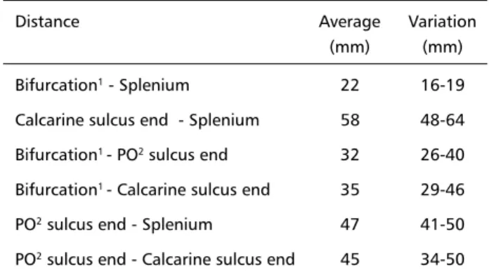
Table 1. Distance measured in the posterior interemispheric fis-sure in 26 anatomic specimen.
Distance Average Variation
(mm) (mm)
Bifurcation1 - Splenium 22 16-19
Calcarine sulcus end - Splenium 58 48-64 Bifurcation1 - PO2 sulcus end 32 26-40
Bifurcation1 - Calcarine sulcus end 35 29-46
PO2 sulcus end - Splenium 47 41-50
PO2 sulcus end - Calcarine sulcus end 45 34-50
1Bifurcation: emergence of the parieto-occiptal sulcus at the calcarine
RESULTS
It was observed that the calcarine sulcus emerged directly from the parahippocampal gyrus and was present as a complete sulcus in all of the hemispheres
examined. In 12 hemispheres, no side branches were observed (46%), in 10 there was just one (38%) and in 4 there were two side branches to the calcarine (15%). In relation to these side branches, 14 (77%) were observed with a perpendicular trajectory to the calcarine and 4 (23%) were in parallel trajectory. Eighty eight percent of the side branch sulci were identified posteriorlly to the emergence of the PO sulcus.
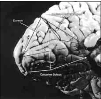
Table 1 summarizes the results of the measured distan-ces on the medial surface of the occipital lobe (Fig 1).
At the lateral surface of the occipital lobe, there was a determined pattern of sulci observed in most of the specimen. Twenty-four hemispheres (92%) showed sulci with evident transverse trajectories and in 16 hemispheres it was possible to identify marked longitudinal sulci (61%). All of the hemispheres in which transverse sulci were identified presented a sulcus localized on the inferior third of the lobe, and it was labeled as inferior transverse occipital sulcus. This one, in all of the specimens, presented as a com-plete sulcus. It was possible to identify a localized sulcus on the middle or superior third of the lobe in eighteen hemispheres (69%) – this was labeled su-perior transverse occipital sulcus – and this one was complete in only 10 hemispheres (55%). In just one case a third transverse sulcus was observed local-ized between the previously mentioned - labeled middle transverse occipital sulcus. Among the 16
Fig 1. Distance meansured on the medial surfarce of the occipital lobe: 1. spleninum to calcarine sulcus end; 2. splenium to calca-rine-PO* bifurcation; 3. splenium to PO* end; 4. PO* sulcus end to calcarine sulcus end; 5. PO*-calcarine sulcus bifurcation to PO* end. *Parieto-occipital.
hemispheres in which it was possible to identify lon-gitudinal sulci, the lunar sulcus was identified in 12 (46% of the specimen). Most of the cases showed patterns of the transverse sulci that permitted a di-vision of the occipital lobe into three gyri- inferior, middle and superior occipital (Fig 2).
Also, distances from the occipital pole to the pre-occipital notch were checked. This distance was lar-gely heterogenic between the hemispheres, ranged from 30 to 61 mm (average 44). In all of them, the preoccipital notch corresponded to a deep indenta-tion on the inferolateral border of the cortical sur-face, formed by the posterior portion of the inferior
temporal gyrus where it reaches the inferior occipi-tal gyrus.
Table 2 shows the measured depths of the sulci in the first section made at the level of the PO sulcus and at the second section, ten millimeters posterior to the previous. There was a tendency that all of the sulci were deeper anteriorlly, becoming shallower as they approached the pole. The occipital horn of the lateral ventricle was identified in 14 hemispheres (53%) at the level of the first section; and in only one specimen at the level of the second section. The ana-tomy of the occipital horn was consistent in all of the specimen: half moon shaped, in such way that the medial wall corresponded to the calcar avis and laterally formed by a convex wall, composed of ta-petum fibers. There were no observed floor nor pres-ence of collateral eminpres-ence or bulb of the corpus calosum (Fig 3). The average measurement from the end of the occipital horn to the splenium was 18 mm, varying from 15 to 32 mm.
DISCUSSION
There are a lot of situations when neurosurgeons need to work in the occipital lobe region, not only for direct intervention of primary lesions, but also for approach to deeper structures underlying the lobe12,13,16. Technology already offers modern
intra-operatory localization tools - such as
neuronavega-Table 2. Depth of the occipital lobe sulcus measured (in milimetrs) at the level of the parieto-occipital sulcus emergence (section 1) and ten millimetres posterior to the first one (section 2), in 26 cerebral hemispheres.
Sulci section 1 section 2
Average Variation Average Variation
Calcarine 20 15-25 15 9-15
Parieto-occipital 21 15-25 -
-STO2 15 10-18 14 8-18
ITO1 13 08-17 08 5-10
1ITO: Inferior transverse occiptal. 2STO: Superior trasverse occiptal.
tion – but the knowledge of anatomy itself is the greatest ally of the surgeon for the planning and execution of the operative act. Differently from other regions of the brain, whose anatomy has already been meticulously detailed6, there exist
controver-sies about the occipital region, those of which occasionally can hinder the surgical approaches to this lobe4.
The calcarine sulcus is the most important anato-mical reference of the posterior interhemispheric
fis-sure region8. The data obtained from the present
study showed that it invariably appears from the pa-rahippocampal gyrus and it is a sulcus with a few number of side branches. This information can fa-cilitate its identification in surgeries of this area. The measured distances from the medial surface permit the configuration of an anatomical map of this re-gion, having as landmarks the splenium, the calcar-ine sulcus and the PO sulcus. These results were com-pared with some measurements done by Ono et al.6.
These authors, in the work about the sulci of the en-cephalon, also found similar measurements to the calcarine and PO sulci. The results were very similar: as an example, the distance of the end of the calcarine sulcus to the splenium - 58 mm in both works; or the extension of the PO sulcus - 34 mm in the work by Ono et al.6 and 35 mm in the present paper.
The results, in relation the lateral surface of the lobe, brought interesting information. Different to what is quoted in most texts of neuroanatomy5-7,10,17,
the occipital lobe has a pattern of sulci with a ten-dency to repeat in the examined specimen. Trans-verse sulci were identified in 92% of the hemispheres, most of them found in the same location. The longi-tudinal sulci were less frequent, but were observed in 62% of the cases. So, this data show that the lobe presents a defined anatomy and this information can be useful to the surgeon who approaches this area. The inferior transverse occipital was the most fre-quently sulcus identified, being complete in most of the specimen. So, it could be used as an anatomical landmark for surgeries to the lateral surface of the occipital region. The superior transverse sulcus was observed in only 62% of the hemispheres, and was interrupted in most of them, decreasing its value as a surgical reference. Oka et al.18 described the
late-ral surface as a region in which there is just one mar-ked sulcus - labeled Lateral Occipital - and divided into two gyri - inferior and superior occipital. The present paper shows that the transverse sulci of the lateral surface delineate three gyri, that can be la-beled superior, middle and inferior occipital.
The preoccipital notch is an anatomical structure poorly defined by specialized neuroanatomy litera-ture5,6,17,19. Most of the texts quote this structure as
posterior boundary of the temporal lobe or the an-terior limit of the occipital lobe5,10,19, most commonly
located 50 mm anteriorlly to the occiptal pole7. In
all of the specimen of the present study, the preoc-cipital notch joined the posterior portion of the in-ferior temporal gyrus to the most anterior portion of the inferior occipital gyrus (localized inferiorly to the inferior transverse occipital sulcus). Its location was quite variable, and it was average positioned 44 mm from the occipital pole. Also, the average distance between the occipital pole and the emer-gence of the PO sulcus was 35 mm. The traditional limits to occipital lobectomies is the resection of 35 mm left and 70 mm to the right, from the occipital pole16. According to the identified parameters in this
study, resection of the right occipital lobe utilizing the traditional criteria includes portions of the pari-etal lobe (in some cases it may be desired, depend-ing on the extension of the lesion).
Timurkaynak et al. in their paper about the ana-tomy of the lateral ventricles, described the occipital horn as a structure with a floor, a medial and a lateral wall11. The present study shows that, at the level of
the occipital lobe, the occipital horn has already an different anatomy. In the specimens where it was pre-sented, all of then showed a half moon shaped cavity and the colateral eminence was not observed form-ing its wall. It was also observed that just in half of the cases there was ventricular cavity at the level of the anterior boundary of the lobe. As such, the surgeon should not expect to find the occipital horn in all per-formed occiptal lobectomies. Besides this, the occipi-tal horn tends to have its end anteriorly to the level of the emergence of the PO sulcus. This information can be useful when working in the posterior interhemis-perhic fissure: if the surgeon penetrates the ventricle, he/she should consider being located at the precu-neus, close to the atrium (obviously, preoperative ra-diological studies could assist to confirm the poste-rior extension of the ventricle).
In conclusion, the occipital lobe is a region of the brain that presents anatomical parameters that tend to repeat themselves in different examined sections. This information can be useful for the surgical plan-ning when approaching this area of the encephalon.
Acknowlegments Acknowlegments Acknowlegments Acknowlegments
REFERENCES
1. Barr LM, Kiernam JA. The humam nervous system: an anatomical 5.Ed. Philadelphia: Lippincott, 1988:143-156.
2. Kandell ER, Schwartz JH, Jessell TH. Essentials of neural science and behavior. New Jersey: Prentice Hall International, 1995:387-407. 3. Smith GE. New studies on the folding of the visual cortex and the
significance of the occiptal sulci in the humam brain. J Anat Physiol 1907;4:198-207.
4. Romero-Sierra C. Neuroanatomy: a conceptual approach. Edimburgh: Churchill Livingstone, 1986:237-278.
5. Carpenter MB. Core text of neuroanatomy. 4.Ed. Baltimore: Williams & Wilkins, 1991:23-54.
6. Ono M, Kubik S, Abernathey CD. Atlas of the cerebral sulci. New York: Thieme Medical Publishers, 1990:62-74.
7. Gray H, Goss CM. Anatomia. 29.Ed. Rio de Janeiro: Editora Guanabara, 1988:683-690.
8. Kubik S, Szarvas B. Anatomy of the calcarine sulcus. In Yasargil MG (ed.) Microneurosurgery III A: AVM of the Brain. Stuttgartd: Thieme Medical Publishers, 1987:350-368.
9. Montemuro DG, Bruni JE. The human brain in dissection. Philad-helphia: Saunders, 1981:129-141.
10. Seeger W. Atlas of topographical anatomy of the brain and surround-ing structures for neurosurgeons, neuroradiologists and neuropatholo-gists. Wien: Spriegel, 1978:340-349.
11. Timurkaynak E, Rhoton A Jr, Barry M. Microsurgical anatomy and operative approaches to the lateral ventricles. Neurosurgery 1986;19:685-723.
12. Dandy WE. Operative experience in cases of pineal tumors. Arch Surg 1936;33:19-46.
13. Drake CG. Aneurysms of the posterior cerebral artery. J Neurosurg 1969; 30: 468-474.
14. Koos WT, Spetzler RF, Lang J. Color atlas of microneurosurgery, Vol 1: Intracranial tumors . New York: Thieme Medical Publishers, 1993:256-276. 15. Lazar ML, Clark WK. Direct surgical management of masses in the
region of the pineal gland. Surg Neurol 1974;2:17-21.
16. Schimidek HH. Surgical management of supratentorial gliomas. In. Schimidek HH (ed) Operative neurosurgical techniques. Vol 1, 3.Ed. Philadelphia: Saunders, 1992:517-535.
17. Larsell OM. Anatomy of the nervous system. New York: Apleton-Cen-tury, 1951:256-278.
18. Oka K, Rhoton Jr.A, Barry M, Rodriguez R. Microsurgical anatomy of the superficial veins of the cerebrum. Neurosurgery 1985;17:711-748. 19. Rohen JW, Yokochi C. Atlas fotográfico de anatomia sistêmica e