New Dasycladales and microbiota from the lowermost Valanginian
of the Mirdita Zone
R
AJKAR
ADOI^I]Abstract. A rich diversified algal microbiota is described from the lowermost Valanginian limestone re-worked in the Upper Cretaceous clastics of the Metohija Cretaceous Unit (Mirdita Zone). Two new dasy-cladalean taxa are introduced: Zujovicella nov. gen. (type species Suppiluliumaella gocanini RADOI^I], 1972)
andFurcoporella vasilijesimici nov. sp.
Microbiota of this bioclastic limestone (containing dominantly corals and other metazoan fragments) consists of algae, microbial epiliths, microproblematica, foraminifera, calcispongie and a few calpionellids of the Calpio-nellopsisZone – Calpionellopsis oblonga (CADISCH), Remaniella cadischiana (COLOM),Tinntinopsella
carpath-ica(MURGEANU& FILIPESCU). Besides the new taxa dasycladales also associated are: Salpingoporella pygmaea
(GÜMBEL),Salpingoporellasp.,Gyroporella lukicaeSOKA^& VELI],Neomerinaeand several indetermined taxa.
Lithocodium aggregatum(ELLIOTT), other encrusting Lithocodioideaand different microbial epiliths are an
im-portant component of this microbiota association.
Foraminiferal assemblage consists of: Coscinophragma cf. C. cribrosum (REUSS), Mohlerina basiliensis
(MOHLER), Nautiloculina bronnimanniARNAUD-VANNEAU & PEYBERNÈS, Neotrocholina valdensis REICHEL,
Neo-trocholina sp. Placopsilina sp., Protopeneroplis trochangulata SEPTFONTAINE, Trocholina alpina (LEUPOLD),
Trocholina delphinensis ARNAUD-VANNEAU, BOISSEAU & DARSAC, Trocholinasp., lituolids, miliolids and other
small benthic taxa.
The analyzed lowermost Valanginian limestone originated from the topmost sequence of the Tithonian–Neo-comian cycle which ended as a consequence of the Main Cimmerian Events, which occerred, as in the Vardar zone, after the lowermost Valanginian. Cretaceous cycle (Mirdita Crtaceous Unit) begins in the Hauterivian.
Key words: Dasycladales, new genus, new species, Udoteaceae, Codiaceae, microproblematica, microbial epiliths, lowermost Valanginian, Late Cimmerian Events, Mirdita Zone.
Apstrakt. Opisana su dva nova taksona dazikladalesa: Zujovicella nov. gen. (type species Suppiluliu-maella gocanini RADOI^I], 1972) iFurcoporella vasilijesimici nov. sp. iz dowovalendiskog kre~waka – va-lutka u gorwokrednim klastitima Metohijske kredne jedinice (Mirdita zona). Prikazana je prate}a algalna mikrobiota (Dasycladales, Udoteaceae, Codiaceae), mibrobijski epiliti, mikroproblematika i kalcispongije.
U raznovrsnoj i bogatoj bioti ovog valendiskog kre~waka dominantan je udio korala, inkrustirajucih algi i mikrobialita.U skupini dazikladalesa, pored vi{e nedeterminisanih vrsta, pomenute su: Salpin-goporella pygmaea, Salpingoporella sp., Gyroporella lukicae. Listu foraminifera ~ine Cosciniphragma cf. C. cribrosum, Mohlerina basiliensis, Nautiloculina bronnimanni, Neotrocholina valdensis, Neotrocholina sp., Placopsilinasp., Protopeneroplis trochangulata, Trocholina alpina, Trocholina delphiensis, Trocholinasp., li-tuolidi, miliolidi i druge sitne bentoske vrste.
Za datirawe ovog valutka najzna~ajnije su kalpionelide – Calpionellopsis oblonga i Remaniella
ca-dischiana.One ukazuju da je ovaj sedimenat, koji poti~e iz najmla|ih slojeva titon–neokomskog ciklusa, deponovan u najni`em dowem valendisu. Na osnovu ovog podatka zakqu~uje se da glavni kimerijski doga|aji u podru~ju Mirdita zone nijesu stariji od najni`eg valendisa. Prema raspolo`ivim podacima kredni ciklus (Metohijska kredna jedinica) otpo~eo je u doba otriva.
Kqu~ne rije~i: Dasycladales, novi rod, nova vrsta, Udoteaceae, Codiaceae, mikroproblematika, mikrobijski epiliti, najstariji valendis, kasni kimerijski doga|aji, Mirdita zona.
Introduction
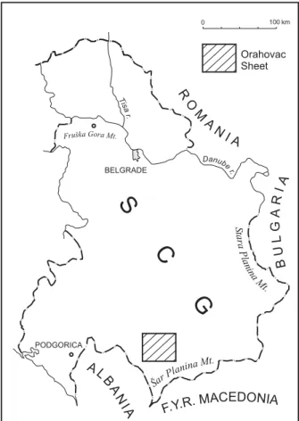
Dasycladales Suppiluliumaella gocanini were describ-ed from Lower Valanginian limestone of the Orahovac region, Mirdita Zone (Fig. 1). In the locality between the villages of Kravoserija and Boka this limestone was not found in situ (RADOI^I], 1972). According to the Geo-logical Map, the sheet Orahovac 1:100 000 (LON^ARE -VI], 1986), this area is covered by Upper Cretaceous carbonate clastics. On the footpath between the men-tioned villages, in a small outcrop of 2–2.5 m thick clastics, horizontally arranged cobblestones in the mi-crobreccia bed were observed. One of them (cobble of cca 15×12 cm) was limestone with large (to 6 cm) coral and other metazoan fragments. On the surface of broken pieces, in the matrix between large fragments (bioclastic packstone-rudstone), some dasycladalean, different coating algal and microbial structures and fo-raminifera, were observed under a lens. The matrix between large metazoan fragments was used for thin slides (17), in some of which sections of Suppiluliu-maella gocanini were found.
The purpose of this paper was the revision of Sup-piluliumaella gocanini, based on which Zujovicellanov. gen. is described, and the presentation rich associated biota including new dasycladalean species Furcoporella vasilijesimici.
Kravoseria Coble
The studied limestone coble bears abundant diversi-fied biota. Corals (different species) were its most im-portant component, calcisponge, hydrozoa, mollusk and other metazoa remains were also present. The micro-biota consisted of algae (Dasycladales, Udoteaceae, Lithocodioidea), microbial epiliths, foraminifera and miroproblematica; while calpionellids were represented by only a few specimens. The corals were an especial-ly alluring substrate for encrusting algae and similar microbial epiliths. Other metazoan remains and dasy-cladales were also the subject of epilithic activity.
The list of foraminifera includes: Coscinophragma cf. C. cribrosum (REUSS), Mohlerina basiliensis (MOH
-LER), Nautiloculina bronnimanni ARNAUD-VANNEAU & PEYBERNÈS,Neotrocholina valdensis, Neotrocholina sp., Placopsilina sp., Protopeneroplis trochangulata SEP -TFONTAINE, Trocholina alpina (LEUPOLD), Trocholina cf. T. delphinensis ARNAUD-VANNEAU, BOISSEAU & DARSAC, Trocholina sp., lituolids, miliolids and other small benthic taxa.
The presence of calpionellids is important for the dat-ing of this coble: Calpionellopsis oblonga (CADISCH) (Fig. 2A), Remaniella cadishiana (COLOM) (Fig. 2B) and Tintinnopsella carpathica MURGEANU & FILIPESCU indicate its lowermost Valanginian age (Calpionellopsis Zone).
Fig. 1. Location map – sheet Orahovac 1: 100 000.
Fig. 2. A. Calpionellopsis oblonga
(CADISCH).×360; thin slide RR2186. B. Remaniella cadischiana(COLOM).
This Valanginian limestone originates from the youngest sediments of the Tithonian–Neocomian cycle, which ended as a consequence of the Main Late Cim-merian Events. A new Cretaceous cycle begins discon-tinuously in Hauterivian (Metohija Cretaceous Unit). Valanginian shallow water limestone in the area of this cretaceous unit was not known in situ. Resedimented benthic biota of this age occurs in some microbreccia and calcarenite beds of the proximal basinal Tithonian– –Neocomian succession, outcropping in the same area. Valanginian limestone with corals as the essential component and Zujovicella gocanini was deposited in the reef thallus area.
Paleontology
Observation on
Suppiluliumaella gocanini
R
ADOI^I], 1972
The genus Suppiluliumaella ELLIOTT, according to the original diagnosis, is characterized by “verticils of long thin primaries with a large conspicuous terminal swelling, each dividing into very short swollen second-aries”.
The calcareous sheet of the holotype (ELLIOTT, 1968, Pl. 3, Fig. 1) is micritized, whereas secondary laterals are not well preserved. The presence of secondaries are discernable only at two or three points in this section. The paratype in Fig. 3 is an oblique section correspon-ding to an inclination of the stalky portion of primar-ies, while the secondary laterals, in central part of the fragment, were cut obliquely – therefore it is not their real length (they are not so short). The impression that the secondary laterals of some Suppiluliumaella species grow out from primaries in the form of a finger-like continuation (SOKA^ & NIKLER, 1983) is not a primary feature – it is only apparent (see BARATTOLO, 1984).
At first sight, Suppiluliumaella gocanini (densely set funnel-like whorls built of very long tubular primary la-terals which touch each other over a considerable part of their length and form terminal swellings; primaries carry at their ends tufts of the secondary laterals) re-sembles only the type-species Suppiluliumaella polyre-me. In fact it differs from any known Suppiluliumael-la species by having:
1. Larger (wider and longer) primary laterals, with apparent terminal swellings;
2. Quite different, trochophorous secondary laterals which could be extended hairy growth. In other species, although not fully preserved, secondary laterals are de-scribed as phloiophorous (ELLIOTT, 1968; BAKALOVA, 1971; DRAGASTAN, 1978, 1989; BARATTOLO, 1984).
These differences are the reason for the doubt as to whether this species belongs to the genus Suppiluliu-maella. Reexamination of Suppiluliumaella gocanini ty-pe material (and some new sections) led to the conclu-sion that this species is not a Suppiluliumaella. The
pri-mary laterals are only apparently terminated by a “swollen” portion. In fact, a tuft of trochophorous sec-ondaries starts from the top of the tubular primary lat-erals, enclosing a fertile ampulla. These characteristics indicate the affiliation of this species to the tribus Dasycladeae.
Order Dasycladales
Family
Dasycladaceae
K
UTZING, 1843
Tribus
Dasycladeae
P
IA, 1920
Subtribus
Neomerinae
(P
IA, 1927) B
ASSOULLETet
al
., 1978
Genus
Zujovicella
n. gen.
Type species. Suppilluliumaella gocanini RADOI^I], 1972
Origin of name. The genus is dedicated to Prof. JOVAN @UJOVI] (1856–1936), founder of geology in Serbia and the author of the first geological map of Serbia printed in Vienna 1886.
Diagnosis. Cylindrical or club-shaped thallus with densely set whorls, unfertile whorls can also be pres-ent. The fertile whorls consist of densely arranged cy-lindrical primary laterals each giving a second whorl segment: tufts of trochophorous secondary laterals and a fertile ampulla situated in the center of the tuft. Sur-face of the calcareous sheet indentated.
Comparisons. The genus Neomeris has a cylindrical to clavate thallus, ampulla and two secondary laterals placed in one plane, the thallus surface is faceted. Cymopolia has an anarticulated thallus and primaries with ampulla and 4–8 secondary laterals enlarged at the outer ends forming a faceted surface
Zujovicella gocanini
(R
ADOI^I], 1972) nov. comb.
Fig. 3; Pl. 1, Figs. l–6; Pl. 2, Figs. l–8
Diagnosis. A large slightly club-shaped thallus of differentiated structure – with a lower unfertile part. A wide central cavity. Densely set funnel-like whorls con-sisting of close-fitted laterals inclined upward at an angle of 65 to 75° from the horizontal. The long and large rounded at the top cylindrical primary laterals bear a large ooidal fertile ampulla and a tuft of 5 or 6 long trochophorous secondary laterals around the ampulla (Figs. 3, 4). Calcareous skeleton compact, mainly around the distal part of the whorls. The sur-face of the skeleton is indentated due to the trocho-phorous form of the second whorl segment.
Dimension (in mm) (based on some additional sec-tions than in 1972; the slightly oblique, deep-tangential section of the holotype is not relevant for the D and d values).
Maximum observed length 11.7. External diameter 2.593–6.500.
Axial diameter (reconstruction) about 20 to 25° of D. Length of primary laterals more than 1.600. Diameter of primary laterals 0.192–0.320. Length of secondary laterals to 1.440 or more. Dimensions of the fertile ampulla 0.240 × 0.450. Number of primaries per whorl 18–32.
Description. It seems that not every whorl in the fer-tile portion of the thallus is ferfer-tile. The ferfer-tile ampulla, placed in the center of the rounded at the top primaries, is protected by secondary laterals of a peculiar tro-chophorous form (Figs. 3, 4; Pl. 2, Fig. 4). Starting as a tubular of 0.048 mm diameter, the secondary laterals become largest (0.160 mm) at the middle of the length, closing the ampulla and distally thinning (Pl. 2, Fig. 4) probably extending in a hairy growth. The cross and oblique sections of the distal tuft portion have a copto-campylodon form (Pl. 2, Fig. 7; Pl. 8, Fig. 10).
The inner part of the thallus – main axis and first seg-ment of the whorls – is usually not calcified. According to a reconstruction, the main axis was narrow, probably
not wider than 25% of the external diameter. Calcifi-cation was limited to the distal area of the whorls, dimin-ishing toward the interior.The membrane of the primar-ies, partly preserved in only some specimens, was very thinly calcified (Pl. 1, Figs. l, 3). The membrane of the fertile ampulla and secondaries was also thinly encrusted (visible as a thin dark line in diverse sections). In fact, the compact calcareous skeleton is formed by a calcified mucilage layer around the secondaries (Pl. 1, Figs. 3, 5; Pl. 2, Figs. 4, 5). Secondary sparry calcite had deposited (before the sheet was coated) mainly in the secondaries and sometimes in the ampulla or inside the primaries. In the slightly oblique deep-tangential section (holotype, Pl. 1, Figs. 1, 3–4), differently preserved second whorl seg-ments are visible. Its different aspect depends also on the cut plane. Hence, for slightly recrystallized segments (= ampullae and basal parts of the secondaries shown in Pl. 1, Fig. 4, arrow 1) it can be wrongly interpreted that the ampulla (“swelling”) bears finger-like short secondaries. A similar apparent relation between the ampulla and “short” secondaries is shown by arrow 2. Tangentially cut primaries and tufts are indicated by arrow 3.
The indentated skeleton surface was usually eroded and often corroded by microbial epiliths. The corroding effect in some specimens appears to have been locally arrested. In such cases it seems that the epiliths even contributed to the preservation of some structure by pro-tecting the skeleton from further erosion and dissolution and moderating the effect of further diagenetic processes.
Neomerinae
div. sp.
Pl. 4, Fig. 5
Neomerinae are a minor component in this Valan-ginian algal association. Only an oblique section of a
Fig. 3. Zujovicella gocanini (RADOI^I]) nov. comb. Oblique
section of the second whorl segment: laterals protecting a fertile ampulla; × 60; thin slide RR2199.
Fig. 4. Zujovicella gocanini (RADOI^I]) nov. comb. Axial
compact cylindrical skeleton (Drimella?) and a small fragment of another species have been found. The frag-ment shown in the same plate Fig. 6, could be related to Neomerinae.
Family
Triploporaceae
(P
IA1920) B
ERGER&
K
AEVER, 1992
Tribus
Salpingoporelleae
B
ASSOULLETet al
., 1979
Genus
Furcoporella
P
IA(
in
T
RAUTH), 1918
The genus Furcoporelawas introduced by PIA from the Middle Eocene in Austria with the species Furco-porella diplopora (PIA, 1918). The simple cylindrical thallus of this species is characterized by spaced whorls of horizontally disposed two orders of laterals. The pri-mary laterals are short, divided into two divergent sec-ondaries enlarged to the exterior. Hitherto, the genus was monospecific. (Based on the characteristic of a new species, upward inclined laterals, diagnosis of thus genus should be amplified).
Furcoporella vasilijesimici
n. sp.
Pl. 3, Figs. 1–3
Origin of name. The species is dedicated to Dr VASILIJE SIMI] (1902–1990), one of the pioneers of modern geology and paleontology in Serbia.
Holotype. longitudinal oblique section shown in Pl. 3, Fig.1, thin slide RR2190, the author’s collection deposit-ed in the Geozavod – Geological Institute in Belgrade.
Isotypes. Specimens in thin slides RR2184 and 2201 figured in Pl. 3, and two poorly preserved specimens in thin slides RR2186 and 2189.
Age. Lowermost Valanginian, based on calpionellids: Calpionellopsis oblonga, Remaniella cadishiana, and Tintinnopsella carpathica.
Diagnosis. Thallus cylindrical with a relatively large axis and spaced whorls consisting of two order of later-als inclined upward, alternating from one whorl to the next. Short primary laterals divided in two vesicular sec-ondaries, slightly swollen at the distal end. Secondary lat-erals are in a somewhat irregular, oblique position. Calci-fication is massive, from the axis to the subcortical area.
Dimension (in mm):
Maximum observed length (holotype) 5.440. Inner diameter 0.740–0.988.
External diameter 1.974–2.024. Distance between whorls cca 0.370. Length of primary laterals 0.080. Length of secondary laterals 0.480.
Distal diameter of secondary laterals 0.096. Number of primary laterals per whorl 16–18.
Description. The peculiarity of this species lies in the position of the secondary laterals which is reflect-ed in more advancreflect-ed corrosion and dissolution in some parts of the skeleton. The upward inclined secondaries
are irregularly disposed, more in the distal area. They are arranged in an oblique to radial plane, which is evi-dent in the transversal or slightly oblique-transversal sections (Pl 3, Figs, 2 and 3).
Comparison. The thallus structure of this species differs from Furcoporella diplopora in having inclined laterals, i.e. funnel like whorls and a different position of the secondaries.
Genus
Salpingoporella
P
IA, 1918
Salpingoporella pygmaea
(G
ÜMBEL)
Pl. 4, Fig. 4
This species is represented by a few specimens only.
Salpingoporella
? sp.
Pl. 4, Fig. 10
Fragment of the oblique section showing a specimen with regularly arranged dense whorls of primary later-als (35 or more per whorl).
Genus
Gyroporella
G
ÜMBEL1872,
emend. B
ENECKE, 1878
According to BASSOULLET et al., (1978), the geno-typeGyroporella vesiculifera is a species with “Aspon-dyl disposition of laterals of one single order of vesi-culiferous type”.
ZANIN-BURI (1965) in a study of carbonate algae from Trias of the Lombardian Pre-alpes presented very well preserved euspondyle specimens of Gyroporella vesiculiferafrom the Norian of the Dolomites (imprints on a rock surface and sections in thin slides: Pl. 46, Figs. 1, 2, and Pl. 47). In these specimens “I pori sono fittamente and omogeneamente disposti tanto che sono in contatto gli uni cogli altri, la loro disposizione e cosi regolare che si puo trovare un perfetto allineamento in file orizzontali, verticali e diagonali.”
As to the disposition of the laterals (aspondyle or euspondyle) see discussion by BARATTOLO et al. (1993). According these authors, the possibility should not be excluded that the genera Gyroporella and Griphoporella PIA are synonyms, which could be resolved by revision of Gyroporella vesiculifera type material.
Gyroporella lukicae
S
OKA^& V
ELI], 1982
Pl. 3, Figs. 4–10
approximately alternating neighbouring whorls which are not always clearly visible”
SENOWBARI-DARYAN et al. (1994), considering that the genus Gyroporella is aspondyle, proposed new combination: Anisoporella lukicae (SOKA^ & VELI]). The comparison with Anisoporella has not been justi-fied. The genus Anisoporella has an entirely different structure: “L’arrangement des rameaux se fait en gen-eral par serie de deux (= double verticilles) come chez Oligoporella. “L’alternance des rameaux est presque la regle, mais les pores sont tantot disposes en quin-quonce, tantot par paires, tantot par groupes de trios. Ces trois modes de distribution se retrouvent alterna-tivement sur le meme manchon, dans un ordre de suc-cession quelconque” (BOTTERON, 1961).
In the Kravoseria limestone coble, Gyroporella luki-cae is a relatively frequent fossil (cylindrical skeleton and numerous fragments), and of very variable dimen-sions. Besides specimens which correspond to the type material, there are larger forms with densely set more numerous laterals in the whorls (Pl. 3, Figs. 8, 10). The peculiarity of the specimen in Pl. 3, Fig. 9 is the supraterminal growth (note: evidently thinner and den-ser laterals at top of the rounded thallus).
Dimensions (in mm; extreme values given in brack-ets):
Dasycladales
div. sp.
The analyzed thin slides contain some unknown dasycladalean, mainly in fragments. They are, distin-guished by symbols KL1 to KL8, illustrated in Pl. 4. Figs. 1–3, 6–9 and 11.
Morphogenus
Coptocampylodon
(E
LLIOTT, 1963)
L
JUBOVI]-O
BRADOVI]& R
ADOI^I], 2003
Three different Coptocampylodon morphospecies occur in this limestone: Coptocampylodoncf. fontis(Pl. 7, Fig. 8), Coptocampylodon sp. 1 (Pl. 7, Fig. 7) and Coptocampylodon sp. 2 (Pl. 4, Figs. 12, 13).
Order Bryopsidales
Family
Udoteaceae
(E
NDLICHER) A
GARDH, 1888
Genus
Pinnatiporidium
D
RAGASTAN, 1990
Pinnatiporidium
sp.
Pl. 9, Figs. 3–6
The transverse section of Nipponophycus ramosus illustrated by SENOWBARY-DARYAN et al. (1994) in Pl.
7, Fig. 3, and the transverse section of Nipponophycus sp. illustrated in the same plate, Fig. 8 have a clearly lobed periphery. These specimens differ from numerous Nipponophycus talli presented in the same paper in Pl. 7. Poorly preserved, recrystallized specimens of this type are found in the studied thin slides. Some oblique sections, such as those illustrated in Pl. 9, Figs. 3–6, indicate a segmented morphology of the thallus (short hemispherical somewhat irregular flabelliform seg-ments) These sections are very similar to the oblique section of a segmented thallus showed by SANOWBARY -DARYAN et al. (1994) in Pl. 5, Fig. 15 as Pinnatipo-ridium sp. cf PinnatipoPinnatipo-ridium cylindricum DRAGASTAN. The genus Pinnatiporidiumis characterized by a mented thallus with superficial constrictions and seg-ments with three distinct zones: central, lateral (with two types of branching) and cortical (DRAGASTAN, 1990). As shown by the illustrated specimens, these structures are generally not observable due to poorly preserved or recrystallized thalli; the central medular zone was not preserved, while relatively large, second order branches are visible only in the section on Pl. 9, Fig. 4, while treads of thin branching of the cortical zone can be soon on the same plate, Fig. 5.
Genus
Felixporidium
D
RAGASTAN, 1999
Felixporidium
? sp.
Pl. 9, Fig. 8
The thallus of Felixporidium, “built of bush-like hemispherical segments or flabellae,” crossed by a large medullary siphon which, in every segment, is followed by two order siphons (filaments) “branched dichotomi-cally and disposed radially”. Felixporidium is related to Pinnatiporidium DRAGASTAN, 1990 by the similar form and structure of the thallus. The differences between the two genera a the generic level is an open question.
In the longitudinal-oblique section of the illustrated specimen, the flabelliform arranged three order laterals appertaining to successive segments are well preserved. Similar flabelliform branching can be discerned on the poorly preserved specimen of Felixporidium alpidicum DRAGASTAN, 1999, illustrated in the original publication on Pl. 6, Fig. 8. This species, according to DRAGASTAN, has an inner morphology which is very close to Pin-natiporidium cylindricum, (type species of the genus).
Genus Nipponophycus YABE & TOYAMA, 1928 Nipponophycus ramosus YABE & TOYAMA, 1928
Pl. 9, Figs. 1–2
The few specimens assigned to this species with a cylindrical branching thallus are strongly recrystallized: the medulare zone is represented by a narrow cavity, Bosnia (type locality) Kravoserija
while the structure of the internal zone is completely obliterated.
Family
Codiaceae
(T
REVISAN) Z
ANARDINI, 1843
Subfamily
Lithocodioidea
B
ANNER, F
INCH&
S
IMMONS, 1990
Lithocodium aggregatum
(E
LLIOTT, 1956),
B
ANNER, F
INCH& S
IMMONS, 1990
Pl. 5, Figs. 1–8; Pl. 6, Figs. 1–4; Pl. 8, Fig. 6
The encrusting alga Lithocodium aggregatum charac-terized by a high degree of variability in both: the thal-lus morphology and, especially, the internal structure (ELLIOTT, 1956; BANNER et al., 1990). The Lithocodium nodules are usually formed by superimposed thalli. This is, certainly, the green alga with the most variable features. Therefore, it seems justified, as noted by BAN -NER et al., that the genus Lithocodium“may subjective-ly be monotypic”.
In the same Lithocodium thallus, different arrange-ments the structure of the medulla and cortex can be ob-served. The specimen illustrated on Pl. 5, Fig. 4 has one type of Lithocodiumstructure developed only in a part of the growth: a coarse medula followed by some order of variously ramifing filaments, ending with short fine cor-tical filaments covered by a thin calcareous sheet (Fig. 7 shows detail of Fig. 4). Laterally, this structure grades into a somewhat reduced subcortical zone similar to those of the thallus in the Fig. 3, or grades in a part of the thallus, apparently ”terminated” without these structures.
The narrow elongate growth (aspect in the section!) on Pl. 5, Fig. 3 has, immediately below the cortical structure, relatively large septate filaments of the me-dulla. Here, the ramification is reduced: large medulla filaments immediately give numerous very short fine cortical (bifurcate?) filaments terminating in a thin well preserved calcareous sheet
Solitary corals are usually encrusted by a Lithoco-dium or other Lithocodioidea crust and more or less corroded (Pl. 6, Figs. 3, 4,); often it is superimposed by a new Lithocodium or another encrusting growth.
The large (cca 30 mm), very irregular, ramified nodu-la incorporating corals, calcispongia and other biocnodu-lasts is, in fact, a mélange of Lithocodium, other Lithocodio-idea and different microbialites (Pl. 6, Figs. 1, 2, 7a, b; Pl. 7, Figs. 1–5). Growth of these organisms is so in-terweaved that its boundaries are impossible to define. Colonia of Koskinobullina socialis, rodophycean alga and encrusting calcispongia (Pl. 7, Figs. 1, 2, 5) were present only in short episodes before further Lithocodium growth. On the surface of Lithocodium and other Litho-codioidea, in some places, thin microbial crusts devel-oped, some of them are illustrated on Pl. 6, Fig. 7, and on Pl. 7, Fig. 3.
Elements encased and more or less corroded by Lithocodium are metazoan, their fragments,
foramini-fera, dasycladales, other algal and problematic struc-tures and different rock grains. The corroding process can results in a more or less disintegrated substratum. It seems that disintegration is usually not an effect of only Lithocodioidea activity but also of some preced-ing microbial chasmoendoliths. For example, in the pores between the skeleton elements of corals and hy-drozoa encrusted by Lithocodioidea, the earlier activity of microbial chasmoendolith is evident.
Lithocodium aggregatum is one of the widely dis-tributed fossils (Upper Jurassic – Albian, BANNERet al., 1990) particularly in the Lower Cretaceous limestone of both Tethyan margins. In the analyzed limestone, Litho-codium aggregatum and similar encrusting growth are important components.
Lithocodioidea
KL11
Pl. 9, Fig. 9
The thin compact crust of the only specimen in the available thin slides is characterized by thin bifurcate filaments, perpendicular or subperpendicular to the sur-face. Similar crust (“aff. KL11”) is shown in Fig. 10 on the same plate
Lithocodioidea
KL12
Pl. 8 Fig. 2
Agglomeration (encased in Lithocodium) of thin-walled, subcircular and irregular rounded cells is the single specimen of this type of structure.
Lithocodioidea
KL13
Pl. 8, Fig. 1
A crust with a peculiar subcrustal structure is part of the superimposed growth on Lithocodium.
Incertae Sedis (algae)
“Tubiphytes” morronensis
C
RESCENTI, 1969
Pl. 9, Fig. 7
The enigmatic fossil “Tubiphytes” morronensis was widespread in the Upper Jurassic and Lower Cretaceous reef areas of both Tethyan margins. Many of those spe-cimens represent a differentiated axial “nubeculariform” part from a coated growth (probably of microbial natu-re). This taxon is not frequent in the Kravoserija lime-stone.
“Gryphoporella” perforatissima
C
AROZZI, 1955
Pl. 3, Fig. 9 p. p.
to this CAROZZI’S species. According to BASSOULLET et al. (1978), dasycladalean affinity of this species is doubtful.
Fossils related to CAROZZI’S species were described as Gryphoporella piae by DRAGASTAN in 1971, then, based on the same specimens, finding similarity with Trinocladus pinarensis KEIJZER, DRAGASTAN in 1978 introduced a new genus Pseudotrinocladus. The doubt in the dasycladalean nature of this species seems justi-fied. This fossil could be related to udoteacean algae.
Microproblematica
KL15
Pl. 8, Fig. 7
The small claviform corpuscles with a thin regular-ly reticulate wall are parts of a larger structure.
Family
Wetheredelidae
V
ACHARD, 1977
Genus
Koskinobullina
C
HERCHI& S
CHROEDER, 1979
Koskinobullina socialis
C
HERCHI& S
CHROEDER, 1979
Pl. 7, Figs. 1, 2 p. p.
Koskinobullina socialis is interpreted as “small vesic-ular individuals forming colonies of varying dimensions which are fixed upon a substrate” “either as solitary indi-viduals or, more frequently, in the form of crust-like agglomeration” (CHERCHI & SCHROEDER, 1984).
Lithocodium and similar structures alternate with crust-like Koskinobullina agglomerations as those shown on Pl. 7, Fig. 1. In the same plate, Fig. 2, the colonia of Koskinobullina socialis is covered by rodophycean alga.
Microbial epiliths and other microbiota
Microbial epilith
KL16
Pl. 6, Fig. 6
Lammelar (stromatolithic) microbial buildup with fine horizontal filaments. The same structure in Fig. 1, also developed on Lithocodium aggregatum nodula, is poorly preserved and is not clearly visible in this pho-tograph.
Microbial epilith
KL17 and KL18
Pl. 8, Figs. 3, 4
The microbial crust coating of an about 7.2 mm long bioclast seems homogeneous without a clear internal structure (except for a few large pores). On the upper side of the bioclast the microbial crust is thicker with a tuberculate surface (Pl. 8, Fig. 3). Meanwhile, on the photograph made at low exposition, two different or-ganisms are evident. In the first thin encrusting
micrit-ic layer (KL17, calcisponge? crust, Fig. 4) perpendmicrit-icu- perpendicu-lar to the surface, wedge-like spicules with upward ori-ented wider ends were noted (wedge-like form of spicules could be apparent!). This even thick layer was encrusted by microbialite of a tuberculate surface (KL18, Fig. 4) in which a fine tubular filamentous tis-sue could be discerned.
The thin crust of atuberculate surface, in Fig. 8 on plate 6, corroding a mollusk shell has large pores only in the figured part of the growth. Excluding this ele-ment of the structure, the tissue of the crust seems sim-ilar to that of the microbialite KL18.
Microbial epilith
KL19
Pl. 7, Fig. 3
On a part of a, Lithocodium mélange nodula, a fine complex crust was developed. It consists of a barely discernible, densely porous micritic tissue covered by thin porous calcitic lamina.
Microbial epilith
KL20
Pl. 6, Fig. 7 a, b
On the same melange nodula, in two parts, a crust of fine, chain-like, slightly interlaced threads were developed. They are longitudinal sections of fine tubules of 0.016 mm diameter. In Fig. 7b transversal sections of tubules are visible.
Microbial epilith
KL21
Pl. 6, Fig. 5
Two growth phases of this microbialite corroding hydrozoan skeleton are evident: epilithic and chasmoen-dolithic in the skeleton pores.
Microbial chasmoendolith
KL22
Pl. 8, Fig. 8
In this case, the pores in the Cladocoropsis? skele-ton has been lined by the microbial chasmoendolit.
Calcisponge
(or
Lithocodioidea
?) KL23
Pl. 8, Fig. 9
A small micritic thallus with aninsufficiently clear internal structure – sponge spicules?
Calcisponges, usually not well preserved, in the ana-lyzed limestone are more abundant than immediately ap-parent. Some of them are illustrated on Plates 7 (Akasp.).
Acknowledgements
The author is gratefull to prof. STJEPKO GOLUBI]
References
BAKALOVA, D., 1971. Nouvelles especes de Dasycladaceae
(Algae) dans les sédiments urgoniens du Prébalkan cen-tral. Bulletin of the Geological Institut, Series Paleonto-logy, 20: 123–127.
BANNER, F.T., FINCH, E.M. & SIMMONS, M.D., 1990. On
LithocodiumElliott (Calcareous algae): Its paleobiological and stratigraphical significance. Journal of Micropaleon-tology, 9 (1): 26–36.
BARATTOLO, F., 1984. Osservazioni su Suppiluliumaella
schro-ederin. sp. (algae verdi, Dasycladali) del Cenomaniano del Matese (Appennino meridionale, Italia). Bolletino della So-cieta dei Naturalisti in Napoli, 92 (1983): 1–27.
BARATTOLO, F., DECASTRO, P. & PARENTE, M., 1993. Some
remarks on Griphoporella curvata (GÜMBEL, 1872) PIA,
1915, dasycladacean green alga from the Upper Triassic. 5th International Symposium on Fossil Algae, 23–45,
Capri, 7–12 April, 1991.
BASSOULLET, J.P., BERNIER, P., CONRAD, M.A., DELOFFRE, R.,
&. JAFFREZO, M., 1978. Les Algues Dasycladales du
Juras-sique et du Crétacé. Geobios, Mémoire spécial, 2: 1–330. BOTTERON, G., 1961. Etude géologique de la région Mont
d-Or (Prealpes romandes). Eclogae geologicae Helvetiae, 54 (1): 29–106.
CHERCHI, A. & SCHROEDER, R., 1984. Koskinobulina socialis
CHERCHI & SCHROEDER, 1979. A colonial microfossil
incertae sedis (algae?) from Jurassic – Cretaceous of the Mediterranean Region. Bolletino della Societa Paleontolo-gica Italiana, 23 (2): 361–374.
DRAGASTAN, O., 1978. Microfacies de la serie calcaire
Cré-tacé inferieur d’Aliman (Dobrogea de Sud). Dari de Seama ale Sedintelor, 64: 107–136.
DRAGASTAN, O., 1989. Calcareous algae (new and revised),
microproblematicae and Foraminiferida of Jurassic–Lower Cretaceous deposits from the Carpathian Area. Rivista Española de Micropaleontología, 21 (1): 5–65.
DRAGASTAN, O., 1990. New Udoteaceae algae of the
Meso-zoic. Rivista Española de Micropaleontología, 22 (3): 481–498.
DRAGASTAN, O., 1999. Jurassic–Cretaceous algae of the
Transylvanides, inner Dacides and Moesian Platform (Ro-mania). Rivista Española de Micropaleontología, 31 (2): 185–218.
ELLIOTT, G.F., 1956. Further records of fossil calcareous
algae from the Middle East. Micropaleontology, 2 (4): 327–334.
ELLIOTT, G. F., 1968. Three new Tethyan Dasycladaceae
(Calcareous Algae). Palaeontology, 11 (4): 491–497. LON^AREVI], C., 1986. Basic geological map of SFR Yugoslavia
1:100000, Sheet Orahovac.Savezni geolo{ki zavod, Beograd. PIA, J. 1918. Thallophyta, Dasycladaceae. In: TRAUTH, F.: Das
Eocanvorkommen bei Radstadt im Pongau. Dankschriften der Kaiserliche Akademie der Wissenschaften (mathema-tisch – naturwissenschatliche Klasse), 95: 209–213. RADOI^I], R., 1972. A new Neocomian Dasycladacea
Sup-piluliumaella gocanini from the environs of Orahovac (Mirdita Zone). Bulletin scientifique, Conseil des
Aca-demies Sciences et des Arts de la RSF de Yougoslavie, 17 (11–12): 73–74.
SENOWBARI-DARYAN, B., BUCUR, I., & ABATE, B. 1994.
Up-per Jurassic Calcareous Algae from the Madonie Mountain, Sicily. Beiträge zur Paläontologie, 19: 227–259, Wien. SOKA^, B. & NIKLER, L., 1983. Calcareous algae from the
Lo-wer Cretaceous of the environs of Nik{i}, Crna Gora (Mon-tenegro). Paleontologia Jugoslavica, 13: 1–57.
SOKA^, B. & VELI], I, 1982. Gyroporella lukicae n. sp.
(Dasycladaceae) from the Lower Aptian of the surround-ings of Jajce. Geolo{ki vjesnik, 35: 37–41.
ZANINBURI, C., 1965. Il Trias in Lombardia. XIII. Le Algae
calcaree delle Prealpi Lombarde. Rivista Italiana di Paleontologia, 71 (2): 449–544.
Rezime
Nove
Dasycladales
i mikrobiota izdowovalendiskog kre~waka Mirdita zone
Prikazane je bogata algalna i druga mikrobiota iz dowovalendiskog kre~waka –valutka na|enog u gorwokrednim klastitima Metohijske kredne je-dinice. Iz ovog valutka bila je opisana dazikla-daleska vrsta Suppiluliumaella gocanini RADOI^I], 1972, koja je sada, na osnovu dodatnog materijala, detaqnije prou~ena. To je rezultiralo uvo|ewem novog roda, Zujovicella, koji je posve}en prof. Jo-vanu @ujovi}u, osniva~u geologije u Srbiji i au-toru prve geolo{ke karte Srbije. Tako|e je opi-sana i nova vrsta roda Furcopporella posve}ena dr Vasiliju Simi}u – Furcoporella vasilijesimici.
Zujovicella
n. gen. (
tipska vrstaSuppiluliumaella gocanini
R
ADOI^I], 1972).
Dijagnoza: Cilindri~an ili kijicast talus sa gustim pr{qenovima fertilnih ali i sterilnih ogranaka. Fertilni pr{qenovi sastoje se od gusto slo`enih cilindri~nih primarnih ogranaka koji nose drugi segment pr{qena: pramenove troho-fornih sekundarnih ogranaka sa fertilnom ampu-lom u sredini. Povr{ina kre~wa~kog omota~a nazubqena.
Rod Zujovicella razlikuje se od rodovaNeomeris i
Cymopoliapo tome {to rodNeomerisima dva sekun-darna distalno pro{irena ogranka po primarnom i fasetiranu povr{inu skeleta, a {to rod
Cymo-polia ima ~lankovit talus sa 4–8 distalno pro{i-renih ogranaka koji tako|e formiraju fasetiranu povr{inu.
Furcoporella vasilijesimici
n. sp.
ogranci daju po dva vesikularna distalno zaobqena ogranka, oni le`e u ravni koso postavqenoj prema aksijalnoj ravni. Kalcifikacija masivna, od cen-tralne ose do distalnog dijela ogranaka.
Furcoporella vasilijesimici razlikuje se od vrste
Furcoporella diplopora po qevkastim pr{qenovima odnosno po polo`aju ogranaka – oni le`e u ravni koja koso sije~e aksijalnu ravan.
Asocijaciju dazikladalesa ~ine jo{
Griphopo-rella lukicae, Salpingoporella pygmaea, Salpingoporel-lasp., Neomerinae, kao i vi{e neodre|enih taksona (fragmenti 10 vrsta). Osim korala i drugih
meta-zoa, zna~ajnu komponentu ovog bioklasti~nog kre~waka ~ine predstavnici Lithocodioidea i mi-krobialiti.
Starost valutka, najni`i dowi valendis, odre-|ena ja na osnovu kalpionelida: Calpionellopsis
oblonga i Remaniella cadischiana
U podru~ju Metohijske kredne jedinice val-endiski kre~waci nijesu poznati in situ. Analizi-rani valutak poti~e iz najmla|ih slojeva titon neokomskog ciklusa. Najstariji sedimenti krednog ciklusa (Metohijska kredna jedinica) otrivske su starosti.
PLATE 1
Figs. 1–6. Zujovicella gocanini (RADOI^I], 1972) nov. comb.
1. Holotype, slightly oblique deep-tangential section; × 15.5; thin slide RR2196. 2. Transversal section, fragment coated by a microbial epilith, arrow: transver-sal section of the tuft with a fertile ampulla in the center; × 17.5; thin slide RR2188.
3–5. Details from Fig. 1; × 35; 3–4: note the different preservation of the lat-erals in successive whorls.
PLATE 2
Figs. 1–8. Zujovicella gocanini (RADOI^I], 1972) nov. comb.
1. Oblique section of the largest specimen; × 7.5; thin slide RR2200. 2. Oblique section; × 20; thin slide RR2187.
3. Oblique section; × 15; thin slide RR2194.
4. Longitudinal-oblique section of the second whorl segments; note: tuft of secon-daries with, between them, a tangentially cut fertile ampulla; × 40; thin slide RR2188.
5. Transversal section; × 15; thin slide RR2199; note: a thin black line of the calcified membrane of secondaries, and, around them, calcified mucilage. On the right an encrusting foraminifer Placopsilina sp. Microbial endolith partly superimposed by Placopsilina(arrow); the axial surface of the calcareous sheet more corroded.
6. Tangential-oblique section through primary laterals; × 30; thin slide RR2193. 7. Different oblique section of tufts (coptocampylodon form); × 20; thin slide
RR2199.
8. Transversal slightly oblique section of a poorly preserved specimen; × 20; thin slide RR2187.
PLATE 3
Figs. 1–3. Furcoporella vasilijesimici nov. sp.
1. Holotype, longitudinsal-oblique section, × 25, thin slide RR2190; arrow 1 = short primary and secondary laterals, arrow 2 = claviform secondary laterals.
2, 3. Transversal slightly-oblique sections; × 20; thin slides RR2184. 2201, ar-rows: position of the secondary laterals in pairs.
Figs. 4–10. Gyroporella lukicae SOKA^ & VELI], 1982.
PLATE 4
Figs. 1, 2. Dasycladales KL1 and KL2. × 30; thin slides RR2201, 2190. Fig. 3. Dasycladales KL3 (Suppiluliumaella?). × 17.5; thin slide RR2194. Fig. 4. Salpingoporella pygmaea (GÜMBEL). × 35; thin slide RR2189. Fig. 5. Neomeris (Drimella?). × 30; thin slide RR2195.
Fig. 6. Dasycladales KL4, Neomerinae? × 30; thin slide 2197. Fig. 7. Dasycladales KL5. × 20; thin slide RR2197.
Fig. 8. Dasycladales KL6. × 30; thin slides RR2190. Fig. 9. Dasycladales KL7. × 20; thin slide RR2197. Fig. 10. Salpingoporella sp. × 60; thin slide RR2195. Fig. 11. Dasycladales KL8. × 20; thin slide RR2197.
PLATE 5
Figs. 1–8. Lithocodium aggregatum (ELLIOTT, 1956) BANNER, FINCH & SIMMONS.
1. × 25; thin slide 2188. 2. × 30; thin slide RR2195. 3, 5, 7. × 20; thin slide RR2190.
4. × 15; RR2190, arrow 1 = detail shown in Fig. 7; arrow 2 = medulla similar to those in Fig. 3.
PLATE 6
Fig. 1. Lithocodium aggregatum(ELLIOTT) BANNER, FINCH& SIMMONS. ×12.5; thin slide RR2191; arrow = boundary with microbialite KL16.
Fig. 2. Lithocodium aggregatum (ELLIOTT) with micritic crust (KL20, not visible in this
photograph); upper righ t= Protopeneroplis trochangulata ARNAUD-VANNEAU &
PEYBERNÈS; × 30; thin slide RR2191.
Fig. 3. Microbial crusts (arrow) englobed in Lithocodium. × 15; thin slide RR2189. Fig. 4. Coral encrusted and corroded by Lithocodium (part of the same melange nodula
shown in Fig. 1). × 15; thin slide RR2189.
Fig. 5. Microbialite (in advanced phase chasmoendolith) KL21. × 30; thin slide RR2199. Fig. 6. Microbialite KL16, (stromatholitic crust on Lithocodium). × 35; thin slide 2201. Fig 7a, b. Microbialite KL20, longitudinal and transversal section of tubules. a = × 35;
b = × 90; thin slide RR2191.
PLATE 7
Fig. 1. Part of Lithocodium melange nodula with episodes of Koskinobullina socialis colonial growth (K). × 60; thin slide RR2191.
Fig. 2. A part of the same nodula with Koskinobullina socialis and rodophycean alga. × 35; thin slide 2191.
Fig. 3. Microbialite KL19, on the melange nodula. × 50; thin slide RR2191.
Fig. 4. Calcisponge encased by Lithocodium melange nodula. × 30; thin slide RR2191. Fig. 5. Encrusting calcisponge encrusted by Lithocodium. × 20; thin slide RR2191. Fig. 6. Aka sp.× 30; thin slide RR2190.
Fig. 7. Coptocampylodon sp. 1. × 30; thin slide RR2189.
Fig. 8. Coptocampylodon fontis (PARTULIUS). × 40; thin slide RR2199.
Fig. 9. Zujovicella gocanini (RADOI^I], 1972) nov. comb. Oblique section of the distal
PLATE 8
Fig. 1. Lithocodioidea KL12. × 30; thin slide RR2191.
Fig. 2. Lithocodioidea KL11 englobed in Lithocodium nodula.× 35; thin slide RR2194. Figs. 3, 4. Lithocodioidea KL17. × 7.5. KL18; × 35; thin slide RR2189. Fig. 4 = detail of Fig. 3 (in the photograph made at low exposition, two different crusts are evi-dent); 1 = KL17 and 2 = KL18.
Fig. 5. Different Lithocodioideaand microbial crusts, a part of the Lithocodium nodula coating coral (Fig. 6). × 40; thin slide RR2201.
Fig. 6. Recrystallized coral? Coated and corroded by Lithocodium (Fig. 5 = topmost part of this nodula); × 7.5; thin slide RR2201.
Fig. 7. Problematica KL15. × 30; thin slide RR2197.
Fig. 8. Microbial chasmoendolits KL22 in pores of a Cladocoropsis? skeleton. × 35; thin slide RR2196.
PLATE 9
Figs. 1, 2. Nipponophycus ramosus YABE & TOYAMA. Longitudinal and transversal section
(coated by microbialite); × 30; thin sections RR2198, 2191.
Figs. 3–6. Pinnatiporidium sp. Transversal, slightly oblique (3, 4) and oblique sections (5, 6) (coated by more or less thin microbial epiliths); × 30; thin slides RR2199, 2189, 2194.
Fig. 7. “Tubiphytes” morronensis CRESCENTI. × 35; thin slide RR2191.
Fig. 8. Felixporidium? sp. Oblique section. × 40; thin slide RR2190. Fig. 9. Lithocodioidea KL11. × 30; thin slide RR2199.
![Fig. 4. Zujovicella gocanini (R ADOI^I] ) nov. comb. Axial section of the top part of the primary laterals bearing a fer-tile ampulla and secondary laterals (reconstruction).](https://thumb-eu.123doks.com/thumbv2/123dok_br/18268022.344184/4.892.561.655.177.461/zujovicella-gocanini-primary-laterals-ampulla-secondary-laterals-reconstruction.webp)
