O
h
r
c
i
r
g
a
in
e
a
s
l
R
e
Gökhan Keleş1, Engin Eren Desteli2, Murat Erdoğan3, Bülent Köksal4, Ebru Kelsaka5, Sabit Numan Kuyubaşı6 1Department of Orthopaedics, Bafra State Hospital, Bafra, 2Department of Orthopaedics, Uskudar State Hospital, Istanbul, 3Orthopaedics, Ondokuz Mayıs University, Samsun, 4Department of Orthopaedics, Yuksek Ihtisas Hospital, Kırıkkale, 5Anaesthesiology, Ondokuz Mayıs University, Samsun, 6Department of Orthopaedics, Ondokuz Mayıs University, Samsun, Türkiye Conservative Treatment of Distal Radius Fractures
Conservative Treatment of Distal Radius Fractures,
Importance of Radial Height
Radius Alt Uç Kırıklarının Kapalı Yöntemle Tedavisi,
Radius Yüksekliğinin Önemi
DOI: 10.4328/JCAM.2820 Received: 15.09.2014 Accepted: 30.12.2014 Printed: 01.09.2016 J Clin Anal Med 2016;7(5): 601-5 Corresponding Author: Engin Eren Desteli, Uskudar Devlet Hastanesi, Ortopedi Bölümü, İstanbul, Türkiye.
E-Mail: erendesteli@yahoo.co.uk Özet
Radius alt uç kırıklarının kapalı veya cerrahi tedavi kararı bazı tip kırıklarda daha zor olabilir. Kliniğimizde kapalı redüksiyon ve alçı ile takip ettiğimiz has-taları klinik ve radyolojik olarak değerlendirerek stabilite ve prognozu etkile-yen başlıca faktörleri değerlendirmeyi amaçladık. Çalışmada Ocak 2007-Ey-lül 2010 tarihleri arasında Klinik değerlendirmede Gartland ve Werley siste-mi, DASH anketi ve radyolojik değerlendirmede; artritik değişiklikler Knirk ve Jupiter artrit değerlendirme sistemi ile ve açılanma Stewart radyolojik de-ğerlendirme kriterleri ile değerlendirildi. Ondokuz Mayıs Üniversitesi Ortope-di Bölümünde kapalı redüksiyon ve alçı ile takip eOrtope-dilmiş 51 hasta dahil eOrtope-dil- edil-di (34 erkek,17 kadın ort. Yaş 39.8 ± 11.3 ). Ortalama takip süresi 17.1 aydı (dağılım, 6-48 ay). Gartland ve Werley klinik değerlendirme sistemine göre 23 hasta (45.1%) 0-2 puan (mükemmel), 19 (37.3%) hasta 3-8 puan (iyi) ve 9 (17.6%) hasta 9-20 puan (orta) olarak değerlendirildi. Kötü sonuç yoktu. Ra-dial yükseklik kaybı fonksiyonel sonuçları olumsuz etkiledi, raRa-dial yükseklik te-davinin prognozuna etki eden başlıca faktörlerden birisi olarak tespit edil-di, radial inklinasyon ,radial kısalık, veya palmar inklinasyon kaybı progno-za olumsuz etki etmedi.
Anahtar Kelimeler
Radius Distal Kırıkları; Kapalı Redüksiyon; Radius Yüksekliği
Abstract
Decision of conservative treatment or surgery still remains a debate for some particular types of radius distal fractures. We aimed to evaluate the clinical and radiological results of a patient group treated in our clinic with closed reduction and plaster cast ixation and tried to evaluate the prime fac-tors afecting stability and prognosis of conservative treatment. The study comprised 51 patients (34 males, 17 females; mean age 39.8 ± 11.3 years) treated with closed reduction and plaster cast ixation for radius distal end fracture at our clinic during the period January 2007 to September 2010. Ob-tained clinical results were scored according to the Gartland and Werley clini-cal evaluation system12 and the Disability Arm, Shoulder and Hand Surgery Questionnaire (DASH), for radiological examination; arthritic changes were evaluated according to the Knirk and Jupiter arthritis scoring system and an-gulation evaluation was made according to the Stewart’s radiological evalu-ation criteria. Mean follow-up period time was 17.1 months (range, 6-48 months). According to the Gartland and Werley clinical evaluation system 23 patients had (45.1%) 0-2 points (excellent), 19 (37.3%) had 3-8 points (good) and 9 (17.6%) had 9-20 points (moderate). No poor results were determined. Loss of radial height had a negative efect on functional outcome, radial height is one of the prime factors afecting the prognosis of the treatment. Impairment of the radial inclination, radial shortening or loss of palmar incli -nation were not observed to negatively afect the prognosis.
Keywords
Introduction
Radius distal end fractures are the most frequently seen of all bone fractures and comprise 8-15% of all fractures [1]. 75-80% of radius distal end fractures are extra-articular stable frac-tures and can be treated conservatively in the Emergency De-partment2 [2]. However, approximately 20% of these fractures are unstable and surgical treatment is required and decision on conservative treatment or surgery still remains a debate for some particular types of fractures [1,2]. In the choice of treatment methods, factors such as the type of fracture, the patient’s age, lifestyle, accompanying health problems, compli-ancy to treatment, physical and mental capacity must be con-sidered [3,4]. Depending on the physician’s experience and the facilities available, several methods including simple plaster cast or percutaneous nailing, various external and internal ixa-tion and grating techniques can be used. The basic principles of treatment are to obtain the most appropriate reduction and the ixation of this reduction [5].
This retrospective study aimed to evaluate the functional and radiological results of a patient group treated in our clinic with closed reduction and plaster cast ixation for distal radius frac-ture and to evaluate the prime factors afecting stability and prognosis of conservative treatment.
Material and Method
Local ethics committee approval from Ondokuz Mayıs Univer-sity and written informed consent was obtained for the study. This retrospective study comprised 51 patients (34 males, 17 females; mean age 39.8 ± 11.3 years) treated with closed re-duction and plaster cast ixation for radius distal end fracture at our clinic during the period January 2007 to September 2010. Patient selection criteria were, i) to have been treated and fol-lowed up at our clinic, ii) aged over 18 years, iii) pre and post-treatment radiographs available, iv) to have had a inal clini-cal evaluation ater at least 6 months polyclinic follow-up, v) to have been diagnosed with distal radius fracture which was treated with closed reduction and plaster casting. During the reduction maneuvre made to patients presenting with a radius distal end fracture, analgesia was administered to patients who could not tolerate the pain (those with cardiovascular diseases, hypertension or those who stated that they would not be able to tolerate pain). A singular dose of intramuscular Diclofenac Na or Metamizol Na was administered as analgesia and intramus-cular Diazepam (10mg) as sedative. With the patient in a su-pine position and the afected arm in abduction with the elbow at 90° lexion, one assistant applied traction above the elbow (opposite traction) and another person applied traction holding the thumb in one hand and the 4 ingers in the other. Ater 2-3 minutes of continuous traction, the reduction manoeuvre was made according to the fracture type and estimated mechanism of the fracture.
An above-the-elbow brace which allowed movement of the metacarpophalangeal joint was applied to all patients. At this stage, the standard position for the wrist was not used but care was taken to apply the brace in the position in which reduction was made.
That the reduction was within acceptable measurements was checked by post-reduction radiographs. The patients were
called to follow-up one day later to inform them about brace complications and compartment syndrome. For patients where the oedema had reduced, radiographs were repeated within the brace and if the reduction was protected, an above-the-elbow plaster cast was applied and radiographs were repeated ater the plaster casting. For patients with continuing oedema, el-evation was explained again and they were called for frequent follow-ups until the oedema had recovered and the above-men-tioned procedure for a circular plaster cast was applied. Ater the application of a circular plaster cast, circulation follow-up and elevation were explained and they were called for follow-up 2 days later for circulation monitoring. Patients with good cir-culation had follow-up radiographs taken ater 4 weeks and the plaster cast was removed to below the elbow. They were fol-lowed up in this way for 2 weeks. At this stage, elbow exercises were recommended to the patients. At the end of 6 weeks, the plaster cast was removed and radiological and clinical exami-nations were made. For rehabilitation, isotonic and isometric wrist, inger and elbow exercises were taught to the patients. At the inal follow-up, the patients were questioned about any complaints. The wrist shape, forearm rotation and wrist move-ments were examined in the clinical examination. Grip strength was measured comparatively with a dynamometer (Jamar, Baseline hydraulic hand dynomometer, Irvington, NY, USA) with the elbow at 90° and the forearm and wrist in a neutral position. In this measurement, the gripping arm is held tight and gripping force is applied at a single time by the patient using maximum strength. The test was applied by making two measurements for each hand alternately. Each force was recorded. There may be 5-10% diference between the dominant and non-dominant hand. The obtained clinical results were scored according to the Gartland and Werley clinical evaluation system12. For the inal evaluation the patients were questioned using the Disability Arm, Shoulder and Hand Surgery Questionnaire (DASH), which consists of 30 questions. Of these 30 questions, 21 evaluate daily activities of the patients, 5 are related to symptoms and 1 to sleep. In this system, no complaint or performing the speci-ied activity without any diiculty is scored as 1 and excessive complaints or inability to perform the activity as 5 [6].
At the same time, anterior-posterior and lateral radiographs of the wrist were taken. In the radiological examination, arthritic changes were evaluated according to the Knirk and Jupiter ar-thritis scoring system [4]. and angulation evaluation was made according to the Stewart et al radiological evaluation criteria [7]. In addition, the patients were evaluated by direct radio-graphs taken ater plaster casting using the above-mentioned scoring systems.
Results
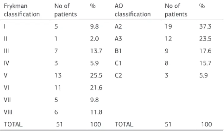
The mean follow-up period of the 51 patients in the study was 17.1 months (range, 6-48 months). Distribution of the patients according to the Frykman and AO Classiications is shown in Table 1.
According to the Gartland and Werley clinical evaluation system the patients were evaluated as 23 (45.1%) at 0-2 points (excel-lent), 19 (37.3%) at 3-8 points (good) and 9 (17.6%) at 9-20 points (moderate). No poor results were determined.
The mean DASH score was 35.9 ± 4.2. When muscle strength
Conservative Treatment of Distal Radius Fractures
was compared with the healthy side, the healthy side was found to be mean 34.1 ± 10.0 kg, and the fractured side, 27.9 ± 9.6 kg. Preoperative mean radial height was 12 mm. Postopera-tive mean radial length was measured to be 11.8 mm. Total of 6 patients had decreased radial heights and mean muscle strengths of these 6 patients was found to be 22.5 this value was found to be statistically signiicantly lower than patients who had post-treatment normal radıal heights. (p<0.05). When the patients were examined according to the Stewart et al radiological criteria, results were obtained of 18 (35.3%) excellent, 28 (54.9%) good and 9 (17.6%) moderate. No poor results were obtained.
Osteoarthritis was determined radiologically in 14 (27.4%) patients according to the Knirk and Jupiter arthritis scoring system. Of 11 patients determined with Grade 1 arthritis, 4 (36.3%) fractures were Frykman Type 7, 5 (45.4%) Frykman Type 8, 1 (9%) Frykman Type 3 and 1 (9%) Frykman Type 4. Of the 3 patients determined with Grade 2 arthritis, 1 fracture was Frykman Type 7 and 2 fractures were Frykman Type 6. In addi-tion, it was determined that as the Frykman degree increased so there was an increase in clinical score, DASH and the radio-logical anatomic score.
32-year old male with let side radius distal end complex frac-ture, Frykman Type 6, AO A3, AP and Lateral Roentgenogram-before closed reduction (Figure 1), ater closed reduction (Fig-ure 2) and ater follow-up period of 10 months (Fig(Fig-ure 3).
Discussion
Radius distal end fractures are fractures which are anatomi-cally in the distal of the distal radius metaphysis. Although these are the most frequently encountered fractures during an orthopaedic career, there is still no consensus on classiication, treatment and the evaluation of treatment results.
To obtain good functional results in radius distal end fractures it is necessary to correct radial shortness, radial inclination, dorsal curve and distal radioulnar joint incongruity.
In a study by Karalezli et al, anatomic results of 40.7% excellent, 44.4% good, 9.9% fair and 5% poor were obtained according to the Stewart score system, clinical results of 30.8% excellent, 47% good, 17.2% fair and 5.0% poor and it was advocated that to achieve a good functional result, it is necessary to obtain a good anatomic result [8]. In the current study, when the results obtained were evaluated, it was determined that as the ana-tomic result worsened so the clinical result also worsened. In addition, patients who did not have good functional and ana-tomic results had a high DASH score.
In the treatment of radius distal fractures, it is still a matter of debate as to which is the most important criteria deining prog-nosis of the result of reduction. According to Palmer, it is the restoration of radial length, while Gartland and Werley reported it to be the presence of residual dorsal tilt [9,10]. Fernandez et al stated that dorsal angulation >25° will negatively afect the prognosis and Pogue et al claimed that more than 20° will lead to poor results [11,12]. Frykman and de Palma reported that it is necessary to provide radial length to be able to obtain good functional results [10,13]. In the current study, that loss of radial height had a negative efect on both grip strength and clinical results, supports the view that radial height is the most important criteria deining the prognosis of the treatment. Im-pairment of the radial inclination, radial shortening or loss of palmar inclination were not observed to negatively afect the prognosis.
In a study by Vural et al where a comparison was made of the Kapandji technique with plaster casting, there was no difer-ence between the groups in respect of radial length and radial inclination but palmar inclination was determined to have been better restored in the Kapandji technique [14]. In the current study, when evaluation was made of the inal follow-up mea-surements of all the patients, the mean radial curve angle was found to be 19.4 ± 4.4°, the mean palmar curve angle 0.3 ± 8.5° and radial height 8.5 ± 2.6 mm. According to these results from the current study, it was concluded that the palmar inclination angle was more diicult to correct than the other angles, al-though as the most obvious limitation of this study is that there was no control group, we are in support of further studies to be made on the subject of by which method the palmar inclination Table 1. Distribution according to Frykman and AO Classiications
Frykman classiication
No of patients
% AO
classiication No of patients
%
I 5 9.8 A2 19 37.3
II 1 2.0 A3 12 23.5
III 7 13.7 B1 9 17.6
IV 3 5.9 C1 8 15.7
V 13 25.5 C2 3 5.9
VI 11 21.6
VII 5 9.8
VIII 6 11.8
TOTAL 51 100 TOTAL 51 100
Figure 1. 32-year old male with let side radius distal end complex fracture, Fryk-man Type 6, AO A3, AP and Lateral Roentgenogrambefore closed reduction.
Figure 2. Ater closed reduction
can be corrected.
It is known that when the treatment of radius distal fractures results in insuicient anatomic restoration, this creates a loss of grip strength. McQueen found more than 2mm shortening in the radius to be an indicator of grip strength loss [15]. In a study by Özdemir et al, while it was observed that as angula-tion increased dorsally, grip strength decreased, no relaangula-tionship was found between the radial angle and grip strength [16]. In the current study, in the correlation between grip strength and radius distal end anatomy, the conclusion was reached that as radial length decreased, so grip strength also decreased. No signiicance was found between grip strength and radial and palmar inclination.
One of the most frequently encountered problems in conserva-tive follow-up of radius distal end fractures is that the reduc-tion is not protected inside the plaster cast. This problem is more evident particularly in unstable fractures [17]. It has be-come known that even if there is successful reduction initially in radius distal end fractures with metaphyseal fragmentation, loss of reduction develops inside the plaster cast in approxi-mately 60% of these cases [18]. In this respect, these frac-tures require careful monitoring in conservative follow-up with plaster cast. Jenkins et al reported that reduction loss of 5% was observed as a result of conservative treatment in unstable intra-articular fractures and this required correction with a new reduction [19]. In a study by Warwick with a 10-year follow-up period, it was reported that plaster casting treatment could not provide suicient radial length and thus external ixators should be preferred [20].
In the current study, anatomic angles were measured following plaster casting and at the inal follow-up. In the light of these values, the radial inclination angle and radial height within the plaster cast were found to be statistically signiicantly high-er than the values measured at the inal follow-up. Howevhigh-er, no signiicant diference was found between the value of the palmar tilt angle in the plaster cast and that at the follow-up following bone union. In addition, while the radial height and radial inclination values in the plaster cast of elderly patients and those with metaphyseal fragmentation were found to be signiicantly higher than the follow-up values, no statistically signiicant diference was determined in the palmar tilt. That no diference was determined between the palmar tilt angles in the plaster cast and at follow-up is thought to be associ-ated with the provision of suicient support from the dorsal of the plaster cast which was applied ater oedema had re-covered. However, osteopenic fractures in elderly patients have a tendency to displacement particularly in the late period and this can be considered to conform with the view of Warwick that plaster casting treatment cannot provide suicient radial length [20,21].
Another subject for debate in radius distal fractures is the ef-fect on prognosis of an ulnar styloid fracture together with a ra-dius fracture. According to Knirk et al, non-union of the styloid afects the prognosis negatively [4]. Smaill et al reported that an ulnar styloid fracture may cause restricted wrist movement [22]. However, in a study by Bradway et al, it was stated that non-union of an ulnar styloid fracture did not afect functional results. [23]. In the current study, 21 (41.1%) patients had an
ulnar styloid fracture as well as the radius distal fracture. At the inal follow-up, union of the ulnar styloid was determined in 6 (28.5%) patients and non-union in 15 (71.5%) patients. Al-though the number of patients was low in the current study, as there was no statistically signiicant diference in the follow-up mean clinical evaluation score and DASH score between the pa-tients with union of the ulnar styloid and those with non-union, the presence of a styloid fracture was not thought to afect the functional results.
Conclusion
The currently accepted view of radius distal end fractures is that they are complex fractures and prognosis varies depending on the type of fracture and the treatment applied. Treatment with closed reduction and plaster cast ixation is extremely cheap and is an easy method to apply in a short time. In the current study, that loss of radial height had a negative efect on both grip strength and clinical results, supports the view that radial height is the most important criteria deining the progno-sis of the treatment. Impairment of the radial inclination, radial shortening or loss of palmar inclination were not observed to negatively afect the prognosis.
Competing interests
The authors declare that they have no competing interests.
References
1. Seitz WH, Froimson Al, Brooks DB, Potsak P, Polando G, Greenwald AS. External ixator pin insertion techniques. Biomechanical analysis and clinical relevance. J Hand Surg Am 1991;16(3):560-3.
2. Rogge R, Adams BD, Goel VK. An analysis of bone stresses and ixation stabil-ity using a inite element model of simulated distal radius fractures. J Hand Surg (Am) 2002;27(1):86-92.
3. Cooney WP, Linsheid RL, Dobyns JH. External pin ixation for unstable Colles fractures. J Bone Joint Surg 1979;61:840-5.
4. Knirk JL, Jupiter JB. Intra-articular fractures of the distal end of the radius in young adults. J Bone Joint Surg 1986:68:647-9.
5. Crenshaw AH. Fractures of shoulder, arm and forearm. In: Canale ST (Ed.). Camp-bell’s operative orthopaedics. 10th ed. Philadelphia: Mosby Publ; 2003.p.2985-3069.
6. Atroshi I, Gummesson C, Anderson B, Dahlgreen E, Johansson A. The disabili-ties of the arm, shoulder and hand (DASH) outcome qestionnaire. Reliability and validity of the swedish version evaluated in 176 patients. Acta Orthop Scan 2000;71(6):613-8.
7. Stewart HD, Innes AR, Burke FD. The hand complications of Colles’ fractures. J Hand Surg 1985;10:103-6.
8. Karalezli K,Demir R, İltar S, Çakır A, Karalezli N, Özeri Z. Radius distal uç kırıklarında konservatif tedavi sonuçlarımız. Gülhane Tıp Dergisi 2004;46(4):315-22.
9. Gartland JJ Jr, Werley WC. Evaluation of healed Colles’ fractures. J Bone Joint Surg 1951;33:895-907.
10. DePalma, AF, Comminuted fractures of the distal end of the radius treated by ulnar pinning. J Bone Joint Surg 1952;34:651-62.
11. Fernandez DL. Reconstructive procedures for malunion and traumatic arthri-tis. Orth Clin of North America 1993;68:341-63.
12. Pogue DJ, Viegas SF, Patterson RM, Peterson PD, Jenkins DK, Sweo TD et al. Efects of distal radius fracture malunion on wrist joint mechanics. J Hand Surg (Am) 1990;15(5):721-7.
13. Frykman G. Fracture of the distal radius including sequelqe Shoulder-handin-ger syndrome disturbance in the distal radioulnar joint and impairment of nerve function. A clinical and experimental study. Acta Orthop Scand 1967;108:1-153. 14. Vural Ö, Okçu G, Özalp RT, Akkaya MG, Yercan HS. Kolles kırığı tedavisinde kapalı redüksiyon alçılı tespit ile Kapandji yönteminin karşılaştırılması. Joint Dis Rel Surg 2008;19(2):55-60.
15. McQueen MM, Hajducka C, Court-Brown C. Redisplaced unstable fractures of the distal radius: a randomised, prospective study of bridging versus non-bridging external ixation. J Bone Joint Surg 1996;78(3):404-9.
16. Özdemir H, Özenci M, GÜL S. Konservatif yöntemle tedavi edilen distal radius kırıklarının erken ve geç dönem sonuçlarının karşılaştırılması. Acta Orthop Trau-matol Turc 2000;34:284-92.
17. Proctor MT, Moore DJ, Paterson JM. Redisplacement ater manipulating of distal radial fractures in children. Journal of Bone Joint Surg 1975;93:453-4.
Conservative Treatment of Distal Radius Fractures
18. Diego L. Fernandez, Andrew K. Palmer. Fractures of the distal radius. In: Green, Hotchkiss, Pederson, editors. Green’s Operative Hand Surgery. Vol.1.Fourth edition. Philadelphia: Churchill Livingstone co.; 1999.p.929-81.
19. Jenkins NH, Jones DG. External ixation of Colles fractures an anatomical study. J Bone Joint Surg 1987;69(B):207-11.
20. Warwick D, Prothero D: Radiological management of the radial shortening in Colles fractures. J Hand Surg 1993;18:50-2.
21. Solgaard S. Classiication of distal radius fractures. Acta Orthop Scand 1984;56:249-52.
22. Smaill GB. Long term follow up Colles fracture. J Bone Joint Surg (Br) 1965;47:80-5.
23. Bradway JK, Amadio PC, Cooney WP. Open reduction and internal ixation of displaced, comminuted intraarticular fractures of the distal end of the radius. J Bone Joint Surg 1989;71:839-47.
How to cite this article: