1 – Chief of the Hand Surgery Department Service of the Hospital Ortopédico de Belo Horizonte, MG, Brazil. 2 – Former Resident at the Hand Surgery Service of the Hospital Ortopédico de Belo Horizonte, MG, Brazil. 3 – Hand Surgeons at the Hospital Ortopédico de Belo Horizonte, Brazil.
Work carried out at the Hand Surgery Service of the Hospital Ortopédico de Belo Horizonte, MG, Brazil.
Correspondence: Arlindo G. Pardini Jr. MD Prof.Otávio Coelho de Magalhães, 111, Bairro Mangabeiras CEP 30210-300 - Belo Horizonte-MG e-mail: [email protected] Received for publication: 05/12/2011, accepted for publication: 07/13/2011
DISTAL RADIUS FRACTURES: LONG TERM FUNCTIONAL
AND RADIOLOGICAL RESULTS OF PERCUTANEOUS
PINNING FIXATION
Arlindo Gomes Pardini Júnior1, Henrique Gubert Freua Bufáiçal2, Afrânio Donato de Freitas3, Antonio Barbosa Chaves3
The authors declare that there was no conflict of interest in conducting this work
This article is available online in Portuguese and English at the websites: www.rbo.org.br and www.scielo.br/rbort ABSTRACT
Objective: To evaluate, functionally and radiologically, the long term outcomes of the management of distal radius fracture treated by closed reduction and percutaneous pin-ning fixation. Methods: From 84 patients submitted to per-cutaneous fixation of the distal radius fracture, we evalu-ated 34, with a medium follow-up of 85.7 months (from 18 to 168 months). Of the 34 patients, 23 were women, and the ages ranged from 28 to 88 years (median 65 years). We analyzed the range of movement, strength, pain and the re-sults of the DASH questionnaire. Radiological evaluation was also carried out, to evaluate healing time and angles of the distal radius. Results: The fractures healed in an aver-age of 41 days. The mean values for wrist flexion, exten-sion, radioulnar deviation, pronation and supination were
within the functional parameters for ROM of the wrist. Most of the patients (76.5%) presented no pain during the examination, and 23 patients presented a DASH value of zero. There was one case of loss of reduction, which was re-operated two weeks after the initial surgery, and one patient developed a sympathetic dystrophy associated with a distal radioulnar joint disturbance. Conclusion: Percutaneous pinning fixation for distal radius fracture results in a long term follow-up with excellent range of movement, little or no pain, acceptable radiographic pa-rameters, and low complication rate, and is an efficient and low cost method.
Keywords - Radius Fractures;Kirschner Wires; Fracture
Fixation.
INTRODUCTION
Fractures of the distal extremity of the radius are a constant subject of studies that seek to define which surgical method would present best results and few-est complications relating to the procedure. These are fractures that have a broad spectrum of presentations and likewise a range of procedures for treating them, going from conservative methods like plaster casts to surgical procedures for unstable fractures: percu-taneous pinning and volar and dorsal plates of a wide variety of models, with or without locking, with or without external fixators and with or without associa-tions with other methods(1, 2).
With the advent of locked plates, several studies have compared their use with the use of other fixation
methods for these fractures, and good results have been reported in the literature(3,4). In the same way,
because this is a more invasive method, it involves a great number of complications than seen in other, more conservative procedures such as percutaneous pinning. Complications such as plate breakage, teno-synovitis, tendon adherence and even torn tendons (both flexors and extensors) have been described in the literature(5-8).
simple to perform and the pins can easily be removed after consolidation(9-13).
The objective of the present study was to conduct long-term functional and radiographic analysis on patients with a fracture of the distal radius who were treated with closed reduction and percutaneous fixa-tion, and to describe the complications encountered during the follow-up period.
This study was approved by the Ethics Committee of the Orthopedics Hospital of Belo Horizonte.
MATERIAL AND METHODS
Between 1996 and 2008, 84 patients were operated to treat unstable fractures of the distal extremity of the radius, by means of percutaneous fixation using Kirschner wires. Only 39 of them made return visits for evaluations. Some patients had died, while oth-ers were experiencing locomotion problems and yet others had changed address or telephone number. All of these patients were therefore excluded from the present study.
Twenty-three women and eleven men were evalu-ated, with a minimum length of follow-up of 18 months and a maximum of 168 months (mean of 85.7 months). Among these, 11 patients were evaluated with more than 10 years of follow-up (Table 1).
Table 1 – Length of postoperative evolution among the patients
operated to treat fractures of the distal extremity of the radius by means of percutaneous fixation.
Length of evolution
18 months 6 patients
4 to 6 years 10 patients
7 to 10 years 7 patients
More than 10 years 11 patients
The patients’ mean age at the time of the examina-tion was 65 years (range: 28 to 88 years), and 25 of them were more than 60 years of age.
In eighteen patients the fracture was in the domi-nant side.
All the patients were assessed by one of the authors (HF), who did not participate in the treatment. All the patients were operated by the same surgeon (AGPJ).
The assessment methods used were the range of motion (ROM), grip strength, radiographic study, pain evaluation and functional analysis (DASH).
The ROM was measured using a goniometer, and measurements were obtained for flexion, extension,
pronation, supination, radial deviation and ulnar de-viation.
Grip strength was measured in both hands, using a Jamar dynamometer, and the mean was taken from three consecutive measurements.
The pain evaluation was done by means of a visual analogue scale.
The functional evaluation was done by means of the DASH test (“Disabilities of the Arm, Shoulder and Hand”).
The radiographic study was done on radiographs that were obtained before the reduction, just after the fixation, during the treatment and at the time of revi-sion, in the posteroanterior and lateral views. The time taken to achieve consolidation and the angles of radial and volar tilt were assessed.
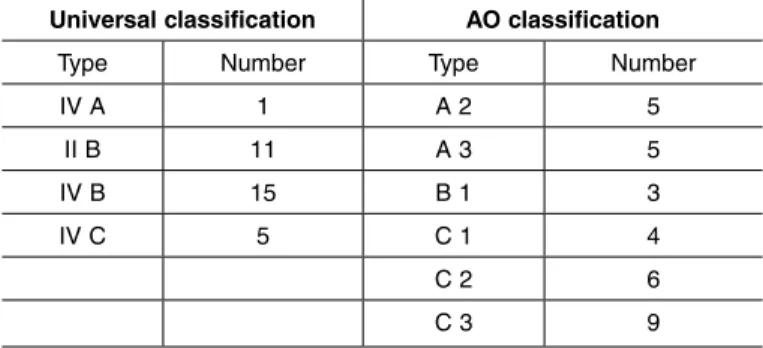
The fractures were classified using the universal and AO systems (Table 2).
Table 2 – Universal and AO classifications for the fractures. Universal classification AO classification
Type Number Type Number
IV A 1 A 2 5
II B 11 A 3 5
IV B 15 B 1 3
IV C 5 C 1 4
C 2 6
C 3 9
Two cases did not have an initial X-ray
SURGICAL TECHNIqUE
All the patients were operated under anesthetic blockage of the brachial plexus. Fracture reduc-tion was achieved in all cases by means of tracreduc-tion and counter-traction performed by two auxiliaries. After manipulation of the fracture by the surgeon, the reduction was checked using an image intensi-fier. For fracture fixation, 1.5 mm Kirschner wires were used.
33
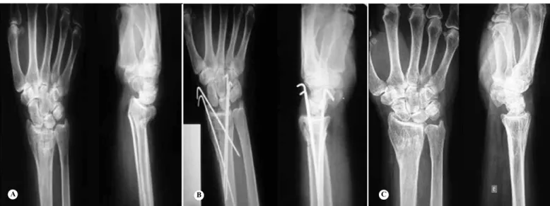
Figure 1 – Patient aged 54 years. (A) – X-ray showing unstable reducible extra-articular fracture. (B) – After reduction and fixation with three
Kirschner wires. (C) – 4.6 years later, showing consolidated fracture with good reconstitution of the angles of the distal extremity of the radius. Asymptomatic patient.
Figure 2 – Patient aged 79 years. (A) – X-ray showing unstable reducible Intra-articular fracture. (B) – X-ray after reduction and fixation with
four Kirschner wires. (C) – 10 years later, showing consolidated fracture with satisfactory distal angles of the radius. Asymptomatic patient.
A B C
A B C
PERCUTANEOUS PINNING FIXATION
short splint was installed to assist in immobilizing the wrist. A clinical control was done on the first postoperative day to check on the sensitivity and per-fusion. One week after the operation, new radiographs in the posteroanterior (PA) and lateral views were ob-tained and the dressing was changed. This procedure was repeated two weeks after the operation, when the patient was then instructed to start hand therapy. The splint was kept in place until around six weeks after the operation, but the pins were removed after around four weeks. At this point, the hand therapy was intensified. The length of time with the pins and splint varied mainly according to the patient’s age, type of fracture, image on control radiographs and bone quality.
STATISTICAL ANALYSIS
The statistical analysis on the study data was per-formed by a biostatistician at the Federal University of Minas Gerais. The t test was applied to analyze sta-tistical differences in ROM. For the DASH and grip strength variables, the Anderson Darling test was used; and for the analysis on the types of fracture according to the AO classification, the Kruskal-Wallis test was applied. The p value was set at p < 0.05.
RESULTS
Among the variables analyzed, we only found a statistically significant difference for flexion, such that patients aged less than 60 years presented mean flexion of 71.11º, versus 59.20º among patients over 60 years.
Twenty-six of the 34 patients (76.5%) did not pres-ent any pain at the time of the examination, while seven patients had low-intensity pain associated with activities in which force was applied and one patient presented moderate-intensity pain, even when resting.
The fractured wrists were evaluated using radio-graphs with two views (PA and lateral). We observed bone consolidation after a period ranging from 27 to 67 days, with a mean of 41 days. The angles of volar tilt (lateral radiograph) and radial tilt (PA radiograph) were measured. The results found were compared with the immediate postoperative radiographs, and these data are shown in Tables 4 and 5. No statisti-cally significant differences were found in relation to these data.
The grip strength was analyzed and we divided the patients into two groups: those who had fractured the dominant side and those who had fractured the non-dominant side. The mean strength in the group that had fractured the dominant side was 24.74 kgf, with standard deviation of 9.53, while the strength in the group that had fractured the non-dominant side was 22.62 kgf, with standard deviation of 9.24. The
Comparison between the patients’ ROM and age.
< 60 years > 60 years 95% CI P-value
N (degrees)Mean SD (degrees)Median N (degrees)Mean SD (degrees)Median Lower Upper
Flexion 9 71.11 10.86 69.00 25 59.20 12.83 61.00 2.122 21.700 0.019
Extension 9 75.44 5.81 74.00 25 70.72 9.83 72.00 -2.401 11.850 0.186
RD 9 20.00 3.12 20.00 25 18.20 4.08 18.00 -1.260 4.860 0.240
UD 9 36.11 5.97 38.00 25 32.00 5.28 32.00 -0.210 8.432 0.061
Pronation 9 81.44 9.00 85.00 25 84.22 4.61 85.00 -7.840 1.689 0.198
Supination 9 81.56 16.03 87.00 25 81.28 10.99 85.00 -2.990 6.000 0.280
SD: standard deviation; RD: radial deviation; UD: ulnar deviation; CI: confidence interval .
Table 4 – Comparison between the angles of radial tilt immediately after the operation and at the final evaluation.
Immediately after operation Final evaluation 95% CI P-value
N Mean (degrees) SD Median (degrees) N Mean (degrees) SD Median (degrees) Lower Upper
Radial tilt 33 22.00 4.76 22.00 34 22.74 3.96 23.50 -1.397 2.867 0.493
Table 5 - Comparison between the angles of volar tilt immediately after the operation and at the final evaluation.
Immediately after operation Final evaluation 95% CI P-value
N Mean (degrees) SD Median (degrees) N Mean (degrees) SD Median (degrees) Lower Upper
Volar tilt 33 -2.03 10.44 0.00 34 -3.82 11.70 2.50 -3.625 7.212 0.511
statistical analysis did not demonstrate any significant difference between the groups.
Regarding the functional evaluation using DASH, we observed that the mean was 2.18, with a standard deviation of 4.78. This value was lower than the mean reported in a study on healthy volunteers (10.1 with standard deviation of 14.7).
The DASH values were analysed in two different categories: comparison of DASH values between in-dividuals older and younger than 60 years; and DASH compared with the type of fracture using the Univer-sal classification.
The analysis on the DASH values comparing between the age groups did not show any statisti-cal significance. The patients younger than 60 years presented a mean of 2.04 with standard deviation of 5.51, while the group older than 60 years had a mean of 2.23 with standard deviation of 4.62.
35
the study included one case of loss of reduction, which was reoperated after two weeks of evolution such that the fracture was fixed again using percutaneous wires; and one patient who developed sympathetic reflex dystrophy associated with changes to the distal ulna, which was resolved after controlling the dystrophy and performing the Bowers procedure (hemi-resection ar-throplasty of the distal ulna).
DISCUSSION
Our study demonstrated that percutaneous fixation with Kirschner wires in fracture of the distal extremity of the radius produces excellent functional and radio-logical results after a long follow-up period. The data analyzed showed that only flexion had a statistically valid difference in comparisons of the results between the groups, but this difference was very small in clini-cal practice.
All the ROM data showed amplitudes within the functional patterns, for flexion, extension and radial and ulnar deviations(14). Oshige et al(15) and Huard
et al(16) advocated using plates because of the great
advantage in using this method so the patient could start physicaltherapy earlier. Several studies have al-ready proven that late evaluations on patients do not show ROM differences between patients operated us-ing plates and percutaneous pinnus-ing(17,18). In addition,
another study showed that, among patients who were operated using a plate, when they were divided into two groups with rehabilitation starting after two or six weeks of immobilization, they did not present any difference in any of the parameters evaluated, in as-sessments three and six months after the operation(19).
Our patients presented low levels of pain, and most of them did not present any pain in the final evaluation. Osteosynthesis with plates is more associated with late postoperative pain than percutaneous wires(17).
The values from the DASH test among our patients were lower than those shown in other studies(20). We
believe that these values were directly connected with the pain levels and profile of our patients(21).
In analyzing the radiographic data on our patients immediately after the operation and at the end of the follow-up, we saw that there was no statistically valid variation in the parameters analyzed, thus showing
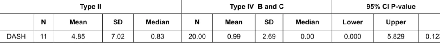
Table 6 – Comparison of DASH values between types II and IV B and C of the universal classification.
Type II Type IV B and C 95% CI P-value
N Mean SD Median N Mean SD Median Lower Upper
DASH 11 4.85 7.02 0.83 20.00 0.99 2.69 0.00 0.000 5.829 0.123
that, on average, the initial reduction was sustained. The critics of percutaneous fixation using Kirschner wires, say that the great problem would be precisely the lack of stability of the method, which would lead to loss of the reduction in the initial phases of consoli-dation(22). Our study did not demonstrate such
com-plication and we attribute this to the type of assembly that we used in most of our cases. Several patterns for placement of Kirschner wires have been described in the literature, with differences in how they are ap-plied and the number used(23). In our study, 67.6% of
the cases were treated using three crossed bicortical Kirschner wires: two through the radial styloid pro-cess and one introduced dorsally through the ulnar edge of the radius. This configuration proved to be more rigid when pins of diameter 1.5 to 2.0 mm were used(24). Another study demonstrated that, although
the configuration that we used was biomechanically more stable, the clinical and radiological results from simple crossed wires (in the radial styloid plus one in the ulnar border of the radius) are similar, which also confirms our results, given that this configuration was used in 14.7% of the cases(25).
Another biomechanical study demonstrated that the difference in deviation of the fracture, comparing percutaneous pinning with volar plates, was only 1.5 mm(26). Thus, even if there is a slight loss of reduction,
thus leading to a reduction that is not exactly anatomi-cal, this reduction will not have a direct implication regarding good results from long-term follow-up, or a high DASH value(27,28).
We believe that the complication rate among our patients, is in accordance with other authors(29).
We had three cases of superficial infection of the Kirschner wires that were resolved through chang-ing the dresschang-ings and administerchang-ing oral antibiotics, one case of sympathetic reflex dystrophy and one case of loss of reduction with the need for a new proce-dure. We did not have any case of lesions of sensory branches of the radial nerve, as also shown by other studies, and by anatomical studies that demonstrated that occurences of such lesions caused by Kirschner wires would be unusual(30). Likewise, we did not
have any case of tendon rupture, either of flexor or
of extensor type. These tears, which generally are not associated with percutaneous pinning, occur more frequently when volar or dorsal plates are used. They are related to the mechanical friction produced by plates and screws, which leads to tenosynovitis that, if undiagnosed, culminates in tendon ruptures(31-36).
A study conducted by Koval et al(37), in which the
tendency for new surgeons to treat fractures of the distal radius with plates was analyzed, showed that the use of this technique increased from 42% to 81% between 1999 and 2007, but without any evidence of improvements in the results. One of the impor-tant factors when analyzing a surgical indication is its cost, especially in a country in which access to
the latest materials is extremely expensive. Thus, use of Kirschner wires becomes an attractive technique because it is inexpensive, considering that each wire costs 8 to 10 reais ( 5 or 6 dollars) on average. On the other hand, locked plates with screws cost from 1800 to 5000 reais (1000 to 3000 dollars) or more.
CONCLUSION
We conclude that using percutaneous fixation with Kirschner wires for fractures of the distal radius re-sults in excellent ROM over the long term, with little or no pain, acceptable radiographic parameters and a low complication rate, in addition to being an inex-pensive and efficient method.
REFERENCES
1. Caporrino FA, Belotti JC, Ulson HJR, Toledo LFQ, Reis FB, Machado JKS. Fraturas da extremidade distal do rádio e da ulna. In: Pardini Júnior AG, Freitas A. Traumatismos da mão. 4a. ed. Rio de Janeiro: MedBook; 2008. p. 411-45. 2. Henry MH. Distal radius fractures: current concepts. J Hand Surg Am.
2008;33(7):1215-27.
3. Osada D, Kamei S, Masuzaki K, Takai M, Kameda M, Tamai K. Prospective study of distal radius fractures treated with a volar locking plate system. J Hand Surg Am. 2008;33(5):691-700.
4. Rozental TD, Blazar PE. Functional outcome and complications after volar plating for dorsally displaced, unstable fractures of the distal radius. J Hand Surg Am. 2006;31(3):359-65.
5. Klug RA, Press CM, Gonzalez MH. Rupture of the flexor pollicis longus tendon after volar fixed-angle plating of a distal radius fracture: a case report. J Hand Surg Am. 2007;32(7):984-8.
6. Cross AW, Schmidt CC. Flexor tendon injuries following locked volar plating of distal radius fractures. J Hand Surg Am. 2008;33(2):164-7.
7. Valbuena SE, Cogswell LK, Baraziol R, Valenti P. Rupture of flexor tendon following volar plate of distal radius fracture. Report of five cases. Chir Main. 2010;29(2):109-13.
8. Yukata K, Doi K, Hattori Y, Sakamoto S. Early breakage of a titanium volar locking plate for fixation of a distal radius fracture: case report. J Hand Surg Am. 2009;34(5):907-9.
9. Clancey GJ. Percutaneous Kirschner-wire fixation of Colles fractures. A pro-spective study of thirty cases. J Bone Joint Surg Am. 1984;66(7):1008-14. 10. Rodríguez-Merchán EC. Plaster cast versus percutaneous pin fixation for
com-minuted fractures of the distal radius in patients between 46 and 65 years of age. J Orthop Trauma. 1997;11(3):212-7.
11. Mah ET, Atkinson RN. Percutaneous Kirschner wire stabilisation following closed reduction of Colles’ fractures. J Hand Surg Br. 1992;17(1):55-62. 12. Glickel SZ, Catalano LW, Raia FJ, Barron OA, Grabow R, Chia B. Long-term
outcomes of closed reduction and percutaneous pinning for the treatment of distal radius fractures. J Hand Surg Am. 2008;33(10):1700-5.
13. Rosati M, Bertagnini S, Digrandi G, Sala C. Percutaneous pinning for fractures of the distal radius. Acta Orthop Belg. 2006;72(2):138-46.
14. Palmer AK, Werner FW, Murphy D, Glisson R. Functional wrist motion: a bio-mechanical study. J Hand Surg Am. 1985;10(1):39-46.
15. Oshige T, Sakai A, Zenke Y, Moritani S, Nakamura T. A comparative study of clinical and radiological outcomes of dorsally angulated, unstable distal radius fractures in elderly patients: intrafocal pinning versus volar locking plating. J Hand Surg Am. 2007;32(9):1385-92.
16. Huard S, Blanchet N, Leclerc G, Rochet S, Lepage D, Garbuio P, et al. [Frac-tures of the distal radius in patients over 70 years old: Volar plates or K-wires?]. Chir Main. 2010;29(4):236-41.
17. Kreder HJ, Hanel DP, Agel J, McKee M, Schemitsch EH, Trumble TE, et al. Indirect reduction and percutaneous fixation versus open reduction and internal fixation for displaced intra-articular fractures of the distal radius: a randomised, controlled trial. J Bone Joint Surg Br. 2005;87(6):829-36.
18. Grewal R, Perey B, Wilmink M, Stothers K. A randomized prospective study on the treatment of intra-articular distal radius fractures: open reduction and internal fixation with dorsal plating versus mini open reduction, percutaneous fixation, and external fixation. J Hand Surg Am. 2005;30(4):764-72. 19. Lozano-Calderón SA, Souer S, Mudgal C, Jupiter JB, Ring D. Wrist mobilization
following volar plate fixation of fractures of the distal part of the radius. J Bone Joint Surg Am. 2008;90(6):1297-304.
20. Hunsaker FG, Cioffi DA, Amadio PC, Wright JG, Caughlin B. The American academy of orthopaedic surgeons outcomes instruments: normative values from the general population. J Bone Joint Surg Am. 2002;84(2):208-15.
21. Souer JS, Lozano-Calderon SA, Ring D. Predictors of wrist function and health status after operative treatment of fractures of the distal radius. J Hand Surg Am. 2008;33(2):157-163.
22. Barton T, Chambers C, Lane E, Bannister G. Do Kirschner wires maintain reduction of displaced Colles’ fractures? Injury. 2005;36(12):1431-4.
23. Rayhack JM. The history and evolution of percutaneous pinning of displaced distal radius fractures. Orthop Clin North Am. 1993;24(2):287-300.
24. Naidu SH, Capo JT, Moulton M, Ciccone W 2nd, Radin A. Percutaneous pinning of distal radius fractures: a biomechanical study. J Hand Surg Am. 1997;22(2):252-7.
25. Kurup HV, Mandalia V, Shaju A, Beaumont A. Bicortical K-wires for distal radius fracture fixation: how many? Acta Orthop Belg. 2007;73(1):26-30.
26. Knox J, Ambrose H, McCallister W, Trumble T. Percutaneous pins versus volar plates for unstable distal radius fractures: a biomechanic study using a cadaver model. J Hand Surg Am. 2007;32(6):813-7.
27. Grewal R, MacDermid JC. The risk of adverse outcomes in extra-articular distal radius fractures is increased with malalignment in patients of all ages but mitigated in older patients. J Hand Surg Am. 2007;32(7):962-70.
28. Chung KC, Kotsis SV, Kim HM. Predictors of functional outcomes after surgi-cal treatment of distal radius fractures. J Hand Surg Am. 2007;32(1):76-83.
29. Turner RG, Faber KJ, Athwal GS. Complications of distal radius fractures. Orthop Clin North Am. 2007;38(2):217-28.
30. Labronici PJ, Franco JS, Hoffmann R, Silva AF, Passos MARF, Lourenço PRBT, Fernandes HJA, Reis FB. Estudo da relação anatômica do nervo sen-sitivo radial após fixação percutânea com fios de Kirschner. Rev Bras Ortop. 2008;43(3):90-95.
31. Hove LM, Nilsen PT, Furnes O, Oulie HE, Solheim E, Mölster AO. Open reduc-tion and internal fixareduc-tion of displaced intraarticular fractures of the distal radius. 31 patients followed for 3-7 years. Acta Orthop Scand. 1997;68(1):59-63.
32. Kambouroglou GK, Axelrod TS. Complications of the AO/ASIF titanium distal radius plate system (pi plate) in internal fixation of the distal radius: a brief report. J Hand Surg Am. 1998;23(4):737-41.
33. Orbay JL, Fernandez DL. Volar fixation for dorsally displaced fractures of the distal radius: a preliminary report. J Hand Surg Am. 2002;27(2):205-15.
34. Lowry KJ, Gainor BJ, Hoskins JS. Extensor tendon rupture secondary to the AO/ASIF titanium distal radius plate without associated plate failure: a case report. Am J Orthop (Belle Mead NJ). 2000;29(10):789-91.
35. Schnur DP, Chang B. Extensor tendon rupture after internal fixation of a distal radius fracture using a dorsally placed AO/ASIF titanium pi plate. Arbeitsge-meinschaft für Osteosynthesefragen/Association for the Study of Internal Fixa-tion. Ann Plast Surg. 2000;44(5):564-6..
36. Al-Rashid M, Theivendran K, Craigen MA. Delayed ruptures of the extensor tendon secondary to the use of volar locking compression plates for distal radial fractures. J Bone Joint Surg Br. 2006;88(12):1610-2.