Early Functional Postoperative Therapy of
Distal Radius Fracture with a Dynamic
Orthosis: Results of a Prospective
Randomized Cross-Over Comparative Study
Fabian M. Stuby1, Stefan Döbele1*, Susanne-Dorothea Schäffer1, Simon Mueller2, Atesch Ateschrang1, Matthias Baumann3☯‡, Derek Zieker4☯‡
1BG Trauma Center, University of Tuebingen, Schnarrenbergstrasse 95, D-72076, Tuebingen, Germany,2 Muon-Stat Statistical Services, Klugestrasse 28, D-70197, Stuttgart, Germany,3Paracelsus Hospital Ruit, Hedelfinger Strasse 166, D-73760, Ostfildern, Germany,4Department of General Surgery, University of Tübingen, Hoppe-Seyler-Strasse 3, D-72076 Tübingen, Germany
☯These authors contributed equally to this work.
‡These authors are joint senior authors on this work. *[email protected]
Abstract
Introduction
This study was conducted according to GCP criteria as a prospective randomized cross-over study. The primary goal of the study was to determine clinical findings and patient sat-isfaction with postoperative treatment. 29 patients with a distal radius fracture that was sur-gically stabilized from volar and who met the inclusion criteria were enrolled over a 12-month period. Each patient randomly received either a dorsal plaster splint or a vacuum-fit flexible but blocked orthosis applied postoperatively in the operating theatre to achieve postoperative immobilization. After one week all patients were crossed over to the comple-mentary device maintaining the immobilization until end of week 2. After week 2 both groups were allowed to exercise wrist mobility with a physiotherapist, in the orthosis group the de-vice was deblocked, thus allowing limited wrist mobility. After week 4 the dede-vices were re-moved in both groups. Follow-up exams were performed after postoperative weeks 1, 2, 4 and 12.
Results and Discussion
Results were determined after week 1 and 2 using SF 36 and a personally compiled ques-tionnaire; after weeks 4 and 12 with a clinical check-up, calculation of ROM and the DASH Score. Comparison of the two groups showed a significant difference in ROM for volar fleion after 4 weeks, but no significant differences in DASH Score, duratfleion of disability or x-ray findings. With regard to satisfaction with comfort and hygiene, patients were significantly more satisfied with the dynamic orthosis, and 23 of the 29 patients would prefer the flexible vacuum orthosis in future.
OPEN ACCESS
Citation:Stuby FM, Döbele S, Schäffer S-D, Mueller S, Ateschrang A, Baumann M, et al. (2015) Early Functional Postoperative Therapy of Distal Radius Fracture with a Dynamic Orthosis: Results of a Prospective Randomized Cross-Over Comparative Study. PLoS ONE 10(3): e0117720. doi:10.1371/ journal.pone.0117720
Academic Editor:Masahiko Sumitani, The University of Tokyo Hospital, JAPAN
Received:May 31, 2014
Accepted:December 20, 2014
Published:March 30, 2015
Copyright:© 2015 Stuby et al. This is an open access article distributed under the terms of the
Creative Commons Attribution License, which permits unrestricted use, distribution, and reproduction in any medium, provided the original author and source are credited.
Data Availability Statement:All relevant data are within the paper and its Supporting Information files.
Funding:The study was financed by a grant from OPED GmbH Medizinpark 1, D-83626 Valley, Germany. The funders had no role in study design, data collection and analysis, decision to publish, or preparation of the manuscript.
Trial Registration
German Clinical Trials Register (DRKS)DRKS00006097
Introduction
The distal radius fracture is one of the most common injuries of the human skeleton. It has an incidence of 31 per 104 person-years and in some countries it is the most common fracture oc-curring in elderly, namely almost 500 per 100.000 [1]. The literature reports that this fracture accounts for 25% of all fractures in adolescent and for 18% of all fractures in the elderly. In the United States alone more than 640.000 radius fractures were documented in 2001 [2]. While the incidence of this fracture has not changed considerably since 1997, the frequency of surgical therapy and thus the number of in-hospital treatments for this injury has increased consider-ably [1,3]. For non-dislocated fractures conservative therapy with immobilization of the wrist in a plaster cast also called plaster of Paris for 6 weeks [4,5] is usually described. For displaced fractures the recent literature mostly recommends surgery, largely performed with plates [6], whose increasing availability also contributes to the increase in surgical treatment for this frac-ture [7,8]. But there are also tendencies to treat these fractures conservatively, as the results in elderly patients are described as comparable[9].
While surgical treatment has meanwhile become the gold standard [10,11] and depending on the type of fracture is usually performed from volar [12,13] under utilization of locking plates [14], to date hardly any studies have been conducted with regard to postoperative thera-py and especially the effect of immobilization during the postoperative period. Some of the au-thors do not mention postoperative immobilization at all, some seem to completely dispense postoperative immobilization [15] and others perform postoperative immobilization in a cir-cumferential cast or a splint for 2 weeks [16,17], three weeks [9] or even up to four weeks [14].
For treatment of injuries of the lower extremities various removable, sometimes vacuum-fit dynamic stabilizing systems are already available, some of which have also been evaluated [18]. In one study involving conventional therapy patients reported removable orthoses to provide clearly better wearing comfort [19].
Nevertheless, to date no data have been published on dynamic postoperative immobilization of the forearm and wrist following surgical stabilization of a distal radius fracture or the wear-ing comfort of such an orthosis and its effect on postoperative outcome in a comparison with customary plaster cast immobilization.
We thus decided to conduct a prospective, randomized cross-over study to examine these is-sues. The primary endpoint was patient satisfaction based on the 7 parameters of aesthetics, handling, sense of hygiene, activity limitations, physical resilience, accuracy of fitting and post-operative pain with the particular form of immobilization. The secondary endpoints were mo-bility of the wrist and finger function as determined by the range of motion (ROM) and the DASH Score, the period of time during which the patient was disabled and the radiological outcome.
Materials and Methods
The protocol for this trial and supporting CONSORT checklist are available as supporting in-formation; seeS1andS2Protocol,S1 CONSORT ChecklistandS1 Raw Data.
not alter the authors' adherence to PLOS ONE policies on sharing data and materials. The funders had no role in study design, data collection and analysis, decision to publish, or preparation of the manuscript. There are no financial or non-financial competing interests. This includes: employment, consultancy, patents, products in development, marketed products, etc. One of the authors (Simon Mueller) is an employee by a commercial company:
“Muon-Stat Statistical Services”. Muon-Stat Statistical
This study was approved by the ethics committee of Tuebingen University under the No. 196/2011 B02 and conducted in conformity with the principles of good clinical practice. The study was performed at the BG Trauma center Tuebingen.
Inclusion criteria were:
• age 18 to 80 years
• isolated, displaced, distal radius fracture type AO 23 A2, A3, B1, B2, B3, C1,C2
• surgical therapy with volar locking plate
Exclusion criteria were:
• open fracture
• non-displaced fracture
• blunt trauma of the soft tissues G2, G3
• associated fracture of the ulna (except Processus styloideus ulna fracture)
• pathological fracture
• patient unable to comply with postoperative therapy
• other concomitant fractures, polytrauma
• bilateral fractures
Between August 2011 and August 2012 all patients meeting the above-mentioned inclusion criteria were asked to participate in the study.
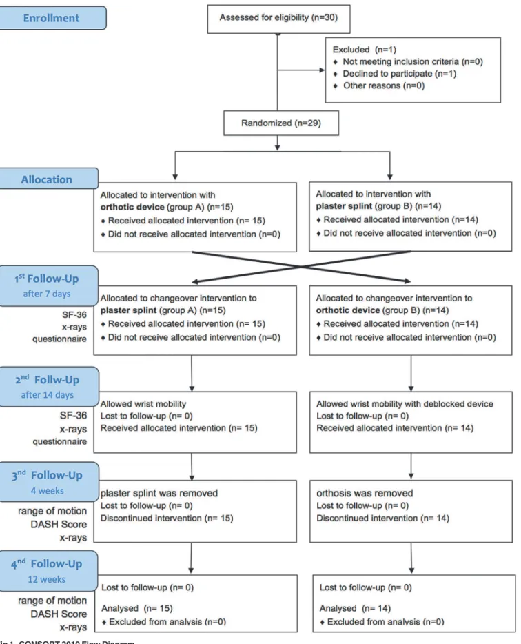
Thirty patients consented to participate (Fig. 1).
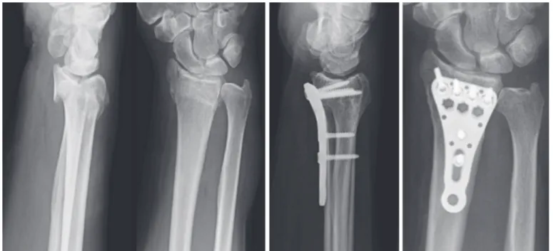
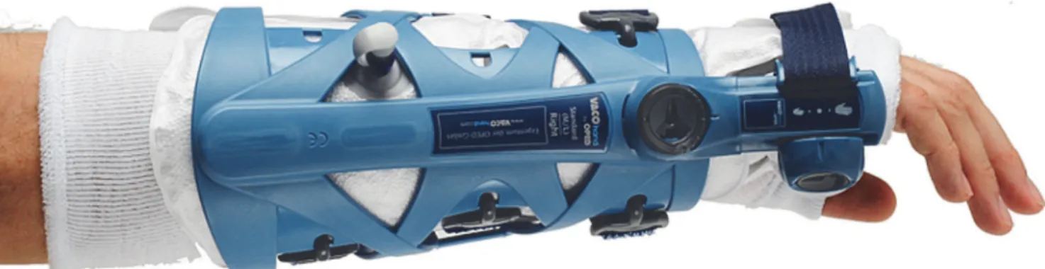
All patients first underwent closed reduction with extension and application of semicircum-ferential splint. Following preoperative preparation and planning ostheosynthesis was per-formed with a volar, distal radius locking plate (either Königsee, 3.5mm (Fig. 2) or Synthes LCP 3.5mm (Fig. 3)). Postoperative immobilization was determined in advance with random assignment to a VacoHand orthosis (Figs.4,5,6) or a dorsal forearm plaster splint (plaster of Paris) (Fig. 7).
The randomization was performed by labeling 30 participating certificates before the trial started. 15 were marked with„plaster splint“and 15 were marked with„VacoHand“. The
sheets were folded to conceal the writing and subsequently blended. Afterwards they were ran-domly inserted into an envelope which was sealed. The envelopes were blended once again and thereafter they were consecutively numbered with number 1–30. Each sealed envelope was
filed into a folder together with the hand out of information material each patient received. The patients who agreed to participate received this folder and the envelope was opened.
„Vacohand“indicating the patient to be randomized into group A,„plaster splint“allocated
the patient to group B. A control for age or other factors except the described inclusion criteria was not performed beforehand.
The VacoHand, as from now on named orthosis, was designed by OPED and is a vacuum fitted orthosis with a wrist hinge which can be used either in a blocked position or in an un-blocked, flexible mode allowing several degrees of range of motion.
Fig 1. CONSORT 2010 Flow Diagram.
for another seven days in both groups. The patients underwent a second check-up 14 days postoperative (time-point 2). From this time on, the patients were allowed to exercise wrist mo-bility out of the splint under the supervision of a physical therapist in case of the plaster of Paris group. The group treated with the orthosis which was deblocked at time-point 2 was
Fig 2. Example of AO 23 C2 fracture lateral and ap (anterior-posterior) view preoperative and 15 weeks postoperative x-rays stabilized with Königsee locking plate.
doi:10.1371/journal.pone.0117720.g002
Fig 3. Example of AO 23 A3 fracture lateral and ap view preoperative and 14 weeks postoperative stabilized with Depuy/Synthes locking plate.
allowed to exercise limited wrist movement in the orthosis at all times themselves in addition to the physiotherapy sessions. At the third follow-up examination four weeks postoperative (time-point 3) the plaster splint or orthosis was removed. The final examination was conducted at 12 weeks postoperative (time-point 4). The study design is also shown in the flow chart for a better understanding (Fig. 1).
At the first and second postoperative check-ups the patients were surveyed concerning their physical health using the SF-36 questionnaire and an additional individual questionnaire. At
Fig 4. Vacuum fitted flexible orthosis (VacoHand OPED) deblocked in dorsal extension.
doi:10.1371/journal.pone.0117720.g004
Fig 5. VacoHand Orthosis deblocked in palmar flexion.
the third and fourth postoperative check-ups wrist mobility was determined according to range of motion and DASH Score.
The Short Form 36 (SF 36) is a validated health survey questionnaire to compare clinical outcomes of certain patient groups with normative data [20].
The DASH score (Disabilitys oft heArm,Shoulder andHand) produces values between 0 and 100 for each module, in which a high DASH score indicates severe disability [21].
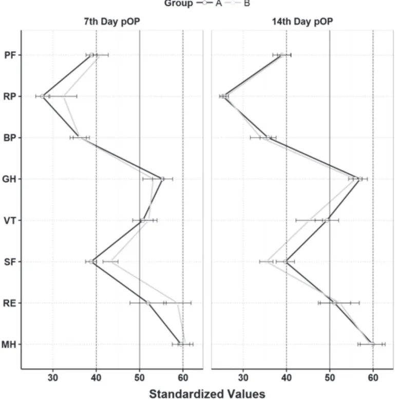
The additional questionnaire used to assess postoperative pain, limitations of activity, physi-cal resilience, handling of orthosis, sense of hygiene and accuracy of fitting was designed individ-ually for this study. It contained 5 graduations for each oft he above mentioned items / questions reaching from 1 = absolutely correct, 2 = mainly correct, 3 = I do not know, 4 = mainly wrong, 5 = absolutely wrong. The results of these 7 parameters were subsumed as patient satisfaction.
Fig 6. VacoHand Orthosis view from ulnar with visible hinge, blocked position 0°, during the study we used 20° dorsal extension as blocked position.
doi:10.1371/journal.pone.0117720.g006
Fig 7. Forearm plaster splint (plaster of Paris) not attached.
At all four postoperative check-ups the patient underwent a clinical examination and x-ray examination of the wrist in anterior-posterior (ap) and lateral projections.
Statistical Analysis
Demographical data, DASH-Scores and SF-36-Scores were presented as means with standard deviation, ROM as medians and interquartile range. Comparison between different demo-graphical groups were made using the Fisher’s Exact Test for counting data and the (Welch)
t-test for continuous variables [22]. The Wilcoxon Rank Sum Test was used for between group comparison of clinical outcome with SF36, DASH Score, and ROM. To match the subjective values of the questionnaire the Cochran–Armitage–Trend-Test was used for between group
comparison and for within group time effects the Wilcoxon Signed Rank Test was used. A 2-sample test for equality of proportions was applied on the variables Right Handed and Fracture occurred on dominant side.
Statistical analysis was performed with R (R Core Team, 2013) (Version 3.0) in addition with R packages coin (Version 1.0.21) and HH (Version 2.3-37). Statistical results with a p-value<0.05 were considered as statistically significant.
Registry of the study
The study has been registered at the German Clinical Trials Register (DRKS) with the registra-tion number: DRKS00006097.
Because of a change of the principal investigator there was a delay in registering this study (after enrolment of participants started). The authors confirm that all ongoing and related trials for this intervention are registered.
A written consent was obtained from all participants involved in this study.
Results
In a 1-year period (August 15th2011 until August 30th2012) we recruited 30 patients with dis-tal radius fractures, one of these was not available for follow up examination (Fig. 1). Therefore 29 patients (21 female, 8 male) took part in the study. Overall mean age was 48,28 (20–75
years) SD ±15,85, of these the female patients had a mean age of 50,14 SD ±15,3 years while the male patients were younger with a mean age of 43,38 SD ±17,5 years (Table 1).
In group A 11 of the 15 patients and in group B 13 of 14 patients were right handed
(Table 1).
In group A the fracture occurred in 8 of 14 patients to the dominant side, one patient did not prefer one side as dominant. In group B the dominant side was affected in 7 of the 14 patients.
The fractures of group A were classified as 7 x AO type A, 2 x AO type B and 6 x AO type C, in group B we found 7 x AO type A, 1 x AO type B and 6 x AO Typ C.
There were no statistical differences in the distribution of the baseline characteristics. The operative treatment in all cases was performed with open reduction and volar plating with anatomical locking plates (23 x Königsee, 6 x Depuy/Synthes), intraoperative or directly postoperative complications did not occur.
The postoperative pain was assessed with a questionnaire, concerning this we could not de-tect any significant differences between the groups at time-point 1 (p = 0,127) or time-point 2 (p = 0,820).
group B when the change of treatment from the plaster splint to the orthosis was performed compared to group A were the treatment was changed vice versa. The patients did not indicate any differences in physical resilience in either group (p = 0,360).
The handling of the immobilizing device was judged as significantly superior for the ortho-sis especially after the switch at time-point 2 with p = 0,009 in group A and p = 0,001 in group B.
The sense of hygiene was becoming significantly better in both groups when changing the treatment at time-point 1. In group B there was a more radical change concerning sense of hy-giene when changing from plaster splint to orthosis (p = 0,001) which was not as distinctive in group A (p = 0,004)
Concerning accuracy of fitting (p = 0,025) and aesthetics (p = 0,001) the orthosis was esti-mated as significantly superior by the patients compared to the plaster splint. In addition there was a significant change in estimation of aesthetics when group B changed the orthotic device at time-point 1 (p = 0,015) (Table 2).
In the analysis of the subgroups we could not detect any influences of side dominance, age of patient or fracture type onto the rating concerning the handling, the sense of hygiene, the ac-curacy of fitting and aesthetical decision.
We could not detect any differences concerning activity limitations, physical resilience or postoperative pain between the two groups.
In the final evaluation performed with Fishers Exact Test for Count Data 23 of the 29 tients would prefer the orthosis in case they would suffer another fracture (p = 0,017) All 14 pa-tients of group B and 9 papa-tients of group A decided in favour of the orthosis.
The clinical evaluation at time-point 3 (4 weeks) and 4 (12 weeks) comprised the investiga-tion of ROM and radiological findings (Figs.8and9).
Table 1. Demographic Data.
Overall Group A Group B P-Value
N 29 (100%) 15 (52%) 14 (48%)
-Male 8 (28%) 5 (33%) 3 (21%) 0.682
Female 21 (72%) 10 (67%) 11 (79%)
Age (years) 48.28±15,85 50.93±15.18 45.43±16.61 0.361
Male 43.38±17.50 48.00±16.78 35.67±19.14 0.499
Female 50.14±15.30 52.40±15.04 48.09±15.78 0.529
Right Handed 24 (83%) 11 (73%) 13 (93%) 0.369
Fracture occurred on dominant side 15 (52%) 8 (53%) 7 (50%) 1.000
Fracture classification
A 14 (48%) 7 (47%) 7 (50%) 1.000
B 3 (10%) 2 (13%) 1 (7%)
C 12 (42%) 6 (40%) 6 (43%)
Decision VacoHand
Yes 23 (79%) 9 (60%) 14 (100%)
No 6 (21%) 6 (40%) 0 (0%) 0.017
15 patients were randomly allocated to group A (first week with orthosis,Fig. 4) 10 of these being female and 5 male with a mean age 50,93 SD±15,18 years.
14 patients were randomly allocated to group B (first week with dorsal forearm plaster splint,Fig. 7), 11 of these were females and 3 male with a mean age 45,43 SD±16,61 years.
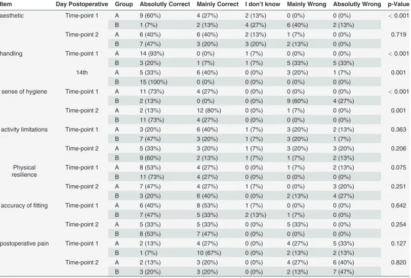
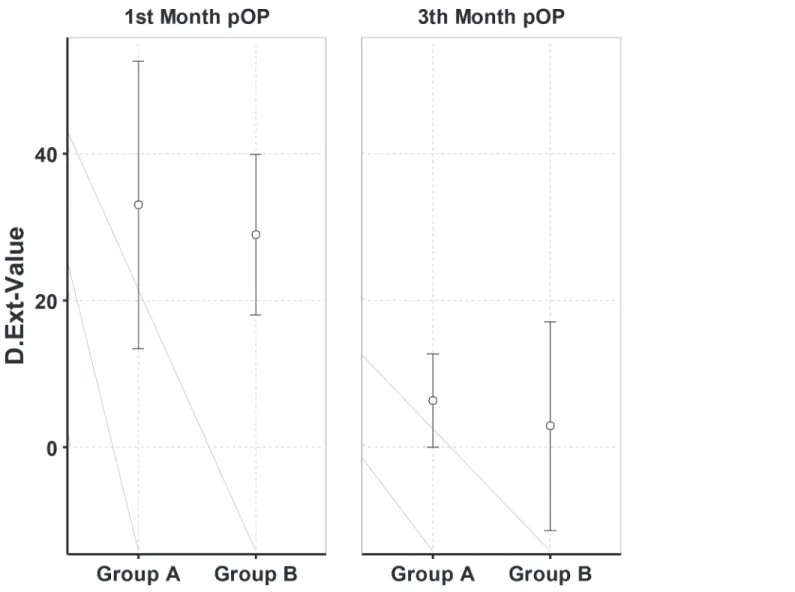
We detected a significant difference between the two groups with a significantly (p = 0,044) reduced ability of palmar flexion at time-point 3 and in addition a reduced, yet not significant dorsal extension in the group which was treated with the plaster splint after week one and thus was only treated by physiotherapy out of the splint in the weeks 3 and 4. The patients treated with the orthosis which was used in a blocked, neutral position for the first two weeks and sub-sequently used unblocked in weeks 3 and 4 showed a significant advantage in functionality measured as ROM related to palmar flexion at time-point 3 and non significant palmar flexion and dorsal extension at time-point 4.
At time-point 4 one patient out of group A could not clench his fist completely, but we did not find a significant difference concerning the ROM of the wrist joint.
The recovery of ROM seemed to be faster in group B compared with group A (Table1and
2) though we could not find significant differences concerning this matter.
The radiological examinations at time-point 2, 3, and 4 did not show any secondary dis-placement or implant loosening. At time-point 4 (12 weeks) all fractures displayed signs of ra-diological consolidation. (Figs.2and3)
The ROM of the forearm concerning pronation and supination and the ROM of the elbow joint was comparable in both groups.
Table 2. Questionaire on subjective experience.
Item Day Postoperative Group Absolutly Correct Mainly Correct I don’t know Mainly Wrong Absolutly Wrong p-Value
aesthetic Time-point 1 A 9 (60%) 4 (27%) 2 (13%) 0 (0%) 0 (0%) <0.001
B 1 (7%) 2 (13%) 4 (27%) 6 (40%) 2 (13%)
Time-point 2 A 6 (40%) 6 (40%) 2 (13%) 1 (7%) 0 (0%) 0.719
B 7 (47%) 3 (20%) 3 (20%) 2 (13%) 0 (0%)
handling Time-point 1 A 14 (93%) 0 (0%) 1 (7%) 0 (0%) 0 (0%) <0.001
B 3 (20%) 1 (7%) 1 (7%) 5 (33%) 5 (33%)
14th A 5 (33%) 6 (40%) 0 (0%) 3 (20%) 1 (7%) 0.001
B 15 (100%) 0 (0%) 0 (0%) 0 (0%) 0 (0%)
sense of hygiene Time-point 1 A 11 (73%) 4 (27%) 0 (0%) 0 (0%) 0 (0%) <0.001
B 2 (13%) 0 (0%) 0 (0%) 9 (60%) 4 (27%)
Time-point 2 A 2 (13%) 12 (80%) 0 (0%) 1 (7%) 0 (0%) 0.001
B 11 (73%) 4 (27%) 0 (0%) 0 (0%) 0 (0%)
activity limitations Time-point 1 A 3 (20%) 6 (40%) 1 (7%) 3 (20%) 2 (13%) 0.363
B 7 (47%) 3 (20%) 1 (7%) 3 (20%) 1 (7%)
Time-point 2 A 5 (33%) 3 (20%) 1 (7%) 3 (20%) 3 (20%) 0.206
B 9 (60%) 2 (13%) 1 (7%) 1 (7%) 2 (13%)
Physical resilience
Time-point 1 A 8 (53%) 4 (27%) 0 (0%) 1 (7%) 2 (13%) 0.075
B 11 (73%) 4 (27%) 0 (0%) 0 (0%) 0 (0%)
Time-point 2 A 7 (47%) 4 (27%) 1 (7%) 0 (0%) 3 (20%) 0.251
B 3 (20%) 6 (40%) 0 (0%) 2 (13%) 4 (27%)
accuracy offitting Time-point 1 A 6 (40%) 8 (53%) 1 (7%) 0 (0%) 0 (0%) 0.642
B 7 (47%) 5 (33%) 2 (13%) 1 (7%) 0 (0%)
Time-point 2 A 5 (33%) 5 (33%) 0 (0%) 5 (33%) 0 (0%) 0.254
B 8 (53%) 7 (47%) 0 (0%) 0 (0%) 0 (0%)
postoperative pain Time-point 1 A 2 (13%) 4 (27%) 0 (0%) 4 (27%) 5 (33%) 0.127
B 1 (7%) 10 (67%) 0 (0%) 2 (13%) 2 (13%)
Time-point 2 A 2 (13%) 3 (20%) 0 (0%) 4 (27%) 6 (40%) 0.820
B 3 (20%) 3 (20%) 0 (0%) 2 (13%) 7 (47%)
We also tried to find out about time of disability to work which was 45.8 days in group A and 39.6 days in group B, but due to different types of professions (pensioners, students, depen-dent employee, and freelancer) and missing values it made no sense to perform a
statistical calculation.
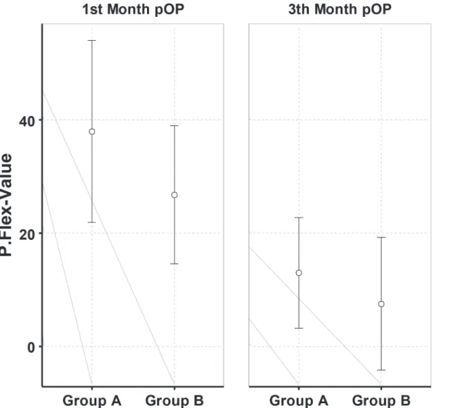
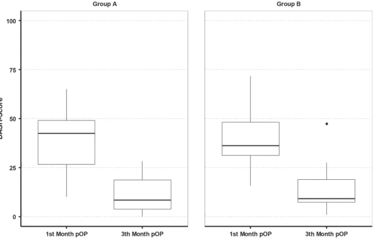
The SF36 did not show any significant differences between the two groups (Fig. 10). The mean DASH score of all patients at time-point 3 was 38,53 ±15,69 and at time-point 4 it was 12,82 ±10,86. Significant differences between the groups could not be detected (Fig. 11).
As complications of our treatment we identified one patient with a CRPS out of group B and one patient with slight paraesthesia in group A.
Discussion
To date the scientific literature contains no prospective comparative study of patient satisfac-tion and funcsatisfac-tional outcome with various orthosis for postoperative treatment. We only found a meta-analysis of 8 studies comparing dynamic versus static fixation of distal radius fractures with an external fixateur device with respect to complications and malunion [23], reporting of
Fig 8. Mean and standard deviation of dorsal extension at time-point 3 and time-point 4 postoperative.
superior but non-significant range of motion and better radiological outcome in the dynamic group [24–31]. Some investigators retrospectively compared dynamic splinting after surgically
treated distal radius fractures versus nonsurgical treatment coming to the conclusion, that dy-namic splinting is directly responsible for gains in ROM [32]. With regard to treatment of con-tractures after distal radius fractures one study did not find a reduction in the rate of
contracture by using a dynamic orthosis and thus concluded them to be difficult to justify for this diagnosis [33].
For therapy of fractures of the upper extremity, the plaster cast or synthetic casts are still as-sumed to be the therapy most frequently used. Although there seem to be advantages of the synthetic materials [34] the plaster cast is still preferred in a lot of hospitals.
Despite the availability of a flexible vacuum-fitted orthosis for the upper extremity it is so far rarely used in hospitals. The reason for this might be the absence of evidence of effects and results of a flexible orthosis in case of postoperative treatment of forearm fractures.
Fig 9. Mean and standard deviation of palmar flexion at time-point 3 and time-point 4 postoperative.
The main focus of this study was patient satisfaction with regard to the two different devices used for immobilization, which was investigated using the described questionnaires. These questionnaires only comprised of 5 steps and were self designed. Retrospectively we should
Fig 10. Mean and standarddeviation of the 8 SC-36 Scales.SF-36 Measurement Model: Physical Health: Physical Functioning (PF), Role-Physical (RP), Bodily Pain (BP), and General Health (GH); Mental Health: Vitality (VT), Social Functioning (SF), Role-Emotional (RE), and Mental Health (MH). Scales of our sample were standardized on German normative postoperativ evaluation [21]. The normative German postoperative evaluation has mean 50 and standarddeviation 10.
have used a scale from 1 to 10 for each item to receive a more detailed graduation and a better reproducibility.
The study design does not include a wash-out period and therefore does not totally correlate to the design of a standard cross-over trial. Thus we could not completely avoid carry–over
ef-fects, but due to the injury related and therefore essential therapy we were not able to fulfill all the requirements.
The study design did not include a one year follow up, because we did not expect a signifi-cant difference concerning the function postoperatively and because the main target point of the study was patient satisfaction and comfort. Therefore we are not able to judge how the dif-ferences in range of motion which already seemed to have leveled after 12 weeks would per-form after a one year period.
For the answers concerning patient satisfaction with the device this design was sufficient. For the evaluation of the questions concerning final ROM and radiological results another ex-amination after 12 month would have been reasonable, but as mentioned before these were only secondary target points in this study.
The therapy regimen for postsurgical treatment of a distal radius fracture employed in our Level I Trauma Center and University Clinic calls for two weeks of immobilization after opera-tive stabilization followed by two weeks of mobilization in the splint under attendance of a physiotherapist. The splint is removed after four weeks and light mobility is prescribed for the subsequent two weeks without strain.
Fig 11. DASH Score at time-point 3 and time-point 4.
The study was conducted according to this therapy regimen and the study design adapted to ensure fair comparison of the two treatment methods, because the deblocked and thus flexible orthosis would permit mobilization after two weeks.
We purposely did not add a third group without any postoperative immobilization as this would have contradicted our well-established treatment regimen and we believe that this might lead to secondary displacement or implant loosening especially in patients with reduced bone quality.
Of course in other hospitals the postoperative treatment of distal radius fractures is handled differently [9,14,16], sometimes even with no immobilization at all [15]. With increasing age of our future patients and thus reduced bone quality the confidence into postoperative stability might decrease and therefore postoperative immobilization to safeguard the operative result might be used more often in future even by those not doing it presently.
Using the SF 36 questionnaire and the DASH Score and several individually developed questionnaires we detected no significant differences concerning pain, overall functional ability and resilience. In relation to change of activity limitations when changing the treatment after week 1 the group changing from orthosis to plaster splint reported a significantly worse result compared to the other group. Also we found a significantly superior evaluation of accuracy of fitting, aesthetics, and hygienic feeling in favour of the orthosis. The main question and target point of the study, namely which treatment (orthosis or plaster splint) would be preferred in future after this cross-over study, was clearly and thus significantly (p = 0,017) answered in fa-vour of the orthosis. Of the 29 study participants, 23 would request the vacuum fitted orthosis as postoperative fracture immobilization in case of another distal radius fracture.
As the orthosis was deblocked after the second week the patients were able to perform some limited movements themselves at all times in addition to the physiotherapy treatment both groups received. We assume that this additional exercises accounts for the superior functional outcome compared to the plaster splint group after 4 weeks and the better, yet not significant ROM after 12 weeks.
From a financial point of view, the flexible orthosis is more economical for hospitals in Ger-many at this time, because it calls for neither material nor personnel to apply or remove it as the plaster splint does. Currently, the social health insurance carriers and not the hospital itself pay for the flexible orthosis. One negative point is certainly the fact that this is thus an added expense for German social health insurance carriers, whereby a comparison of costs for the provision of a plaster room, personnel and material will not be entirely transparent and thus not comparable. The costs of the plaster splint for example are taken over by the hospital itself.
The study demonstrates that there are some advantages in functional outcome after 4 weeks and in postoperative management when using a functional orthosis, and that patient satisfac-tion clearly is superior with the vacuum-fitted flexible orthosis we used in this study. After twelve weeks the functional advantages seem to have almost leveled. Consequently, the study data suggests the flexible orthosis to be an alternative to the plaster splint if postoperative im-mobilization is wanted.
All data underlying the findings described in this manuscript are fully available without re-striction. They were submitted with the manuscript and are available via PLOSONE.
Supporting Information
S1 CONSORT Checklist. (DOC)
S2 Protocol. Trial study protocol (English). (DOCX)
S1 Raw Data. Raw data and statistical analysis. (PDF)
Author Contributions
Conceived and designed the experiments: FMS SD SDS SM AA MB DZ. Performed the experi-ments: FMS SDS AA MB DZ. Analyzed the data: FMS SD SDS AA SM MB DZ. Contributed re-agents/materials/analysis tools: FMS SD SDS AA MB DZ. Wrote the paper: FMS SD SDS AA MB DZ. Statistical analysis: FMS SD SDS SM. Revision: FMS SD. Registration of the study: FMS SD.
References
1. de Putter CE, Selles RW, Polinder S, Hartholt KA, Looman CW, et al. (2013) Epidemiology and health-care utilisation of wrist fractures in older adults in The Netherlands, 1997–2009. Injury 44: 421–426. doi:10.1016/j.injury.2012.10.025PMID:23199760
2. Chung KC, Spilson SV (2001) The frequency and epidemiology of hand and forearm fractures in the United States. J Hand Surg Am 26: 908–915. doi:10.1053/jhsu.2001.26322PMID:11561245
3. Wilcke MKT, Hammarberg H, Adolphson PY (2013) Epidemiology and changed surgical treatment methods for fractures of the distal radius: a registry analysis of 42,583 patients in Stockholm County, Sweden, 2004–2010. Acta Orthop 84: 292–296. doi:10.3109/17453674.2013.792035PMID:
23594225
4. Khudairy Al A, Hirpara KM, Kelly IP, Quinlan JF (2013) Conservative treatment of the distal radius frac-ture using thermoplastic splint: pilot study results. Eur J Orthop Surg Traumatol 23: 647–650. doi:10. 1007/s00590-012-1042-8PMID:23412175
5. O'Connor D, Mullett H, Doyle M, Mofidi A, Kutty S, et al. (2003) Minimally displaced Colles' fractures: a prospective randomized trial of treatment with a wrist splint or a plaster cast. J Hand Surg Br 28: 50–
53. PMID:12531669
6. Mehling I, Müller LP, Rommens PM (2012) [Comparative biomechanical studies on implant systems for the treatment of distal radius fractures: what are the conclusions for clinical practice?]. Handchir Mikro-chir Plast Chir 44: 300–305. doi:10.1055/s-0032-1323761PMID:23027335
7. Egol KA, Kubiak EN, Fulkerson E, Kummer FJ, Koval KJ (2004) Biomechanics of locked plates and screws. J Orthop Trauma 18: 488–493. PMID:15475843
8. Osada D, Kamei S, Masuzaki K, Takai M, Kameda M, et al. (2008) Prospective study of distal radius fractures treated with a volar locking plate system. J Hand Surg Am 33: 691–700. doi:10.1016/j.jhsa. 2008.01.024PMID:18590852
9. Arora R, Lutz M, Deml C, Krappinger D, Haug L, et al. (2011) A prospective randomized trial comparing nonoperative treatment with volar locking plate fixation for displaced and unstable distal radial fractures in patients sixty-five years of age and older. J Bone Joint Surg Am 93: 2146–2153. doi:10.2106/JBJS. J.01597PMID:22159849
10. Köck H, Bandl W-D, Chan T (2005) [Experiences and results with the locked compression plate for 603 fractures of the distal radius]. Handchir Mikrochir Plast Chir 37: 303–308. doi:10.1055/s-2005-865897
PMID:16287014
11. Chen NC, Jupiter JB (2007) Management of distal radial fractures. J Bone Joint Surg Am 89: 2051–
2062. doi:10.2106/JBJS.G.00020PMID:17768207
12. Protopsaltis TS, Ruch DS (2008) Volar approach to distal radius fractures. J Hand Surg Am 33: 958–
965. doi:10.1016/j.jhsa.2008.04.018PMID:18656773
13. Jakubietz MG, Gruenert JG, Jakubietz RG (2012) Palmar and dorsal fixed-angle plates in AO C-type fractures of the distal radius: is there an advantage of palmar plates in the long term? J Orthop Surg Res 7: 8. doi:10.1186/1749-799X-7-8PMID:22340861
15. Fowler JR, Ilyas AM (2013) Prospective evaluation of distal radius fractures treated with variable-angle volar locking plates. J Hand Surg Am 38: 2198–2203. doi:10.1016/j.jhsa.2013.08.116PMID:
24206983
16. Karantana A, Downing ND, Forward DP, Hatton M, Taylor AM, et al. (2013) Surgical treatment of distal radial fractures with a volar locking plate versus conventional percutaneous methods: a randomized controlled trial. J Bone Joint Surg Am 95: 1737–1744. doi:10.2106/JBJS.L.00232PMID:24088965
17. Loveridge J, Ahearn N, Gee C, Pearson D, Sivaloganathan S, et al. (2013) Treatment of distal radial fractures with the DVR-A plate—the early Bristol experience. Hand Surg 18: 159–167. doi:10.1142/ S0218810413500184PMID:24164118
18. Stöckle U, König B, Tempka A, Südkamp NP (2000) [Cast immobilization versus vacuum stabilizing system. Early functional results after osteosynthesis of ankle joint fractures]. Unfallchirurg 103: 215–
219. PMID:10800385
19. Collinge C, Kuper M, Larson K, Protzman R (2007) Minimally invasive plating of high-energy metaphy-seal distal tibia fractures. J Orthop Trauma 21: 355–361. doi:10.1097/BOT.0b013e3180ca83c7PMID:
17620992
20. Jenkinson C, Coulter A, Wright L (1993) Short form 36 (SF36) health survey questionnaire: normative data for adults of working age. BMJ 306: 1437–1440. PMID:8518639
21. Gummesson C, Atroshi I, Ekdahl C (2003) The disabilities of the arm, shoulder and hand (DASH) out-come questionnaire: longitudinal construct validity and measuring self-rated health change after sur-gery. BMC Musculoskelet Disord 4: 11. doi:10.1186/1471-2474-4-11PMID:12809562
22. Ellert U, Bellach BM (1999) [The SF-36 in the Federal Health Survey—description of a current normal sample]. Gesundheitswesen 61 Spec No: S184–S190. PMID:10726419
23. Cui Z, Yu B, Hu Y, Lin Q, Wang B (2012) Dynamic versus static external fixation for unstable distal radi-us fractures: an up-to-date meta-analysis. Injury 43: 1006–1013. doi:10.1016/j.injury.2011.11.018
PMID:22178307
24. Atroshi I, Brogren E, Larsson G-U, Kloow J, Hofer M, et al. (2006) Wrist-bridging versus non-bridging external fixation for displaced distal radius fractures: a randomized assessor-blind clinical trial of 38 pa-tients followed for 1 year. Acta Orthop 77: 445–453. doi:10.1080/17453670610046389PMID:
16819684
25. Hayes AJ, Duffy PJ, McQueen MM (2008) Bridging and non-bridging external fixation in the treatment of unstable fractures of the distal radius: a retrospective study of 588 patients. Acta Orthop 79: 540–
547. doi:10.1080/17453670710015553PMID:18766489
26. Hove LM, Krukhaug Y, Revheim K, Helland P, Finsen V (2010) Dynamic compared with static external fixation of unstable fractures of the distal part of the radius: a prospective, randomized multicenter study. J Bone Joint Surg Am 92: 1687–1696. doi:10.2106/JBJS.H.01236PMID:20660231
27. Krishnan J, Wigg AER, Walker RW, Slavotinek J (2003) Intra-articular fractures of the distal radius: a prospective randomised controlled trial comparing static bridging and dynamic non-bridging external fixation. J Hand Surg Br 28: 417–421. PMID:12954249
28. McQueen MM (1998) Redisplaced unstable fractures of the distal radius. A randomised, prospective study of bridging versus non-bridging external fixation. J Bone Joint Surg Br 80: 665–669. PMID:
9699834
29. McQueen MM, Hajducka C, Court-Brown CM (1996) Redisplaced unstable fractures of the distal radi-us: a prospective randomised comparison of four methods of treatment. J Bone Joint Surg Br 78: 404–
409. PMID:8636175
30. Sommerkamp TG, Seeman M, Silliman J, Jones A, Patterson S, et al. (1994) Dynamic external fixation of unstable fractures of the distal part of the radius. A prospective, randomized comparison with static external fixation. J Bone Joint Surg Am 76: 1149–1161. PMID:8056795
31. Uchikura C, Hirano J, Kudo F, Satomi K, Ohno T (2004) Comparative study of nonbridging and bridging external fixators for unstable distal radius fractures. J Orthop Sci 9: 560–565. doi: 10.1007/s00776-004-0828-xPMID:16228671
32. Berner SH, Willis FB (2010) Dynamic splinting in wrist extension following distal radius fractures. J Orthop Surg Res 5: 53. doi:10.1186/1749-799X-5-53PMID:20691093
33. Jongs RA, Harvey LA, Gwinn T, Lucas BR (2012) Dynamic splints do not reduce contracture following distal radial fracture: a randomised controlled trial. J Physiother 58: 173–180. doi: 10.1016/S1836-9553(12)70108-XPMID:22884184