Dentoskeletal effects of Class II malocclusion treatment with the
Twin Block appliance in a Brazilian sample: A prospective study
Luciano Zilio Saikoski1, Rodrigo Hermont Cançado2, Fabrício Pinelli Valarelli3, Karina Maria Salvatore de Freitas2
How to cite this article: Saikoski LZ, Cançado RH, Valarelli FP, Freitas KMS. Dentoskeletal effects of Class II malocclusion treatment with the Twin Block ap-pliance in a Brazilian sample: A prospective study. Dental Press J Orthod. 2014 Jan-Feb;19(1):36-45. doi: http://dx.doi.org/10.1590/2176-9451.19.1.036-045.oar
» Patients displayed in this article previously approved the use of their facial and in-traoral photographs.
Contact address: Rodrigo Hermont Cançado
Rua do Amparo, nº 100 - Centro - Diamantina-MG — Brazil CEP: 39100-000 - E-mail: [email protected]
» The authors report no commercial, proprietary or financial interest in the products or companies described in this article.
1 MSc in Orthodontics, Ingá College (UNINGÁ).
2 Adjunct professor, Department of Orthodontics, postgraduate program,
(UNINGÁ).
3 Adjunct professor, Department of Orthodontics, Ingá College (UNINGÁ).
Submitted: April 03, 2012 - Revised and accepted: August 06, 2012
Objective:The aim of this study was to assess the dentoskeletal effects of Class II malocclusion treatment performed with the Twin Block appliance. Methods: The experimental group comprised 20 individuals with initial mean age of 11.76 years and was treated for a period of 1.13 years. The control group comprised 25 individuals with initial mean age of 11.39 years and a follow-up period of 1.07 years. Lateral cephalograms were taken at treatment onset and completion to assess treatment outcomes. Intergroup comparison was performed by means of the chi-square and independent t tests.
Results: The Twin Block appliance did not show significant effects on the maxillary component. The mandibular com-ponent showed a statistically significant increase in the effective mandibular length (Co-Gn) and significant improvement in the maxillomandibular relationship. The maxillary and mandibular dentoalveolar components presented a significant inclination of anterior teeth in both arches. The maxillary incisors were lingually tipped and retruded, while the man-dibular incisors were labially tipped and protruded. Conclusions: The Twin Block appliance has great effectiveness for correction of skeletal Class II malocclusion in individuals with growth potential. Most changes are of dentoalveolar na-ture with a large component of tooth inclination associated with a significant skeletal effect on the mandible.
Keywords:Angle Class II malocclusion. Skull circumference. Functional orthodontic appliances. Prospective studies. Treatment outcome.
Objetivo:avaliar os efeitos dentoesqueléticos do tratamento da má oclusão de Classe II com o aparelho Twin Block
comparado a um grupo controle. Métodos: o grupo experimental foi composto por 20 pacientes com idade inicial
média de 11,76 anos e que foram tratados por um período de 1,13 anos. O grupo controle foi composto por 25 indiví-duos com idade inicial média de 11,39 anos e que foram acompanhados por um período de 1,07 anos. Telerradiografias em norma lateral foram obtidas ao início e final do tratamento para avaliar as alterações decorrentes do tratamento. A comparação intergrupos foi realizada por meio do teste qui-quadrado e do teste t independente. Resultados: o
apa-relho Twin Block não apresentou alterações significativas no componente maxilar. O componente mandibular revelou um aumento estatisticamente significativo do crescimento efetivo da mandíbula (Co-Gn) e uma melhora significativa da relação existente entre as bases ósseas. Os componentes dentoalveolar superior e inferior apresentaram um significa-tivo componente de inclinação dos dentes anteriores em ambas as arcadas. Os incisivos superiores foram inclinados para lingual e retruídos, ao passo que os incisivos inferiores foram inclinados para vestibular e protruídos. Conclusões: o
aparelho Twin Block apresenta grande efetividade na correção da má oclusão de Classe II esquelética em pacientes em fase de crescimento. A maior parte das alterações ocorridas é de natureza dentoalveolar, com um grande componente de inclinação dentária associado a um significativo efeito esquelético na mandíbula.
Palavras-chave:Má oclusão de Angle Classe II. Circunferência craniana. Aparelhos ortodônticos funcionais. Estudos
INTRODUCTION
Functional appliances have been widely used for treatment of skeletal Class II malocclusion. Even though a few clinicians do not recognize the great efectiveness of these appliances, scientiic evidence about the fact that these appliances promote changes
in jaw growth remains undeined.1,2
Some authors believe that there is little evidence to support the fact that functional appliances
signii-cantly alter mandibular growth.3,4 Conversely, other
authors suggest that these appliances may have a sig-niicant inluence over mandibular growth, when used
in proper timing.5,6,7
The main changes caused by functional appliances are of dentoalveolar nature, including distalization of the maxillary posterior segment, lingual inclination of max-illary incisors, mesialization of the mandibular posterior
segment and buccal inclination of mandibular incisors.8
The main vertical changes comprise restriction of verti-cal development of maxillary molars and stimulation of
vertical development of mandibular molars.8
However, most of the aforementioned results have been obtained from retrospective studies, and a rela-tively small number of studies which aimed at assessing
dentoskeletal changes were considered as prospective.9-12
Thus, this study prospectively assessed the dentoskeletal efects of the Twin Block appliance for treatment of the Class II malocclusion .
MATERIAL AND METHODS
Sample
This study was approved by the Institutional Review Board of Ingá College and all subjects in the sample signed an informed consent form before treatment on-set. Sample size calculation was performed to determine the minimum number of individuals in each group.
It was calculated considering α = 5% (type I error),
β = 20% (type II error), estimated variability (s) of 1.513
and a minimum diference of 2 mm to be detected (d) between the control and experimental groups. The re-sults revealed a sample of 17 individuals in each group (accounting for occasional losses), with a test power of 80%. A sample of 19 individuals in each group allows a test power of 85%.
The prospective sample comprised 20 dental casts
ob-tained at treatment onset (T1) and 40 lateral cephalograms
obtained at onset (T1) and completion (T2) of orthopedic
treatment of 20 individuals with Class II division 1 mal-occlusion. Twenty-ive dental casts and 50 lateral cepha-lograms obtained from 25 individuals with Class II di-vision 1 malocclusion, who did not receive treatment, comprised the control group. The cephalograms and dental casts in the control group were obtained from the iles of the Department of Orthodontics of School of Dentistry — University of São Paulo/Bauru.
The experimental group comprised 20 individu-als, 11 males and 9 females, with initial mean age of 11.76 ± 1.64 years presenting Class II division 1 maloc-clusion at treatment onset and who were treated with the modiied Twin Block functional orthopedic appli-ance. The mean treatment time was 1.13 ± 0.40 years and the inal mean age was 12.89 ± 1.56 years. With regard to the initial severity of anteroposterior relation-ship between the permanent irst molars assessed on the dental casts, 9 individuals presented full Class II, 3 pre-sented ¾ of Class II, 7 prepre-sented ½ Class II and 1 pre-sented ¼ of Class II.
The control group comprised 25 untreated indi-viduals, 14 males and 11 females, with Class II divi-sion 1 maloccludivi-sion, with initial mean age of 11.39 ± 1.35 years. The mean follow-up time was 1.07 ± 0.17 years and the inal mean age was 12.46 ± 1.38 years. As for the initial severity of anteroposterior relation-ship between the permanent irst molars assessed on the dental casts, 4 individuals presented full Class II, 6 presented ¾ of Class II, 9 presented ½ Class II and 6 presented ¼ of Class II.
The inclusion criteria for the experimental group were: 1) presence of Class II division 1 malocclusion assessed on the dental casts and clinically conirmed (no cephalometric criterion was used to determine that individuals presented skeletal Class II with ANB values greater than 4 degrees); 2) crowding in the mandibular arch not greater than 4 mm; 3) no pre-vious orthodontic treatment; 4) presence of clinically observable facial convexity.
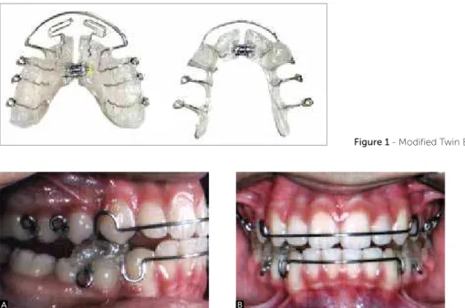
Description of the modified Twin-Block appliance Maxillary portion — composed of an acrylic base covering the hard palate, open at the midpalatal
su-ture line with a Dentaurum® 6.5 mm expanding
and control the inclinations of maxillary incisors. The appliance has simple coils on the palatal region of maxillary central and lateral incisors for tongue pressure control and teeth uprighting. The appliance retention is achieved in posterior teeth with Benac clasps, which allow activation and present good flex-ibility due to the great amount of wire employed for fabrication. The acrylic blocks are placed on the oc-clusal surface of posterior teeth with enough height to allow disocclusion of anterior teeth. The anterior portion of planes present an angle of 70 degrees, which, in combination with the mandibular planes, keeps the mandible protruded (Figs 1 and 2).
The mandibular portion is composed of an acrylic base on the lingual alveolar ridge, with anterior Haw-ley bow to control the inclination of incisors. The
presence of a Dentaurum® 5.5-mm expanding screw
on the midline allows correction of small lingual in-clinations of posterior teeth. Benac clasps are used for appliance retention on the posterior portion, and, if the bow is not sufficient in the anterior portion, an acrylic coverage should be applied on the edges of mandibular incisors. The planes are located ahead, at the region of the first premolars, and are extended up to the canines in order to achieve greater strength. They are fabricated at 70 degrees to fit with the max-illary portion of the appliance, keeping the mandible in a more anterior position. Plane height is compat-ible with the upper plane, without contact with teeth
in the maxillary arch (Figs 1 and 2). The individuals were instructed to use the modiied Twin Block for an approximate period of 20h/day.
Lateral cephalograms
Aiming to verify the dentoskeletal changes of the modiied Twin Block appliance, lateral cephalograms obtained at treatment onset and completion were as-sessed and compared to the control group. All radio-graphic images were obtained with the lips at rest and in maximum intercuspation, with the aid of the Broadbent cephalostat to standardize head positioning. All cepha-lograms in the sample were performed in three difer-ence machines and the magniication of each appliance was determined in order to allow greater accuracy of re-sults. The diferent machines presented distinct magni-ication percentages which ranged from 6% to 10.94%.
Cephalometric tracing and achievement of measurements
The cephalograms were digitized at a resolution of 9600 x 4800 dpi in a Microtek ScanMaker i800 scan-ner (Microtek International, Inc., Carson, CA, USA) connected to a Pentium microcomputer. The images were transferred to the Dolphin Imaging Premium 10.5 sotware (Dolphin Imaging & Management Solutions, Chatsworth, CA, USA) through which the cephalo-metric points of interest were marked and measure-ments involving the planes and lines were obtained.
Figure 1 - Modified Twin Block appliance.
Figure 2 - Twin Block appliance in use - A) Right lateral view. B) Frontal view. C) Left lateral view.
Intergroup comparison
The Kolmogorov-Smirnov test was applied to analyze if cephalometric data in the experimental and control groups presented normal distribution. The results revealed that the cephalometric variables presented normal distribution in both groups and in all periods analyzed (P > 0.05). Thus, paramet-ric tests were used for intergroup comparison. The compatibility between experimental and control
groups in relation to the initial (T1) and final mean
ages (T2) and the treatment/follow-up time was
as-sessed by the independent t test. The chi-square test was used to verify the compatibility between groups with regard to gender distribution and anteroposte-rior severity existing between molars.
The independent t test was used for intergroup
comparison at the initial (T1) and final periods (T2)
and to assess changes between the initial and final
periods (T2-T1) in both groups. Bonferroni
correc-tion was used for false-positive control (type I er-ror), and differences were considered statistically significant at P < (0.05/24) = 0.002.
All statistical tests were performed by means of the Statistica for Windows 7.0 software (Stat Soft Inc., Tulsa, Oklahoma, USA).
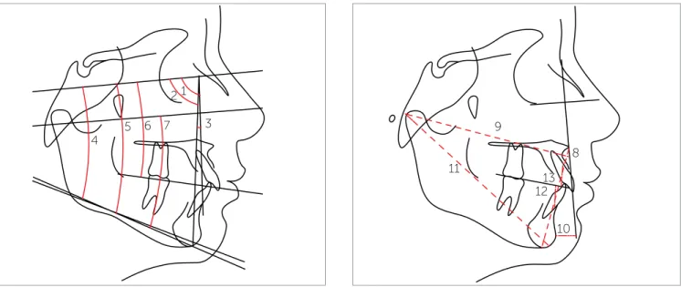
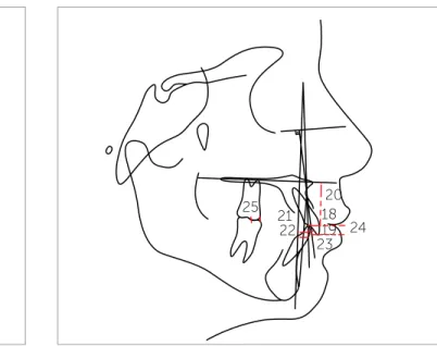
Cephalometric measurements employed (Figs 3, 4, 5 and 6)
The following cephalometric measurements were used in this study:
1. Maxillary component: SNA, A-Nperp and Co-A. 2. Mandibular component: SNB, P-Nperp and
Co-Gn.
3. Maxillomandibular relationship: ANB and Wits. 4. Growth pattern: SN.GoGn, SN.GoMe,
SN.Ocl, FMA and LAFH.
5. Maxillary dentoalveolar component: 1.NA, 1-NA, 1-Aperp, 1.PP and 1-PP.
6. Mandibular dentoalveolar component: 1.NB, 1-NB, 1-AP and IMPA.
7. Dental relationships: overjet, overbite and molar relationship.
STATISTICAL ANALYSIS
Method error
To evaluate the intra-examiner error, all measurements were repeated by the same investigator on 30 lateral cepha-lograms randomly selected ater a three-week interval. Ap-plication of the mathematical formula proposed by Dahl-berg (Se2 = Σd2/2n) allowed estimation of casual errors.14
Systematic errors were assessed by the dependent t test.15,16
Figure 3 - Skeletal angular cephalometric measurements: 1) SNA; 2) SNB;
3) ANB; 4) SN.GoMe; 5) SN.GoGn; 6) SN.Ocl; 7) FMA.
Figure 4 - Linear skeletal cephalometric measurements: 8) A-Nperp;
9) Co-A; 10) P-Nperp; 11) Co-Gn ;12) Wits; 13) LAFH.
1 2
3 9
8
13 12
10 11
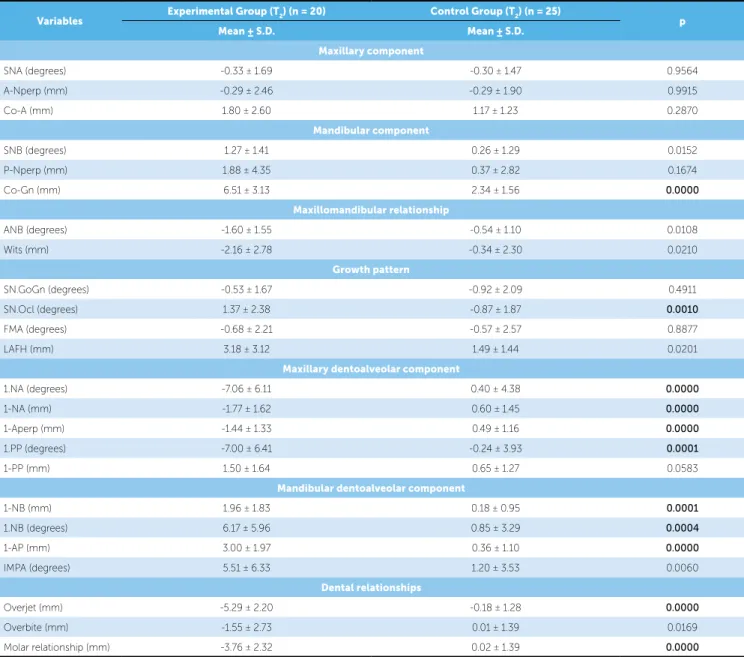
Comparison of dentoskeletal changes (T2-T1) be-tween the experimental and control groups revealed that, in relation to the mandibular component, the experi-mental group exhibited a signiicantly greater increase in mandibular length (Co-Gn). As for the growth pattern component, the Sn.Ocl variable exhibited signiicantly greater increase in the experimental group in comparison to the control group. With regard to the maxillary den-toalveolar component, the experimental group presented greater and signiicant lingual inclination and retrusion of maxillary incisors in comparison to the control group. In the mandibular dentoalveolar component, the ex-perimental group exhibited greater and signiicant buc-cal inclination and protrusion of mandibular incisors in comparison to the control group. In the analysis of dental relationships, the experimental group exhibited signii-cantly greater reduction in overjet and molar relationship when compared to the control group (Table 6).
DISCUSSION
The use of removable functional orthopedic appli-ances in growing individuals with skeletal Class II has demonstrated to have some advantages promoted by treatment of Class II malocclusion in two stages
(func-tional orthopedics and ixed appliance).11,17 Reduction
in overjet at early ages, better relationship between the jaws, reduction in facial convexity and shorter treatment time with ixed appliances are factors that encourage
treatment of Class II malocclusion in two stages.9
RESULTS
Three variables (SNA, SN.GoGn and LAFH) pre-sented systematic error (P < 0.05) and the amplitude of casual errors ranged from 0.32 (ANB) to 2.39 (LAFH).
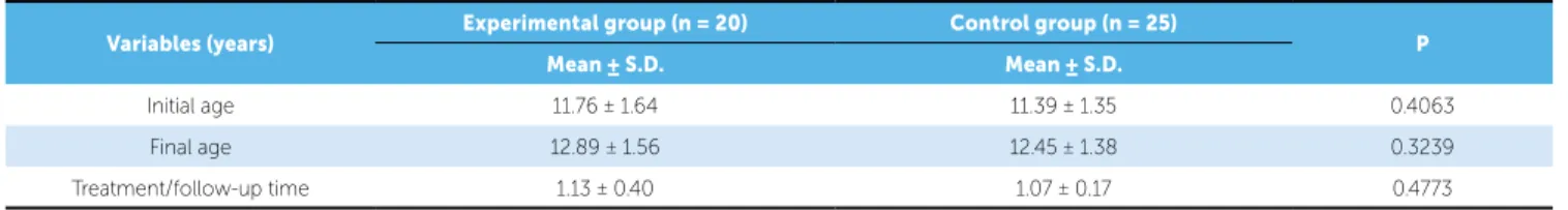
The experimental and control groups were compat-ible in initial and inal age, treatment/follow-up time, gender distribution and severity of anteroposterior re-lationship existing between molars (Tables 1, 2 and 3).
At treatment onset (T1), the experimental and
con-trol groups presented moderate cephalometric compat-ibility, with the variables ANB and Wits in the max-illomandibular relationship component presenting the worst relationship between jaws in the experimental group (P < 0.002). In the maxillary dentoalveolar com-ponent, the 1-Aperp variable revealed that maxillary incisors in the experimental group were signiicantly more buccally inclined and protruded in the maxilla (P < 0.002). As for the dental relationship component, the overjet variable signiicantly increased in relation to the control group (P < 0.002) (Table 4).
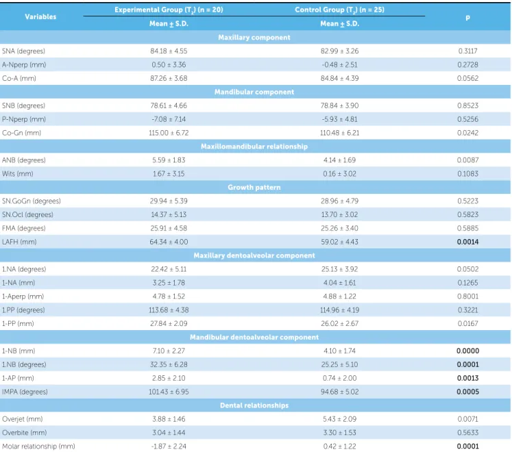
At treatment completion (T2), the growth pattern,
eval-uated by the LAFH variable, was signiicantly greater in the experimental group in comparison to the control group. In the mandibular dentoalveolar component, the experimental group presented signiicantly more protruded and buccally inclined mandibular incisors in comparison to the control group. In the evaluation of dental relationships, the experi-mental group presented signiicantly smaller molar relation-ship in comparison to the control group (Table 5).
Figure 5 - Angular dental cephalometric measurements: 14) 1.NA; 15) 1.PP;
16) 1.NB; 17) IMPA.
Figure 6 - Linear dental cephalometric measurements: 18) 1-NA; 19) 1-Aperp;
20) 1-PP; 21) 1-NB; 22) 1-AP; 23) overjet; 24) overbite; 25) molar relationship.
14
25 21
22 23
24 20 18 19 15
Variables (years) Experimental group (n = 20) Control group (n = 25) P
Mean ± S.D. Mean ± S.D.
Initial age 11.76 ± 1.64 11.39 ± 1.35 0.4063
Final age 12.89 ± 1.56 12.45 ± 1.38 0.3239
Treatment/follow-up time 1.13 ± 0.40 1.07 ± 0.17 0.4773
Table 1 - Evaluation of compatibility between groups considering initial age, final age and treatment/follow-up time (independent t test).
Table 2 - Comparison of sex distribution in the two groups (chi-square
test).
Group
Sex
Total
Female Male
Experimental 9 11 20
Control 11 14 25
Total 20 25 45
c2= 0.005; df = 1; P = 0.9465
Table 3 - Result of the chi-square test for comparison between experimental
and control groups with regard to the severity of existing anteroposterior molar relationship.
Severity Experimental group
(n = 20)
Control group (n = 25)
¼ Class II 1 6
½ Class II 7 9
¾ Class II 3 6
Full Class II 9 4
c2= 6.2663; df = 3; P = 0.0993
Table 4 - Results of the independent t test for comparison between experimental and control groups at the initial period (T1).
Variables Experimental Group (T1) (n = 20) Control Group (T1) (n = 25) p
Mean ± S.D. Mean ± S.D.
Maxillary component
SNA (degrees) 84.51 ± 3.51 83.30 ± 3.16 0.2270
A-Nperp (mm) 0.78 ± 2.98 -0.18 ± 2.70 0.2618
Co-A (mm) 85.45 ± 3.38 83.67 ± 4.80 0.1686
Mandibular component
SNB (degrees) 77.33 ± 4.10 78.59 ± 3.49 0.2723
P-Nperp (mm) -8.95 ± 6.64 -6.30 ± 4.83 0.1287
Co-Gn (mm) 108.49 ± 6.60 108.14 ± 6.27 0.8570
Maxillomandibular relationship
ANB (degrees) 7.19 ± 2.27 4.69 ± 1.66 0.0001
Wits (mm) 3.84 ± 2.65 0.50 ± 2.34 0.0001
Growth pattern
SN.GoGn (degrees) 30.46 ± 5.24 29.88 ± 4.95 0.7049
SN.Ocl (degrees) 13.00 ± 5.20 14.57 ± 2.99 0.2095
FMA (degrees) 26.58 ± 4.85 25.83 ± 4.06 0.5743
LAFH (mm) 61.16 ± 4.03 58.49 ± 4.55 0.0461
Maxillary dentoalveolar component
1.NA (degrees) 29.48 ± 6.75 24.73 ± 6.29 0.0190
1-NA (mm) 5.03 ± 2.10 3.44 ± 1.87 0.0107
1-Aperp (mm) 6.23 ± 1.74 4.40 ± 1.05 0.0001
1.PP (degrees) 120.68 ± 5.63 115.20 ± 5.80 0.0027
1-PP (mm) 26.35 ± 1.86 25.37 ± 2.76 0.1837
Mandibular dentoalveolar component
1-NB (mm) 5.14 ± 2.44 3.92 ± 1.97 0.0713
1.NB (degrees) 26.18 ± 6.98 24.40 ± 6.34 0.3760
1-AP (mm) -0.15 ± 2.12 0.37 ± 2.16 0.4213
IMPA (degrees) 95.92 ± 8.16 93.47 ± 6.59 0.2715
Dental relationships
Overjet (mm) 9.16 ± 2.10 5.61 ± 2.61 0.0000
Overbite (mm) 4.59 ± 2.50 3.29 ± 1.73 0.0464
Table 5 - Results of the independent t test for comparison between experimental and control groups at the final period (T2).
Variables
Experimental Group (T2) (n = 20) Control Group (T2) (n = 25)
p
Mean ± S.D. Mean ± S.D.
Maxillary component
SNA (degrees) 84.18 ± 4.55 82.99 ± 3.26 0.3117
A-Nperp (mm) 0.50 ± 3.36 -0.48 ± 2.51 0.2728
Co-A (mm) 87.26 ± 3.68 84.84 ± 4.39 0.0562
Mandibular component
SNB (degrees) 78.61 ± 4.66 78.84 ± 3.90 0.8523
P-Nperp (mm) -7.08 ± 7.14 -5.93 ± 4.81 0.5256
Co-Gn (mm) 115.00 ± 6.72 110.48 ± 6.21 0.0242
Maxillomandibular relationship
ANB (degrees) 5.59 ± 1.83 4.14 ± 1.69 0.0087
Wits (mm) 1.67 ± 3.15 0.16 ± 3.02 0.1083
Growth pattern
SN.GoGn (degrees) 29.94 ± 5.39 28.96 ± 4.79 0.5223
SN.Ocl (degrees) 14.37 ± 5.13 13.70 ± 3.02 0.5823
FMA (degrees) 25.91 ± 4.58 25.26 ± 3.40 0.5885
LAFH (mm) 64.34 ± 4.00 59.02 ± 4.43 0.0014
Maxillary dentoalveolar component
1.NA (degrees) 22.42 ± 5.11 25.13 ± 3.92 0.0502
1-NA (mm) 3.25 ± 1.78 4.04 ± 1.61 0.1265
1-Aperp (mm) 4.78 ± 1.52 4.88 ± 1.22 0.8001
1.PP (degrees) 113.68 ± 4.38 114.96 ± 4.19 0.3221
1-PP (mm) 27.84 ± 2.09 26.02 ± 2.67 0.0167
Mandibular dentoalveolar component
1-NB (mm) 7.10 ± 2.27 4.10 ± 1.74 0.0000
1.NB (degrees) 32.35 ± 6.28 25.25 ± 5.10 0.0001
1-AP (mm) 2.85 ± 2.10 0.74 ± 2.00 0.0013
IMPA (degrees) 101.43 ± 6.95 94.68 ± 5.02 0.0005
Dental relationships
Overjet (mm) 3.88 ± 1.46 5.43 ± 2.09 0.0071
Overbite (mm) 3.04 ± 1.44 3.30 ± 1.53 0.5633
Molar relationship (mm) -1.87 ± 2.24 0.42 ± 1.22 0.0001
Conversely, some authors have demonstrated that treatment of Class II malocclusion performed in one stage in the permanent dentition (fixed appliance) is more efficient in comparison to treatment performed in two stages, given that similar occlusal results are
obtained in significantly shorter treatment time.18,19,20
Investigations into the actual dentoskeletal changes obtained with the Twin Block appliance in the first treatment stage did not reveal any re-striction of anterior maxillary displacement (Table 6). This result suggests that treatment of Class II malocclusion with the Twin Block did not pres-ent any significant extraoral effect, as reported in
previous studies.17,21
Evaluation of the mandibular component revealed a statistically signiicant increase of 4.17 mm in the man-dibular length (Co-Gn) with anterior displacement of the Gonion, two changes that are desirable in the treat-ment of individuals with skeletal Class II malocclu-sion (Table 6). It was not possible to determine if the increase in the Co-Gn variable was caused by an in-crease in mandibular length or mandibular reposition-ing. Some authors have also evidenced similar changes
in relation to mandibular length.9,11,17,22 However, the
Table 6 - Results of the independent t test for comparison of changes (T2-T1) between experimental and control groups.
Variables Experimental Group (T2) (n = 20) Control Group (T2) (n = 25) p
Mean ± S.D. Mean ± S.D.
Maxillary component
SNA (degrees) -0.33 ± 1.69 -0.30 ± 1.47 0.9564
A-Nperp (mm) -0.29 ± 2.46 -0.29 ± 1.90 0.9915
Co-A (mm) 1.80 ± 2.60 1.17 ± 1.23 0.2870
Mandibular component
SNB (degrees) 1.27 ± 1.41 0.26 ± 1.29 0.0152
P-Nperp (mm) 1.88 ± 4.35 0.37 ± 2.82 0.1674
Co-Gn (mm) 6.51 ± 3.13 2.34 ± 1.56 0.0000
Maxillomandibular relationship
ANB (degrees) -1.60 ± 1.55 -0.54 ± 1.10 0.0108
Wits (mm) -2.16 ± 2.78 -0.34 ± 2.30 0.0210
Growth pattern
SN.GoGn (degrees) -0.53 ± 1.67 -0.92 ± 2.09 0.4911
SN.Ocl (degrees) 1.37 ± 2.38 -0.87 ± 1.87 0.0010
FMA (degrees) -0.68 ± 2.21 -0.57 ± 2.57 0.8877
LAFH (mm) 3.18 ± 3.12 1.49 ± 1.44 0.0201
Maxillary dentoalveolar component
1.NA (degrees) -7.06 ± 6.11 0.40 ± 4.38 0.0000
1-NA (mm) -1.77 ± 1.62 0.60 ± 1.45 0.0000
1-Aperp (mm) -1.44 ± 1.33 0.49 ± 1.16 0.0000
1.PP (degrees) -7.00 ± 6.41 -0.24 ± 3.93 0.0001
1-PP (mm) 1.50 ± 1.64 0.65 ± 1.27 0.0583
Mandibular dentoalveolar component
1-NB (mm) 1.96 ± 1.83 0.18 ± 0.95 0.0001
1.NB (degrees) 6.17 ± 5.96 0.85 ± 3.29 0.0004
1-AP (mm) 3.00 ± 1.97 0.36 ± 1.10 0.0000
IMPA (degrees) 5.51 ± 6.33 1.20 ± 3.53 0.0060
Dental relationships
Overjet (mm) -5.29 ± 2.20 -0.18 ± 1.28 0.0000
Overbite (mm) -1.55 ± 2.73 0.01 ± 1.39 0.0169
Molar relationship (mm) -3.76 ± 2.32 0.02 ± 1.39 0.0000
comparison to untreated individuals. This characteris-tic of functional appliances is known in the literature as
the mortgage of mandibular growth.2,23 Improvement
in mandibular retrognathism was also observed in in-dividuals in the experimental group, who presented a greater increase in the SNB variable (1.01 degrees) when compared to the control group (Table 6). This change probably contributed to reduce facial convex-ity in individuals in the experimental group.
A probable lingual movement of the roots of man-dibular incisors may promote alveolar remodeling, changing the position of point B to a more posterior po-sition and, as a consequence, reducing the SNB variable. The mandibular incisors presented signiicant buccal
inclination and protrusion, yet evidenced an increase in the SNB angle (Table 6). Previous studies also found similar changes in the evaluation of cephalometric
ef-fects promoted by the use of functional appliances.11,21
Evaluation of the maxillomandibular relationship component revealed that mandibular growth and/or repositioning did not promote signiicant changes in ANB and Wits variables with consequent reduction in skeletal discrepancy between jaws in individuals in the experimental group (Table 6). This result does not agree with previous studies, since several studies in the litera-ture demonstrate the great efectiveness of functional appliances in achieving a better relationship between
With regard to growth pattern, there was a non-sig-niicant increase in LAFH (1.69 mm) in individuals in the experimental group compared to the control group, with consequent clockwise rotation of the occlusal plane, as ob-served by the signiicant increase in the SN.Ocl variable (Table 6). These efects were probably caused by selective wear of the acrylic in contact with the mandibular poste-rior teeth, allowing greater vertical development of these teeth, which contributes for correction of Class II
relation-ship, curve of Spee and deep bite in the individuals.26,27,28
The maxillary and mandibular dentoalveolar compo-nents presented a signiicant component of inclination of anterior teeth in both arches. The maxillary incisors were lingually inclined and retruded, while the mandibular inci-sors were buccally inclined and protruded (Table 6). These dentoalveolar changes signiicantly contributed for
correc-tion of the overjet.9,17,22 However, excessive inclination of
incisors should be carefully controlled, since they
substan-tially reduce the potential of changes of orthopedic nature.9
In the evaluation of dental relationships, there was a signiicant reduction of 5.11 mm in the overjet and of 3.78 mm in molar relationship in comparison to the control group. These changes contribute to correct the anteroposterior discrepancy in individuals with Class II malocclusion (Table 6). These results represent a desir-able consequence of treatment of skeletal Class II mal-occlusion, and are established by the combination of dentoalveolar and skeletal changes that occurred in the
experimental group.29,30
CONCLUSION
1. Woodside DG. Do functional appliances have an orthopedic efect? Am J Orthod Dentofacial Orthop. 1998;113(1):11-4.
2. Chen JY, Will LA, Niederman R. Analysis of eicacy of functional
appliances on mandibular growth. Am J Orthod Dentofacial Orthop. 2002;122(5):470-6.
3. Björk A. The principles of the Andresen method of orthodontic
treatment: a discussion based on cephalometric x-ray analysis of treated cases. Am J Orthod. 1951;37(6):437-58.
4. Pancherz H. A cephalometric analysis of skeletal and dental changes
contributing to Class II correction in activator treatment. Am J Orthod. 1984;85(2):125-34.
5. DeVincenzo JP. Changes in mandibular length before, during and after
successful orthopaedic correction of Class II malocclusions using a functional appliance. Am J Orthod Dentofacial Orthop. 1991;99(3):241-57. 6. Windmiller EC. The acrylic-splint Herbst appliance: a cephalometric
evaluation. Am J Orthod Dentofacial Orthop. 1993;104(1):73-84.
7. Olibone VLL, Guimarães AS, Atta JY. Inluência do aparelho propulsor
Twin Block no crescimento mandibular: revisão sistemática da literatura. Rev Dental Press Ortod Ortop Facial. 2006;11(1):19-27.
8. Hirzel HC, Grewe JM. Activators: a practical approach. Am J Orthod.
1974;66(5):557-70.
9. Lund DI, Sandler PJ. The efects of Twin Blocks: a prospective controlled study. Am J Orthod Dentofacial Orthop. 1998;113(1):104-10.
10. Gill DS, Lee RT. Prospective clinical trial comparing the efects of conventional Twin-block and mini-block appliances: Part 1. Hard tissue changes. Am J Orthod Dentofacial Orthop. 2005;127(4):465-72; quiz 517. 11. Illing HM, Morris DO, Lee RT. A prospective evaluation of Bass, Bionator
and Twin Block appliances. Part I--The hard tissues. Eur J Orthod. 1998;20(5):501-16.
12. Brunharo IHVP, Quintão CA, Almeida MAO, Motta A, Barreto SYN. Alterações dentoesqueléticas decorrentes do tratamento com aparelho ortopédico funcional Twin Block em pacientes portadores de má oclusão de Classe II esquelética. Dental Press J Orthod. 2011;16(5):40.e1-8. 13. Antonarakis GS, Kiliaridis S. Short-term anteroposterior treatment efects
of functional appliances and extraoral traction on class II malocclusion. A meta-analysis. Angle Orthod. 2007;77(5):907-14.
14. Dahlberg G. Statistical methods for medical and biological students. New York: Interscience; 1940.
15. Baumrind S, Frantz RC. The reliability of head ilm measurements. 1. Landmark identiication. Am J Orthod. 1971;60(2):111-27.
ReFeRenCeS
16. Houston WJ. The analysis of errors in orthodontic measurements. Am J Orthod. 1983;83(5):382-90.
17. Jena AK, Duggal R, Parkash H. Skeletal and dentoalveolar efects of Twin-block and bionator appliances in the treatment of Class II malocclusion: a comparative study. Am J Orthod Dentofacial Orthop. 2006;130(5):594-602. 18. Dolce C, McGorray SP, Brazeau L, King GJ, Wheeler TT. Timing of Class II
treatment: skeletal changes comparing 1-phase and 2-phase treatment. Am J Orthod Dentofacial Orthop. 2007;132(4):481-9.
19. Ghafari J, Shofer FS, Jacobsson-Hunt U, Markowitz DL, Laster LL. Headgear versus function regulator in the early treatment of Class II, division 1 malocclusion: a randomized clinical trial. Am J Orthod Dentofacial Orthop. 1998;113(1):51-61.
20. Tulloch JF, Phillips C, Proit WR. Beneit of early Class II treatment: progress report of a two-phase randomized clinical trial. Am J Orthod Dentofacial Orthop. 1998;113(1):62-72.
21. DeVincenzo JP, Huffer RA, Winn MW. A study in human subjects using a new device designed to mimic the protrusive functional appliances used previously in monkeys. Am J Orthod Dentofacial Orthop. 1987;91(3):213-24.
22. Mills CM, McCulloch KJ. Treatment efects of the twin block appliance: a cephalometric study. Am J Orthod Dentofacial Orthop. 1998;114(1):15-24. 23. Johnston LE Jr. Functional appliances: a mortgage on mandibular
position. Aust Orthod J. 1996;14(3):154-7.
24. Kalha A. Early treatment with the twin-block appliance is efective in reducing overjet and severity of malocclusion. Evid Based Dent. 2004;5(4):102-3.
25. O’Brien K, Wright J, Conboy F, Sanjie Y, Mandall N, Chadwick S, et al. Efectiveness of early orthodontic treatment with the Twin-block appliance: a multicenter, randomized, controlled trial. Part 1: Dental and skeletal efects. Am J Orthod Dentofacial Orthop. 2003;124(3):234-43. 26. Clark WJ. The Twin Block technique. A functional orthopedic appliance
system. Am J Orthod Dentofacial Orthop. 1988;93(1):1-18. 27. Clark WJ. The Twin Block technique. Part 2. Funct Orthod.
1992;9(6):45-9.
28. Clark WJ. The Twin Block technique. Part 1. Funct Orthod. 1992;9(5):32-4, 36-7.
29. Lee RT. How orthodontic functional appliances work. Prim Dent Care. 2000;7(2):67-73.