Human Bone Marrow Mesenchymal Stem Cell Behaviors on
PCL/Gelatin Nanofibrous Scaffolds Modified with A Collagen
IV-Derived RGD-Containing Peptide
Ali Mota, Ph.D.1, Abbas Sahebghadam Loti, Ph.D.1, 2*, Jalal Barzin, Ph.D.3, Mostafa Hatam, Ph.D.2, Behzad Adibi, Ph.D.1, Zahra Khalaj, M.Sc.2, Mohammad Massumi, Ph.D.2
1. Department of Clinical Biochemistry, Faculty of Medical Sciences, Tarbiat Modares University, Tehran, Iran 2. National Institute of Genetic Engineering and Biotechnology (NIGEB), Tehran, Iran
3. Iran Polymer and Petrochemical Institute (IPPI), Tehran, Iran
*Corresponding Address: P.O.Box: 14115-111, Department of Clinical Biochemistry, Faculty of Medical Sciences, Tarbiat Modares University, Tehran, Iran
Email: Loti_ab@modares.ac.ir
Received: 2/Dec/2012, Accepted: 10/Feb/2013 Abstract
Objective: We introduce an RGD (Arg-Gly-Asp)-containing peptide of collagen IV origin that possesses potent cell adhesion and proliferation properties.
Materials and Methods: In this experimental study, the peptide was immobilized on an electrospun nanofibrous polycaprolactone/gelatin (PCL/Gel) hybrid scaffold by a chemical bonding approach to improve cell adhesion properties of the scaffold. An io-dine-modified phenylalanine was introduced in the peptide to track the immobilization process. Native and modified scaffolds were characterized with scanning electron microscopy (SEM) and fourier transform infrared spectroscopy (FTIR). We studied the osteogenic and adipogenic differentiation potential of human bone marrow-de-rived mesenchymal stem cells (hBMSCs). In addition, cell adhesion and prolifera-tion behaviors of hBMSCs on native and peptide modified scaffolds were evaluated by 3-(4,5-dimethylthiazol-2-yl)-2,5-diphenyltetrazolium bromide (MTT) assay and 4',6-diamidino-2-phenylindole (DAPI) staining, and the results compared with tissue culture plate, as the control.
Results: FTIR results showed that the peptide successfully immobilized on the scaffold. MTT assay and DAPI staining results indicated that peptide immobilization had a dramatic effect on cell adhesion and proliferation.
Conclusion: This peptide modiied nanoibrous scaffold can be a promising biomaterial for tissue engineering and regenerative medicine with the use of hBMSCs.
Keywords:Nanoibers, Polycaprolactone, RGD Peptide, Surface Modiication, Mesen-chymal Stem Cell
Cell Journal(Yakht eh), Vol 16, No 1, Spring 2014, Pages: 1- 10
Citation: Mota A, Sahebghadam Loti A, Barzin J, Hatam M, Adibi B, Khalaj Z, Massumi M. Human bone marrow mesenchymal stem cell behaviors on PCL/Gelatin nanoibrous scaffolds modiied with a collagen IV-derived RGD-containing peptide. Cell J. 2014; 16(1): 1-10.
Introduction
In recent years a wide range of polymeric bioma-terials with various properties have been developed for bioengineering and tissue engineering applica-tions. These biomaterials should be biodegradable, bioactive and biocompatible (1). The biomaterials architecture is also important and affects the
cell-matrix interaction (2), which is of signiicance in
cellular behaviors such as cell adhesion,
prolifera-tion, and differentiation (3). Cell adhesion is the
irst event in the cellular response to biomaterials
cells (6). Approaches to improve the biomimetic properties of such biomaterials include materials
modiication by hybridization with bioactive com
-pounds and/or immobilization of bioactive mo
-tives to enhance and control interaction between cells and synthetic biomaterials (7).
Electrospinning is a simple and highly
through-put technique for producing hybrid nanoibers with
suitable high porosity and surface area structure to mimic nanoscaled patterns of natural extracellu-lar matrix (ECM) to control cell behavior (2). In recent years several attempts have resulted in the
production of polymeric nanoibrous scaffolds us
-ing electrospinn-ing for biomedical applications (8,
9). Hybridization of biomaterials with bioactive
substances such as gelatin (10, 11) collagen (12,
13), and ibroin (14) have been used in numerous
studies to modulate the biomimetic properties of polymeric materials for a variety of applications.
Immobilization of cell recognition peptides on
biomaterials (15, 16) or peptide amphiphils (17, 18) accelerates improvement of the biomaterial’s surface chemistry. Cell-cell and cell-ECM interac-tions are mediated by cell adhesion receptors. The integrin family is among the most versatile group of cell adhesion receptors. They are involved not only in cell anchoring but also in numerous other processes such as proliferation, differentiation, and homeostasis (19). The RGD (Arg-Gly-Asp) sequences are the most prominent cell recognition sequences present in many ECM proteins (20). It has been demonstrated that approximately half of the integrin family binds to ECM proteins through the RGD sequences. Therefore, the RGD peptides are by far the most effective peptides for improv-ing cell adhesion to biomaterials surfaces.
We introduced and synthesized a novel
RGD-containing peptide of collagen IV origin and
im-mobilized the peptide on the surface of a hybrid polycaprolactone/gelatin (PCL/Gel) nanoibrous
scaffold. The morphology and structure of the PCL/
Gel nanoibrous scaffold were evaluated by scan
-ning electron microscopy (SEM). We characterized the RGD-modiied scaffold with fourier transform
infrared spectroscopy (FTIR). Behaviors of hu-man bone marrow-derived mesenchymal stem cells (hBMSCs) including cell adhesion and proliferation
on the RGD-modiied PCL/gel nanoibrous scaffold were studied by 3-(4,5-dimethylthiazol-2-yl)-2,5-diphenyltetrazolium bromide (MTT) assay and
compared with non-modiied scaffold and tissue
culture plate (TCP) as the control. Cell adhesion to
scaffolds was visualized by 4',6-diamidino-2-phe
-nylindole (DAPI) staining of the cell nucleus.
Materials and Methods
In this experimental study, Fmoc (9-luorenyl
-methoxycarbonyl)- protected amino acids with various protected side chains,
rink-amide-MB-HA resin, and O-(benzotriazole-1-yl)-N, N, N´, N´-tetramethyluronium-hexaluorophosphate
(HBTU) were purchased from GL Biochem
(Shanghai, China). HPLC grade triluoroacetic
acid (TFA), acetonitrile (ACN) and water, N-ethyl
-diisopropylamine (DIEA) and triisopropylsilane (TIPS) were products of Merck Chemicals (Ger-many). Gelatin type B powder (300 g Bloom) from porcine skin was prepared from Sigma (USA). PCL (Mn 80,000) and 1-ethyl-3-(3-dimethylami-nopropyl) carbodiimide (EDAC) were obtained from Sigma-Aldrich (St. Louis, MO, USA).
Dul-becco Modiied Eagle’s Medium (DMEM), fetal
bovine serum (FBS), phosphate buffered saline (PBS) and trypsin/EDTA were products of Gibco
(Invitrogen, USA).
3-(4,5-dimethylthiazol-2-yl)-2,5-diphenyltetrazolium bromide (MTT) and 4',6-di
-amidino-2-phenylindole (DAPI) were purchased from Promega (USA). All other chemicals and sol-vents were obtained from Merck unless otherwise
stated and used without further modiications.
Peptide synthesis
A 10-mer RGD-containing peptide of collagen IV origin with a sequence of
KK-[GPRGDPG]-F(4-I) and a theoretical mass of m/z 1183.1 was synthesized by standard Fmoc chemistry using an Advanced Chemtech 90 peptide synthesizer (USA). The peptide was synthesized on
rink-amide-MBHA resin with a theoretical loading of
≈0.55 mmol/g. The total loading of the resin was
used for all calculations. Standard Fmoc-protect-ed amino acids (4 equivalents) were usFmoc-protect-ed in each coupling reaction. Coupling was performed for 15 minutes with a mixture of DIEA (2.5 equivalents) and HBTU (0.98 equivalent). A solution of 20% (v/v) piperidin in DMF was used for the depro-tection reaction. Coupling and deprodepro-tection
reac-tions were conirmed by the Kaiser test. After inal
cleav-age cocktail mixture of 95% TFA, 2.5% TIPS and
2.5% deionized water for 2 hours on ice was used
for cleavage of the peptide from the resin. Cleaved peptide was precipitated by cold diethylether and
resolubileized in deionized water. A modiied io
-dinated phenylalanine (4-I-phenylalanine) at the carboxyl end of the peptide was used in order to
conirm the immobilization of the peptide on the surface of the PCL/Gel nanoibrous scaffold.
Peptide puriication and characterization
The purity of crude peptide was analyzed by
reverse-phase high performance liquid chromatog-raphy (RP-HPLC; Agilent, USA). RP-HPLC was done on an octadecylsilica (C18) column (4.6 mm
id×250 mm length, 5 μm) with TFA/water (0.1% v/v) and TFA/ACN (0.1% v/v) as the mobile phase with a linear gradient from 0 to 60% of TFA/ACN over 30 minutes and low rate of 1 mL/minute at
25˚C. RP-HPLC results were used to scale up and
purify the peptide. The peptide was puriied by pre
-parative HPLC (Agilent, USA) with >95% purity
with the same mobile phase as RP-HPLC. Puriied peptide was lyophilized and stored at -20˚C until future use in the experiments. For characterization of the synthesized peptide, mass spectrum of the
peptide was obtained by quadropole LC-MS
(Agi-lent, USA) with an electrospray ionization system.
Electrospinning
2, 2, 2-triluoroethanol (TFE) was used as a com
-mon solvent for PCL and gelatin. PCL granules and gelatin powder were dissolved separately in TFE by stirring overnight at room temperature to prepare 10% w/v solutions. A PCL/Gel blended solution at a ratio of 1:1 was loaded into a 10 mL syringe. The syringe was attached to the pump and
the blended solution delivered by a steady low of
1 mL/hour to a 22G stainless steel blunt needle by a plastic tube. A high voltage of 22 kV was applied to the needle and the solution was electrospun for 4
hours. The electrospun hybrid nanoibers were col
-lected on a rotating drum collector on aluminum foil that was placed at a distance of 15 cm from the tip of the needle. The collector rotation speed was set at 100 rpm and shuttling speed at 5 mm/ minute. The electrospinning process was done at
25˚C and 50% humid atmosphere. The electrospun nanoibers were dried in a vacuum overnight and
named PCL/Gel.
Scaffold cross-linking
Electrospun PCL/Gel nanoibrous scaffolds were
cross-linked using a solution of EDC/NHS in etha
-nol/water (80:20 v/v) as previously described (21). The cross-linking process was done by placing the air-dried PCL/Gel hybrid scaffolds together with a supporting aluminum foil in a 250 mM solution of
EDC/NHS for different time periods. The extent
of cross-linking was assessed for the presence of free amine group content by the ninhydrin assay. A sample of each scaffold was incubated with 0.3 M ninhydrin solution in ethanol for 5 minutes at
95˚C. The irst sample that become negative ac
-cording to the ninhydrin test after a certain period of time was considered to be a complete cross-linking process. This complete cross-linked scaf-fold was used in the next step.
Peptide immobilization
Cross-linked scaffolds were rinsed 3 times with
PBS to remove excess EDC/NHS and the scaf
-folds were immersed immediately 1 μmol/mL of
the peptide in PBS and incubated for 24 hours with mild stirring at 25˚C (22). Functional ized scaffolds were then washed 3 times with PBS and dried un-der vacuum for 24 hours.
Fourier transforms infrared spectroscopy (FTIR) For surface characterization and chemical analysis of scaffolds and conirmation of the immobilization of the peptide on nanoibers, FTIR spectroscopy of PCL/Gel and RGD-modiied PCL/Gel scaffolds were
performed over a range of 4000 to 400 cm-1 at a
reso-lution of 4 cm-1 using a Bruker (Bruker, Tensor 27,
USA) FTIR system. To assign a certain peak to the iodine tag, FTIR spectra of phenylalanine and 4-iodo-phenylalanine as controls were also recorded.
Scanning electron microscopy (SEM) analysis
The morphology and structure of electrospun PCL/
Gel and RGD-modiied PCL/Gel nanoibers were analyzed by SEM. Air-dried samples were sputter
coated with gold in a KYKY sputter coater (KYKY-SBC-12, China) prior to analysis. SEM images were recorded with KYKY-EM-3200 SEM system (Chi-na) at an accelerating voltage of 25 kV. The
distribu-tion of electrospun nanoibers diameter was analyzed
by measuring at least 100 ibers using Image J soft
Human bone marrow-derived mesenchymal stem cells (hBMSCs) culture
hBMSCs were isolated from the iliac crests of healthy donors at Royan Institute (Royan Institute Cell Bank). All samples were collected following-donors’ informed consents. The study was approved by the Ethical Committee of Tarbiat Modares Uni-versity. hBMSCs were grown on DMEM medium supplemented with 10% FBS and 1% penicil lin (10000 U/mL)/streptomycin (10 mg/mL). Cells
were subcultured to 80% conluency and incubated
at 37˚C in a humidiied atmosphere of 5% CO2. Cells were used for osteogenic and adipogenic differentiation and cell adhesion and proliferation studies at passage 4.
Osteogenic differentiation
To induce osteogenic differentiation, upon
reach-ing 80% conluency, hBMSCs were detached us
-ing 0.05% trypsin/EDTA and plated in 4-well culture dishes at a density of 5000 cells/well and were treated with osteogenic medium (DMEM supplemented with 10% FBS, 0.1 mmol/L dexa-methasone, 10 mmol/L glycerol phosphate, and 0.2 mmol/L ascorbic acid) for 14 days. The me-dium was changed twice weekly. The cells were
ixed with 4% paraformaldehyde for 10 minutes
and washed with phosphate buffered saline (PBS).
Osteogenesis was assessed by alizarin-red staining for visualizing deposition of calcium (9).
Adipogenic differentiation
For adipogenic differentiation, hBMSCs were seeded in 4-well culture plates at a density of 5000 cells/well. The cells were treated with adi-pogenic medium (DMEM supplemented with 10% FBS, 0.5 mmol/L 3-isobutyl-1-methylxanthine, 1
μmol/L dexamethasone, 1.7 μmol/L insulin and 200 μmol/L indomethacin), for 2 weeks. Medium was replaced twice weekly. The cells were ixed
with 4% paraformaldehyde for 10 minutes and washed with 70% ethanol. Adipogenesis was
as-sessed by oil red O staining for visualizing deposi
-tion of fat droplets in differentiated cells (9).
Cell adhesion and proliferation studies
The interaction between cells and scaffolds on
PCL/Gel, RGD-modiied PCL/Gel and TCP as
control were studied using a colorimetric MTT
as-say. Cross-linked and RGD-immobilized PCL/Gel
scaffolds were cut with a paper hole punch,
trans-ferred into 96-well plates (Nunc, Denmark), and then sterilized with 70% ethanol for 30 minutes.
Ethanol was aspirated and the scaffolds were dried
and further sterilized for 15 minutes under UV prior to cell seeding. hBMSCs were trypsinized and
plated at 5000 cells/well. The cells were incubated for 1, 2, 3 and 4 hours for cell adhesion and 72 hours for the cell proliferation and viability assay at 37˚C in a 5% CO2 and 95% humidiied atmosphere in DMEM supplemented with 10% FBS. At each time point, the medium was discarded and the scaffolds were washed 3 times with PBS to remove non-adhered cells. The adhered cells were then incubated with MTT solu-tion (10% (v/v) MTT stock (1 mg/mL) in DMEM) at 37˚C for 2 hours. Afterwards, the attached cells were washed twice with PBS. The developed purple
formazan precipitates were solubilized and extracted
with DMSO. The absorbance of each well was
meas-ured at 570 nm using a plate reader (Tecan, Switzer
-land). hBMSCs cultured on a blank plate were used as
the control. The absorbance of solubilized formazane
crystals is directly attributed to the number of adhered live cells. To verify the presence of cell adhesion on scaffolds, DAPI staining was done. In brief, in an-other set the scaffolds were washed with PBS 3 times
and the attached cells were ixed with 4% paraformal
-dehyde for 10 minutes. The scaffolds were washed 3 times with PBS for 5 minutes. DAPI (1:1000 dilution) was added onto the scaffolds and incubated for 5
min-utes. Fluorescent images were recorded by a Nikon
TE2000 microscope.
Statistical analysis
Statistical analysis was performed with SPSS, ver-sion 17 (SPSS Inc., USA). All cell culture experi-ments were performed in triplicate. Mean ± standard deviations were used for adhesion and proliferation data analyses. To compare mean values in different
scaffolds analysis of variance (ANOVA) was used.
Comparisons were performed by Tukey’s post HOC
test. A p value of <0.05 with 95% conidence interval was considered as statistically signiicant.
Results
Electrospinning
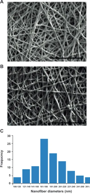
We successfully fabricated PCL/Gel scaffolds of
the desired porosity and nanoiber properties. SEM
-ogy of the PCL/Gel nanoibers (Fig 1A). The SEM images in igure 1B show no dramatic changes in
the morphology and diameter of the nanoibers af
-ter cross-linking and peptide immobilization. These nanoibers had a randomly oriented structure where
the majority of mats had nano-scale diameters be-tween 160 to 200 nm (190 ± 38 nm, Fig 1C).
Nanoiber diameters (nm)
Fraqucncy
30
25
20
15
10
5
0
100-120 121-140 141-160 161-180 181-200 201-220 221-240 241-260 261-280
Fig 1: Scanning electron microscopy (SEM) images of nanoi -brous scaffolds electrospun from 10% (w/v) PCL/Gel blended solution (A) before and (B) after cross-linking. Arrow shows some artifacts due to precipitation of phosphate buffered saline (PBS). Distribution of iber diameter in electrospun PCL/Gel scaffolds (C). Scale bar: 10 μm. Resolution: ×7500.
Peptide characterization
We successfully synthesized an RGD-containing
pep tide with cell attachment properties and an iodine
tag to track peptide immobilization. As shown in igure
2A, the crude peptide HPLC chromatogram showed
some impurities which were omitted after puriication
with preparative HPLC (Fig 2B). Mass spectrometry analysis showed the main peak with the
correspond-ing m/z ratio of 592.4 which closely approximated the
theoretical mass of the peptide (1183.1), assuming the peptide obtain two positive charge in the process of sample preparation (Fig 2C).
A
Retention time (minute)
900
700
500
300
100
-100
0 2 4 6 8 10 12 14 16
Absorbance
B
Retention time (minute)
900
700
500
300
100
-100
0 2 4 6 8 10 12 14 16
Absorbance
Fig 2: A. Analytical and B. preparative HPLC chromato-gram for analyzing and puriication of the synthesized pep -tide. C. Mass spectrum for analyzing peptide mass.
A
B
C
Scaffold cross-linking
In the cross-linking process, the carboxyl groups present on gelatin were activated when
the scaffold was treated with EDC/NHS and
amide bonds formed between the carboxyl and amine groups in gelatin. The ninhydrin test is an indicator of the presence of free amine groups. As shown in figure 3 the scaffolds were stained dark blue before cross-linking. After complete cross-linking, the ninhydrin test was negative (Fig 3).
Fig 3: Ninhydrin staining of PCL/Gel scaffold. A. On the native scaffold, ninhydrin produced a dark blue color when reacting with free amine groups present in gela-tin lysine residues. B. After cross-linking these amine groups coupled to free carboxyl groups and the scaffolds became colorless.
RGD immobilization and scaffold characterization
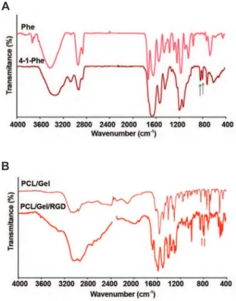
The FTIR spectrum of phenylalanine and 4-I-phenylalanine are shown in figure 4A. FTIR spectrum of 4-I-phenylalanine showed char-acteristic peaks at 798 cm-1 and 812 cm-1 that
could be ascribed to the C-I bond since the C-I bond led to the appearance of some peaks close to 800 cm-1. These significant peaks were not
present in the FTIR spectrum of the PCL/Gel (Fig 4B). The characteristic peaks attributed to
the C=0 and N-H bond were present at 3500
cm-1 (C=O) and 3300 cm-1 (N-H). The peak
at 1650 cm-1 was assigned to the amide I that
corresponded to gelatin in the PCL/Gel (23). The FTIR for PCL/Gel scaffold modified with RGD peptide revealed a characteristic peak at 798 cm-1. The other peak at 812 cm-1 was also
present with some delocalization to 840 cm-1 (Fig
4B). The presence of these peaks implied that
the RGD peptide immobilized on the PCL/Gel
scaffold.
A
B
Fig 4: A. Fourier transform infrared spectroscopy (FTIR) of Phe and 4-Iodo-Phe standards. Arrows show the char-acteristic peaks assigned to C-I bond stretching. B. FTIR spectroscopy of PCL/Gel and RGD modiied PCL/Gel. The assigned peaks are close to 800 cm-1 on the RGD modiied PCL/Gel scaffold spectrum.
Osteogenic and adipogenic differentiation potential
Figure 5A shows the characteristic lipid drop-lets stained by oil-red-O staining. According to the
results of alizarin red staining (Fig 5B), hBMSCs
cultured in osteogenic differentiation medium
de-posited a mineralized matrix after 14 days of incu
-bation. The results of this experiment showed that hBMSCs could differentiate into osteocytes and adipocytes.
A B
Cell adhesion and proliferation studies
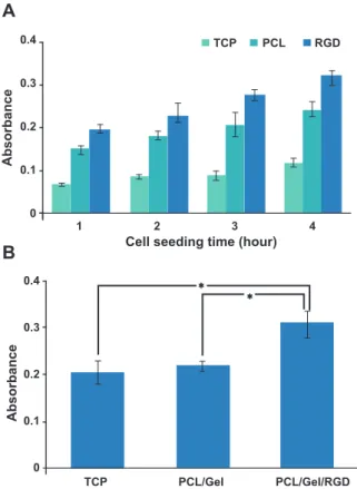
Figure 6 shows the status of hBMSC adhesion on different scaffolds. Cell adhesion and proliferation were measured by the MTT assay. It was clearly ob-vious that cell adhesion on TCP did not seem to
in-crease signiicantly at different time points (p>0.05).
hBMSCs adhered rapidly on both PCL/Gel and RGD
modiied PCL/Gel scaffolds (Fig 6A). The adhesion
increased gradually in a time-dependent manner.
Im-mobilization of RGD peptides on PCL/Gel nanoi
-brous scaffolds resulted in increased adhesion behav-ior of the scaffold. There was more adhered cells on the PCL/Gel scaffold grafted by RGD compared to those
cultured on TCP and non-grafted PCL/Gel (p≤0.05). Adhesion of cells on RGD-modiied PCL/Gel also
gradually increased at different time points. TCP did not show a time-dependent adhesion. We found that cell adhesion on TCP slightly increased over time.
Cell seeding time (hour)
Absorbance
0.4
0.3
0.2
0.1
0
1 2 3 4 TCP PCL RGD
A
Absorbance
0.4
0.3
0.2
0.1
0
TCP PCL/Gel PCL/Gel/RGD
B
*
*
Fig 6: A. MTT assay results of hBMSC adhesion on differ-ent scaffolds at 1, 2, 3 and 4 hours after cell seeding. There are signiicant differences in cell adhesion between scaffolds at each time point. B. MTT assay for hBMSC proliferation on different scaffolds. After 72 hours of cell seeding RGD modiied PCL/Gel scaffold demonstrated a dramatic effect on hBMSC proliferation according to the MTT assay (*; p≤0.5). No signiicant difference was found between PCL and TCP in their effects on cell proliferation (p>0.5).
While PCL/Gel did not show signiicant ef
-fects on hBMSCs proliferation (p>0.05), it was
clearly apparent that immobilization of RGD had a very signiicant effect on cell proliferation (p≤0.05, Fig 6B). DAPI stained images showed
that a greater number of hBMSCs attached on the
RGD-modiied scaffold compared with TCP and the unmodiied scaffold (Fig 7).
A
B
C
Fig 7: DAPI staining for visualization of attached cells on: A. tissue culture plate (TCP); B. native PCL/Gel scaffold; and C. RGD-modiied PCL/Gel scaffold. Clearly, RGD-modiied PCL/Gel scaffold showed a signiicantly greater number of bound hBMSCs after cell seeding compared with TCP and the unmodiied scaffold.
Discussion
We synthesized a collagen IV-derived
RGD-containing peptide that had cell adhesion and proliferation properties. The peptide was
success-fully immobilized on the surface of electrospun hybrid nanoibrous PCL/Gel scaffolds. The RGD-containing peptide was immobilized on scaffolds
treatment after cross-linking to enhance the adhe-sion of hBMSCs and improve biomimetic proper-ties of the scaffold.
Morphology and surface chemistry of
nano-ibrous scaffolds used in tissue engineering and
regenerative medicine have an important impact on cell behaviors such as adhesion, proliferation, differentiation and cell-matrix interaction (24). The most challenging issue regarding polymeric materials such as PCL are the inadequate and
non-speciic interactions that occur between polymers
and cells (25). Most synthetic polymers such as PCL are hydrophobic and lack functional active groups such as hydroxyl, carboxyl, amine and sul-fate groups to interact with cells. Many surface
modiication strategies have thus been developed
to introduce such functional active groups on the surface of PCL.
Since the PCL surface lacks active groups such as amine or carboxyl groups, it cannot be easily
modiied. Alkaline hydrolysis and aminolysis are
suitable methods for introducing these functional groups. The use of these methods, however, alters the structural and morphological properties of PCL
nanoibers and can lead to formation of unstable
nanoibers in terms of mechanical and morpholog
-ical properties (26). To produce reactive groups, PCL is usually blended or surface-activated with
other biomolecules such as collagen, elastin, ibro
-in, and surface activating peptides. Gelat-in, which is derived from collagen, is cheaper and can be used to overcome this incompetency.
In the present study, we physically blended PCL with gelatin. We found that the cross-linked hy-brid PCL/Gel scaffold had improved cell adhe-sion properties. This resulted from the presence of highly polar and functional groups of gelatin
on the surface of electrospun nanoibers. The ef
-fect of hybrid PCL/Gel on cell behavior has been
studied in previous works. PCL/Gel nanoibrous
scaffolds can enhance differentiation of cerebellar stem cells toward functional nerve cells (27). Hy-brid scaffolds composed of PCL and collagen are constructed and used in vascular reconstruction. Tillman et al. (28) have shown that PCL/collagen electrospun scaffolds maintained high potential and integrity in vivo without an abnormal inlam
-matory response. They concluded that this hybrid electrospun scaffold might have clinical applica-tions. McClure et al. (29) used a three layered
elec-trospun matrix to mimic native arterial structure using three different materials, PCL, elastin and collagen. They predicted that this three layered vascular graft contained overall properties within the range of the native artery.
Electrospinning of PCL was successfully per-formed with polar solvents in order to facilitate the electrospinning process. Gelatin, as a denatured form of collagen, has been used in many types of biodegradable and biocompatible scaffolds. A disadvantage for gelatin itself, or in combination with other biomaterials is its swelling in aqueous medium. Gelatin may be cross-linked to solve this problem by chemical cross-linkers such as glu-taraldehyde and formaldehyde. However, these cross-linkers have been shown to be toxic (30) and disrupt the electrospun morphology (26). EDC is an effective linker which introduces cross-links that do not release any toxic end product or a foreign bond in cross-linked gelatin. It has been shown that EDC as a water-soluble carbodiimide can be used in gelatin cross-linking. Once it is used in an ethanol-water mixture, the swelling of gelatin can be prevented even in the cross-linking process (21).
Cross-linking of scaffolds by EDC involves the activation of a C-terminal, glutamate side chain and aspartate side chain carboxyl groups, and the formation of amide bonds upon coupling with
amine groups of lysine. EDC/NHS leads to the
formation of intermolecular and intramolecular cross-links (26). Grover et al. (31) have shown that cross-linking of gelatin-based scaffolds can im-prove mechanical strength and modulate degrada-tion resistance providing scaffolds with increased structural integrity.
After crosslinking, we immobilized the peptide
from amine groups using excess carboxyl groups that activated in the preceding step. It has been shown that gelatin has approximately 12.3% ex-cess carboxyl (D and E) groups than amine (K) groups (32). Therefore, when gelatin is cross-linked to the degree that the ninhydrin test be-comes negative, the majority of amine groups are coupled with carboxyl groups. Excess carboxyl
groups can be used for peptide immobilization and further functionalization. To immobilize the target
peptide or other biomolecules, different strategies
have been used. In this regard, in the synthesized
order to couple the peptide using lysine side chain amines to carboxyl groups activated in the preced-ing cross-linkpreced-ing step in gelatin molecules. Gabriel et al. (33) have used a three-step procedure to
im-mobilize RGD peptide on polycaprolactone ilm.
This procedure involves amination, reaction with hetero-bifunctional cross-linkers and conjugation
of an RGD-motif-containing peptide to the ilm. In
the present study we have used a simple one step
cross linking and immobilization procedure. With
this procedure we employed an approach that not only cross-linked the scaffold but also provided
activated carboxyl groups for immobilization of
the peptide in the next step without any additional
modiication.
Although this peptide has the same
composi-tion as gelatin, a modiied iodinated phenylalanine must be used to detect the immobilized peptide on
the scaffold. FTIR studies conirmed these ind
-ings (Fig 4).
The results conirmed that immobilization of the peptides on the scaffold signiicantly enhanced
hBMSCs adhesion and proliferation.
Immobili-zation of cell adhesion motifs has been known to
improve the surface chemistry of biomaterials. Re-cently, a large number of RGD-containing peptides with different sequences from ECM proteins have
been synthesized (25). These peptides are immo
-bilized on several polymers by a series of physical
and chemical procedures. Many of these peptides
have the RGD peptide sequence of ibronectin
with some variations. Santiago et al. (34) have
modiied polycaprolactone disks with a three lam
-inin-derived peptide sequence using carbodiimide chemistry. These investigators used aminolysis of polycaprolactone disks to introduce amine groups
on the disks. We synthesized an RGD peptide from the origin of collagen IV. For the irst time, in the current study, a peptide modiication applied to gelatin-based nanoibrous scaffold was used. It has been shown that immobilization of RGD-containing peptides on nanoibrous biomaterials
improved cell adhesion and cell-matrix interaction (35, 36). Zhang et al. (37) studied the interaction
between hBMSCs and a RGD-modiied porous
scaffold. These researchers observed that
immo-bilization of RGD on a PCL scaffold improved
hBMSC attachment and cellular distribution. They also found that integrin-mediated signal
transduc-tion pathways were signiicantly up-regulated by
RGD modiication. Activation of these pathways
resulted in cell survival and growth.
The results of the cell adhesion assay showed
that the PCL/Gel scaffold modiied with designed
peptide had better adhesion potential than PCL/
Gel itself. This modiied scaffold could be used
on biomaterial surfaces that have poor cell
adhe-sion potential. Immobilization of the peptide has
a dramatic effect on cell proliferation, which is important in fabricating biomaterials useful for re-generative medicine.
Conclusion
A collagen IV-derived RGD-containing peptide
was successfully synthesized and directly immo
-bilized on a PCL/Gel scaffold pre-activated in
the preceding cross-linking step. An iodine tag
was used to track immobilization of the peptide. In order to conirm the adhesion properties of the synthesized peptide, we performed a cell adhesion
assay. The results conirmed that the immobiliza
-tion of the peptide on PCL/Gel scaffolds was suc-cessfully accomplished and could increase the cell adhesion and proliferation. Given the results of this study, engraftments of PCL/Gel by this novel peptide could increase the biocompatibility and suitability of PCL/Gel scaffolds for stem cells cul-tures.
Acknowledgments
This study was inancially supported by Tarbiat
Modares University. The authors would like to
thank Dr. Mozhgan Zandi and Mahdi Saeed for
technical support. The authors declare that there is no conflict of interest.
References
1. Hench LL, Polak JM. Third-generation biomedical materi-als. Science. 2002; 295(5557): 1014-1017.
2. Stevens MM, George JH. Exploring and engineering the cell surface interface. Science. 2005; 310(5751): 1135-1138. 3. Hunt JA. Regenerative medicine: materials in a cellular
world. Nat Mater. 2008; 7(8): 617-618.
4. Geiger B, Spatz JP, Bershadsky AD. Environmental sens-ing through focal adhesions. Nat Rev Mol Cell Biol. 2009; 10(1): 21-33.
5. Peña J, Corrales T, Izquierdo-Barba I, Doadrio AL, Vallet-Regí M. Long term degradation of poly(3-caprolactone) ilms in biologically related luids. Polym Degrad Stab. 2006; 91(7): 1424-1432.
6. Elbert DL, Hubbell JA. Surface treatments of polymers for biocompatibility. Annu Rev Mater Sci. 1996; 26: 365-394. 7. Hubbell JA. Bioactive biomaterials. Curr Opin Biotechnol.
8. Jahani H, Kaviani S, Hassanpour-Ezatti M, Soleimani M, Kaviani Z, Zonoubi Z.The effect of aligned and random electrospun ibrous scaffolds on rat mesenchymal stem cell proliferation. Cell J. 2012; 14(1): 31-38.
9. Eslaminejad MB, Bagheri F, Zandi M, Nejati E, Zomorodi-an E. Study of mesenchymal stem cell proliferation Zomorodi-and bone differentiation in composite scaffolds of PLLA and nano hydroxyapatite with different morphologies. Cell J. 2011; 12(4): 469-476.
10. Zhang Y, Ouyang H, Lim CT, Ramakrishna S, Huang ZM. Electrospinning of gelatin ibers and gelatin/PCL compos -ite ibrous scaffolds. J Biomed Mater Res B Appl Biomater. 2005; 72(1): 156-165.
11. Chong EJ, Phan TT, Lim IJ, Zhang YZ, Bay BH, Ram-akrishna S, et al. Evaluation of electrospun PCL/gelatin nanoibrous scaffold for wound healing and layered der -mal reconstitution. Acta Biomater. 2007; 3(3): 321-330. 12. Choi JS, Lee SJ, Christ GJ, Atala A, Yoo JJ. The inluence
of electrospun aligned poly(epsilon-caprolactone)/col-lagen nanoiber meshes on the formation of self-aligned skeletal muscle myotubes. Biomaterials. 2008; 29(19): 2899-2906.
13. Zhang YZ, Venugopal J, Huang ZM, Lim CT, Ramakrishna S. Characterization of the surface biocompatibility of the electrospun PCL-collagen nanoibers using ibroblasts. Biomacromolecules. 2005; 6(5): 2583-2589.
14. Wang S, Zhang Y, Yin G, Wang H, Dong Z. Electrospun polylactide/silk ibroin-gelatin composite tubular scaffolds for small-diameter tissue engineering blood vessels. J Appl Polym Sci. 2009; 113(4): 2675-2682.
15. Jo S, Engel PS, Mikos AG. Synthesis of poly(ethylene glycol)-tethered poly(propylene fumarate) and its modii -cation with GRGD peptide. Polymer. 2000; 41(21): 7595-7604.
16. Quirk RA, Chan WC, Davies MC, Tendler SJ, Shakesh-eff KM. Poly(L-lysine)-GRGDS as a biomimetic surface modiier for poly(lactic acid). Biomaterials. 2001; 22(8): 865-872.
17. Andukuri A, Kushwaha M, Tambralli A, Anderson JM, Dean DR, Berry JL, et al. A hybrid biomimetic nanomatrix composed of electrospun polycaprolactone and bioactive peptide amphiphiles for cardiovascular implants. Acta Bio-mater. 2011; 7(1): 225-233.
18. Tambralli A, Blakeney B, Anderson J, Kushwaha M, An-dukuri A, Dean D, et al. A hybrid biomimetic scaffold com-posed of electrospun polycaprolactone nanoibers and self-assembled peptide amphiphile nanoibers. Biofabri -cation. 2009; 1(2): 025001.
19. van der Flier A, Sonnenberg A. Function and interactions of integrins. Cell Tissue Res. 2001; 305(3): 285-298. 20. Ruoslahti E. RGD and other recognition sequences for
in-tegrins. Annu Rev Cell Dev Biol. 1996; 12: 697-715. 21. Tomihata K, Ikada Y. Cross-linking of gelatin with
carbodi-imides. Tissue Eng. 1996; 2(4): 307-313.
22. Seo HS, Ko YM, Shim JW, Lim YK, Kook JK, Cho DL, et al. Characterization of bioactive RGD peptide immobilized onto poly(acrylic acid) thin ilms by plasma polymerization. Appl Surf Sci. 2010; 257(2): 596-602.
23. Ki CS, Baek DH, Gang KD, Lee KH, Um IC, Park YH. Characterization of gelatin nanoiber prepared from gel
-atin-formic acid solution. Polymer. 2005; 46(14): 5094-5102.
24. Massumi M, Abasi M, Babaloo H, Terraf P, Sai M, Saeed M, et al.The effect of topography on differentiation fates of matrigel-coated mouse embryonic stem cells cultured on PLGA nanoibrous scaffolds. Tissue Eng Part A. 2012; 18(5-6): 609-620.
25. Hersel U, Dahmen C, Kessler H. RGD modiied polymers: biomaterials for stimulated cell adhesion and beyond. Bio-materials. 2003; 24(24): 4385-4415.
26. Zhang YZ, Venugopal J, Huang ZM, Lim CT, Ramakrishna S. Crosslinking of the electrospun gelatin nanoibers. Pol -ymer. 2006, 47(8): 2911-2917.
27. Ghasemi-Mobarakeh L, Prabhakaran MP, Morshed M, Nasr-Esfahani MH, Ramakrishna S. Electrospun poly(epsilon-caprolactone)/gelatin nanoibrous scaffolds for nerve tissue engineering. Biomaterials. 2008; 29(34): 4532-4539.
28. Tillman BW, Yazdani SK, Lee SJ, Geary RL, Atala A, Yoo JJ. The in vivo stability of electrospun polycaprolactone-collagen scaffolds in vascular reconstruction. Biomateri-als. 2009; 30(4): 583-588.
29. McClure MJ, Sell SA, Simpson DG, Walpoth BH, Bowlin GL. A three-layered electrospun matrix to mimic native arterial architecture using polycaprolactone, elastin, and collagen: a preliminary study. Acta Biomater. 2010; 6(7): 2422-2433.
30. Sisson K, Zhang C, Farach-Carson MC, Chase DB, Rabolt JF. Evaluation of cross-linking methods for elec-trospun gelatin on cell growth and viability.Biomacromol-ecules. 2009; 10(7): 1675-1680.
31. Grover CN, Cameron RE, Best SM. Investigating the morphological, mechanical and degradation properties of scaffolds comprising collagen, gelatin and elastin for use in soft tissue engineering. J Mech Behav Biomed Mater. 2012; 10: 62-74.
32. Songchotikunpan P, Tattiyakul J, Supaphol P. Extraction and electrospinning of gelatin from ish skin. Int J Biol Macromol. 2008; 42(3): 247-255.
33. Gabriel M, van Nieuw Amerongen GP, Van Hinsbergh VW, Amerongen AV, Zentner A. Direct grafting of RGD-motif-containing peptide on the surface of polycaprolactone ilms. J Biomater Sci Polym Ed. 2006; 17(5): 567-577. 34. Santiago LY, Nowak RW, Peter Rubin J, Marra KG.
Peptide-surface modiication of poly(caprolactone) with laminin-derived sequences for adipose-derived stem cell applications. Biomaterials. 2006; 27(15): 2962-2969. 35. Irvine DJ, Ruzette AV, Mayes AM, Grifith LG.Nanoscale
clustering of RGD peptides at surfaces using comb poly-mers. 2. Surface segregation of comb polymers in polylac-tide. Biomacromolecules. 2001; 2(2): 545-556.
36. Park KH. Arg-Gly-Asp (RGD) sequence conjugated in a synthetic copolymer bearing a sugar moiety for improved culture of parenchymal cells (hepatocytes). Biotechnol Lett. 2002; 24(17): 1401-1406.


