Curcumin induces human HT-29 colon
adenocarcinoma cell apoptosis by
activating p53 and regulating
apoptosis-related protein expression
1The Key Laboratory, Ministry of Education for Cell Biology and
Tumor Cell Engineering, Xiamen University, Xiamen, China
2Department of Pharmacology and Cancer Biology,
Duke University Medical Center, Durham, NC, USA G. Song1, Y.B. Mao1,
Q.F. Cai1, L.M. Yao1,
G.L. Ouyang1
and S.D. Bao1,2
Abstract
Curcumin, a major yellow pigment and active component of turmeric, has multiple anti-cancer properties. However, its molecular targets and mechanisms of action on human colon adenocarcinoma cells are unknown. In the present study, we examined the effects of curcumin on the proliferation of human colon adenocarcinoma HT-29 cells by the 3-[4,5-dimethylthiazol-2-yl]-2,5-diphenyltetrazolium bromide method and confirmed the curcumin-induced apoptosis by morpholo-gy and DNA ladder formation. At the same time, p53, phospho-p53 (Ser15), and other apoptosis-related proteins such as Bax, 2, Bcl-xL, pro-caspase-3, and pro-caspase-9 were determined by Western
blot analysis. The colon adenocarcinoma cells were treated with curcumin (0-75 µM) for 0-24 h. We observed that p53 was highly expressed in HT-29 cells and curcumin could up-regulate the serine phosphorylation of p53 in a time- and concentration-dependent man-ner. An increase in expression of the pro-apoptotic factor Bax and a decrease in expression of the anti-apoptotic factor Bcl-2 were also observed in a time-dependent manner after exposure of 50 µM curcumin, while the expression of the anti-apoptotic factor Bcl-xL was
unchanged. Curcumin could also down-regulate the expression of pro-caspase-3 and pro-caspase-9 in a time-dependent manner. These data suggest a possible underlying molecular mechanism whereby curcumin could induce the apoptosis signaling pathway in human HT-29 colon adenocarcinoma cells by p53 activation and by the regulation of apoptosis-related proteins. This property of curcumin suggests that it could have a possible therapeutic potential in colon adenocarcinoma patients.
Correspondence
G.L. Ouyang The Key Laboratory Ministry of Education for Cell Biology and Tumor Cell Engineering Xiamen University
422 South Siming Road Xiamen 361005 China
Fax: +886-592-218-6091 E-mail: [email protected] Research supported by the National Natural Science Foundation of China (Nos. 30170463, 30370307, and 30400239).
Received April 26, 2005 Accepted September 1, 2005
Key words
•Curcumin •Apoptosis •HT-29 cells •p53
•Phosphorylation •DNA ladder
Introduction
Curcumin is the major yellowpigment in turmeric which is derived from the herb Cur-cuma longa Linn (1). Consumption of tur-meric or curcumin has been associated with many beneficial effects on human health and
(2,3). In vivo, curcumin suppresses carcino-genesis of the skin (4), the forestomach (5), the colon (6), the breast (7), and the liver (8) in mice, and in vitro, it has been shown to inhibit the growth of a wide variety of tumor cell lines (9). Although the mechanisms of the anti-cancer action of curcumin are not fully understood, in recent years, curcumin-induced apoptosis by targeting mitochon-dria, affecting p53-related signaling or block-ing NF-κB activation, has emerged as the major anti-cancer mechanism (10).
The p53 protein has emerged as a key tumor suppressor protein by playing a central role in cellular stress response pathways. Through these pathways, one of its roles is to survey cellular stress and to induce apoptosis. p53 can promote apoptosis by several mechan-isms (11). The Bcl-2 family has been shown to be a p53 target. Bax, the pro-apoptotic mem-ber, is up-regulated in a number of systems during p53-mediated apoptosis (12). On the other hand, down-regulation of Bcl-2, the anti-apoptotic member, has also been demonstrated during apoptosis (12). Recent studies have proposed that the alteration of the Bcl-xL (a
pro-proliferative member of the Bcl-2 family) and Bax ratio is one of the important factors which decide the fate of a cell (13). However, the mechanism of curcumin-induced apopto-sis involved in the regulation of the balance between these pro- and anti-apoptotic proteins is not fully clear.
Colon cancer is a serious health problem in most developed countries and is one of the leading causes of cancer mortality throughout the world (14). To date, chemoprevention is a major strategy since other therapies have not been effective in controlling either the high incidence or low survival rate of colon cancer (15). The anti-cancer properties of curcumin on colon cancer have been demonstrated in vivo (6,16) and, in vitro, curcumin has been shown to inhibit the growth of human colon cancer cells independent of cyclooxygenase-2 (COX-2) expression (17,18). In order to ex-tend the observations on the potential
inhibi-tion of human colon adenocarcinoma cells by curcumin, in the present study we evaluated the effect of curcumin on HT-29 cells in vitro, and investigated the possible underlying mo-lecular mechanism. The study showed that curcumin can induce the apoptosis signaling pathway in human HT-29 colon adenocarci-noma cells through p53 activation and the regulation of apoptosis-related proteins.
Material and Methods
Antibodies
Mouse anti-p53 monoclonal antibody and rabbit anti-phospho-p53 (Ser15) polyclonal antibodies were purchased from Calbiochem (Darmstadt, Germany). Rabbit anti-Bax, Bcl-xL, Bcl-2, pro-caspase-3, and pro-caspase-9
were purchased from Santa Cruz Biotech-nology (Santa Cruz, CA, USA). Mouse anti-actin monoclonal antibody was purchased from Sigma Chemical Co. (St. Louis, MO, USA). Anti-rabbit IgG and anti-mouse IgG antibodies were purchased from Santa Cruz Biotechnology.
Cell culture and curcumin treatment
The human HT-29 colon adenocarcinoma cell line was obtained from the Institute of Biochemistry and Cell Biology, Chinese Academy of Sciences. The cells were main-tained in Dulbecco’s modified Eagle’s me-dium (Life Technologies, Inc., Grand Is-land, NY, USA) supplemented with 10% (v/ v) heat-inactivated fetal bovine serum, 100 U/mL penicillin, and 0.1 mg/mL streptomy-cin in a humidified atmosphere of 95% air and 5% CO2 at 37ºC. Curcumin (Sigma) was
80% confluent, the cells were starved over-night in serum-free medium and then ex-posed to curcumin at different concentra-tions (0-75 µM) and for different periods of time (0-72 h). Cells grown in medium con-taining an equivalent amount of DMSO with-out curcumin served as control.
Cell viability assay
Cell viability was assessed by the 3-[4,5-dimethylthiazol-2-yl]-2,5-diphenyltetrazolium bromide (MTT, Sigma) method. Briefly, cells were plated onto a 96-well plate at a density of 5 x 10³ cells/well in 200 µL of medium. After
24 h they were treated with a series of curcu-min concentrations (0-75 µM) for 12, 24, 36, 48, and 72 h. After treatment, medium con-taining curcumin was carefully removed, and 100 µL MTT solution (0.25 mg/mL in PBS) was added to each well. After 4 h of incubation at 37ºC, MTT was discarded and 200 µL of extraction buffer (90% DMSO, 10% 0.1 mol/ Lglycine-NaOH, pH 10.0) was added to each well, followed by shaking for 30 min. Absorb-ance (A) at 570 nm was measured with an ELISA plate reader, with the extraction buffer used as a blank. Percent cell viability was calculated as follows: A of the experimental group/A of the control group x 100%.
Cell morphology studies
After treatment with 50 µM curcumin for 24 h, the cells, cultured on glass coverslips, were washed three times with PBS, fixed with 3% paraformaldehyde for 10 min at room temperature, and then washed in PBS again, and the morphological changes were observed. Some cells were incubated with 10 µg/mL Hoechst 33258 (Calbiochem, San Diego, CA, USA) for 10 min. In addition, when the cells had been treated for the indicated time, both adherent cells and floating cells were centri-fuged and the pellet was resuspended in 2 µL acridine orange and ethidium bromide (AO/ EB, AO 100 µg/mL and EB 100 µg/mL in
PBS). Cell morphology was then observed by fluorescence microscopy (Leica DM IRB, Wetzlar, Germany).
DNA fragmentation assay
Briefly, cells (2 x 10⁶) were harvested
and lysed in cold buffer containing 10 mM Tris-HCl, pH 8.0, 150 mM NaCl, 2 mM MgCl2, 1 mM dithiothreitol (DTT), and 0.5%
NP-40 on ice for 40 min. Lysates were cen-trifuged and the pellets were resuspended in cold buffer containing 10 mM Tris-HCl, pH 8.0, 350 mM NaCl, 1 mM MgCl2, and
1 mM DTT on ice for 20 min. Lysates were then extracted once with a phenol: chloroform:isoamyl alcohol mixture (25: 24:1) and DNA was precipitated with 10 mM MgCl2 and 2.5 volumes 100% ethanol
overnight at -20ºC. DNA was collected by centrifugation at 14,000 g for 20 min, resus-pended in TE buffer (10 mM Tris-HCl, pH 8.0, 1 mM EDTA) plus 0.1 mg/mL RNase A, and incubated at 37ºC for 1 h. Proteinase K (1 mg/mL) was added and the mixture was incu-bated at 37ºC for an additional hour. Frag-mented DNA was then electrophoresed in 1.5% agarose gels containing 0.5 µg/mL EB.
Western blot analysis
After harvesting, the experimental cells were washed twice with PBS and lysed in lysis buffer (20 mM Tris-HCl, 100 mM NaCl, 20 mM KCl, 1.5 mM MgCl2, 50 mM ß-GPA, 10
for 1.5 h with primary antibody. After hybrid-ization with the primary antibody, the mem-brane was washed with Tris-buffered saline containing Tween-20 three times, then incu-bated with HRP-labeled secondary antibody for 1 h at room temperature and washed with Tris-buffered saline containing Tween-20 three times. Final detection was performed with ECL enhanced chemiluminescence Western blotting reagents (Amersham, Pharmacia Biotech, Piscataway, NJ, USA). The blot was then stripped in buffer (62.5 mM Tris-HCl, pH 6.8, 2% SDS, 200 mM 2-mercaptoethanol) at 50ºC for 30 min. After extensive washing, the same blot was used to probe for the next protein, beginning from the blocking step.
Results
Effect of curcumin on the growth of HT-29 cells
We examined the effects of different con-centration of curcumin on the viability of
HT-29 cells for 12, 24, 36, 48, and 72 h by the MTT method (Figure 1). Curcumin in-hibited growth in a concentration- and time-dependent manner. At the concentration used here (50 and 75 µM), a significant loss of viability was detectable during the 48 h of treatment. The cell growth-inhibiting rate was about 80% at 50 µM curcumin concen-tration after 72 h of treatment. Furthermore, DNA fragmentation, a hallmark event in cell apoptosis, was also detected in a concentra-tion- and time-dependent manner in HT-29 cells (Figure 2A,B). The results showed a typical ladder pattern of internucleosomal fragmentation in HT-29 cells treated with 25 and 50 µM curcumin for 48 and 72 h.
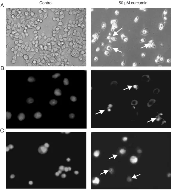
Effect of curcumin on cell morphology of HT-29 cells
To determine curcumin-induced apopto-sis of HT-29 cells, we first examined the changes in cell morphology after exposure to curcumin. Twenty-four hours after expo-sure to 50 µM curcumin, HT-29 cells began to show cell shrinkage, rounding and frag-mentation, thus taking on the typical appear-ance of apoptotic cells when compared to untreated cells (Figure 3A). We also ana-lyzed changes in cell morphology by Hoechst 33258 (Figure 3B) and AO/EB (Figure 3C) staining. The curcumin-treated cells also exhibited morphological changes indicative of apoptosis, including chromatin condensa-tion and nuclear fragmentacondensa-tion.
Effect of curcumin on the serine phosphorylation of p53.
p53 has been shown to be involved in apoptosis induced by a broad range of agents. In order to understand the exact mechanism of curcumin-induced apoptosis in HT-29 cells, we first examined the activation of p53 in the presence of 50 µM curcumin for dif-ferent periods of time or at difdif-ferent curcu-min concentrations for 6 h. Results showed
Figure 1. Effect of curcumin on HT-29 cell viability. Cell viability was determined by the 3-[4,5- dimethylthiazol-2-yl]-2,5-diphen-yltetrazolium bromide assay. Data are reported as the means ± SD of three separate experi-ments.
Figure 2. Curcumin induces DNA fragmentation in HT-29 cells. A, Cells were treated with 50 µM curcumin and the control was treated with medium containing an equivalent amount of DMSO with-out curcumin. At the indicated times, the cells were harvested and lysed. DNA fragmentation was examined by agarose gel electro-phoresis. B, Cells were incubated in medium containing curcumin at
very little or no change of total p53 expres-sion for different exposure times or at differ-ent concdiffer-entrations for 6 h (Figure 4A, B). However, a notable change was observed in p53 phosphorylation. Upon treatment with 50 µM curcumin, the serine phosphorylation level of p53 started to increase as early as after 1 h, and reached a high level at 6 h (Figure 4A). The serine phosphorylation level of p53 also started to increase in a concentra-tion-dependent manner. Upon treatment with 75 µM curcumin, the serine phosphorylation level of p53 reached a maximum level after
6 h as compared to other curcumin concen-trations (Figure 4B).
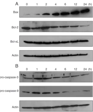
Effect of curcumin on the expression of apoptosis-related proteins
To further understand the mechanism of curcumin-induced apoptosis in HT-29 cells, we also examined the expression of apopto-sis-related proteins. Bcl-xL levels were
mod-erately high in HT-29 cells and remained almost unchanged after curcumin treatment, but Bcl-2 levels decreased after 4 h. On the
other hand, the Bax levels increased signifi-cantly and remained higher after 4 h (Figure 5A). The levels of caspase-3 and pro-caspase-9 zymogen in HT-29 cells treated with 50 µM curcumin were low and de-creased in a time-dependent manner. Simul-taneously, we observed a clear reduction of pro-caspase-9 zymogen expression after 6 h of curcumin treatment (Figure 5B).
Discussion
Curcumin [1,7-bis(4-hydroxy-3-meth-oxyphenyl)-1,6-heptadiene-3,5-dione] is a hydrophobic molecule that passes easily through the plasma membrane into the cyto-sol (19). This phenolic substance inhibits tumor initiation induced by various carcino-gens and has also been shown to inhibit the
growth of many human cancer cell lines in vitro (9). A number of mechanisms of cell proliferation inhibition and apoptosis induc-tion by curcumin have been demonstrated. The present results and other literature re-ports (17,18) show that curcumin strongly reduces the viability of HT-29 colon adeno-carcinoma cells by the induction of apopto-sis. However, the signaling pathways gov-erning apoptosis in mammalian cells are complex and the pro- and anti-apoptotic var-iations regulating cell survival change ac-cording to cell type (20). The cellular and molecular mechanisms underlying curcumin-induced apoptosis in HT-29 cells have not been well defined. Therefore, it is interest-ing to study the mechanism of action of curcumin on HT-29 cells.
In agreement with a previous study (18),
Figure 4. Enhanced serine phosphorylation of p53 in HT-29 cells by treatment with curcumin. A, Cells were incubated in serum-free medium overnight and then further incubated for 0, 1, 2, 4, 6, and 12 h in the presence of 50 µM curcumin. B, Cells were incubated in serum-free medium overnight and then further incu-bated for 6 h in the presence of 0, 10, 25, 50, and 75 µM curcumin, respectively. Equal amounts of cell lysates were resolved by SDS-PAGE and analyzed by Western blot using specific antibodies. The blots were re-probed with anti-actin antibody to confirm equal pro-tein loading.
Figure 5. Expression of the apoptosis-related proteins Bax, Bcl-2, Bcl-xL, pro-caspase-3, and pro-caspase-9 in HT-29 cells by
the present investigation showed that cur-cumin inhibited the growth of HT-29 colon adenocarcinoma cells in a concentration-and time-dependent manner. In our study, however, we observed that a cell growth inhibitory rate of about 80% occurred at the curcumin concentration of 50 µM after 72 h of treatment. In contrast to our results, in a previous report the same cell growth inhibi-tory effect of curcumin was observed at about 75 µM after 72 h of treatment (18). We also observed a typical ladder pattern of internucleosomal fragmentation through DNA fragmentation in HT-29 cells treated with 50 µM curcumin for 48 h. Similar ef-fects of curcumin in inhibiting cell growth have been previously reported for other co-lon cancer lines (21,22).
In a study of the mechanism of action of curcumin on HT-29 cells, Hanif et al. (17) reported that curcumin inhibits the growth of human colon cancer cells independent of COX-2 expression. Goel et al. (18) also reported that curcumin markedly inhibited the mRNA and protein expression of COX-2, but not of COX-1. In the present study, we propose another possible underlying molec-ular mechanism of curcumin-induced HT-29 cell apoptosis via the activation of p53 and the regulation of apoptosis-related pro-teins. Despite its central role in cell sis, the mechanism of p53-mediated apopto-sis after cellular stress remains unclear. Cur-rent evidence indicates that the mode of action of p53-mediated apoptosis involves transactivation of target genes and direct signaling events that are transcription inde-pendent (23). It has been proposed that p53 may induce two sets of genes upon stress signals. One set, such as p21/waf-1 and GADD45, mainly functions in cell growth control, and the other, such as Bax and Bcl-2, acts on apoptosis (24).
In the present study, we observed that p53 was highly expressed in HT-29 cells, but the total p53 protein was almost un-changed. This was in agreement with
previ-ous reports (25,26). However, we found that the serine phosphorylation level of p53 was enhanced prominently when HT-29 cells were treated by curcumin. A recent study implicated that curcumin could impair p53 function required for serine phosphorylation of p53 in colon cancer cells (27). The activa-tion of p53 by curcumin would affect the expression of its downstream effectors, such as the Bcl-2 family proteins. This implies a possible underlying molecular mechanism of curcumin action on HT-29 cells.
It is well recognized that the Bcl-2 family proteins are central regulators of apoptosis and the Bcl-2 family members act like check-points through which survival and death sig-nals pass before they determine the fate of the cell (20). In our study, the high serine phosphorylation level of p53 was shown to be capable of both down-regulating the anti-apoptotic factor, Bcl-2 and up-regulating the pro-apoptotic factor Bax, thereby decreas-ing the Bcl-2/Bax ratio and disposdecreas-ing to apoptosis. Interestingly, curcumin induced apoptosis with an increased serine phospho-rylation level of p53 which transactivates Bax expression. But in these cells, Bcl-xL
levels remained almost unchanged, thereby shifting the Bcl-xL/Bax ratio towards
apop-tosis. The activation of the p53-mediated apoptotic signaling pathway may play an important role in apoptosis by modulating the Bcl-2/Bax or Bcl-xL/Bax ratio, as also
pro-caspase-9 and pro-caspase-3 expression in a time-dependent manner on the HT-29 cells. Our results agree with data reported for other cancer cell lines (30,31).
Taken together, our results show that curcumin induces apoptosis in HT-29 colon adenocarcinoma cells by up-regulating the serine phosphorylation level of p53 and the
level of Bax, while down-regulating the lev-els of Bcl-2, pro-caspase-3, and pro-cas-pase-9. These findings suggest a mechanism of curcumin action on HT-29 cells and should further establish its use as a valid chemopre-ventive and chemotherapeutic agent in co-lon cancer.
References
1. Ammon HPT & Wahl MA (1991). Pharmacology of Curcuma longa. Planta Medica, 57: 1-7.
2. Lin JK & Lin-Shiau SY (2001). Mechanisms of cancer chemopreven-tion by curcumin. Proceedings of the Nachemopreven-tional Science Council, Republic of China. Part B, Life Sciences, 25: 59-66.
3. Joe B, Vijaykumar M & Lokesh BR (2004). Biological properties of curcumin cellular and molecular mechanisms of action. Critical Re-views in Food Science and Nutrition, 44: 97-111.
4. Limtrakul P, Lipigorngoson S, Namwong O et al. (1997). Inhibitory effect of dietary curcumin on skin carcinogenesis in mice. Cancer Letters, 116: 197-203.
5. Singh SV, Hu X, Srivastava SK et al. (1998). Mechanism of inhibition of benzo [a] pyrene-induced forestomach cancer in mice by dietary curcumin. Carcinogenesis, 19: 1357-1360.
6. Kawamori T, Lubet R, Steele VE et al. (1999). Chemopreventive effect of curcumin, a naturally occurring anti-inflammatory agent, during the promotion/progression stages of colon cancer. Cancer Reseach, 59: 597-601.
7. Inano H, Onoda M, Inafuku N et al. (1999). Chemoprevention by curcumin during the promotion stage of tumorigenesis of mammary gland in rats irradiated with gamma rays. Carcinogenesis, 20: 1011-1018.
8. Chuang SE, Kuo ML, Hsu CH et al. (2000). Curcumin-containing diet inhibits diethylnitrosamine-induced murine hepatocarcinogen-esis. Carcinogenesis, 21: 331-335.
9. Aggarwal BB, Kumar A & Bharti AC (2003). Anticancer potential of curcumin: preclinical and clinical studies. Anticancer Reseach, 23: 363-398.
10. Leu TH & Maa MC (2002). The molecular mechanisms for the antitumorigenic effect of curcumin. Current Medicinal Chemistry, 2: 357-370.
11. Hofseth LJ, Hussain SP & Harris CC (2004). p53: 25 years after its discovery. Trends in Pharmacological Sciences, 25: 177-181. 12. Martin DA & Elkon KB (2004). Mechanisms of apoptosis. Rheumatic
Diseases Clinics of North America, 30: 441-454.
13. Gonzalez de Aguilar JL, Gordon JW, Rene F et al. (2000). Alteration of the Bcl-x/Bax ratio in a transgenic mouse model of amyotrophic lateral sclerosis: evidence for the implication of the p53 signaling pathway. Neurobiology of Disease, 7: 406-415.
14. Labianca R, Beretta G, Gatta G et al. (2004). Colon cancer. Critical Reviews in Oncology/Hematology, 51: 145-170.
15. Gustin DM & Brenner DE (2002). Chemoprevention of colon cancer: current status and future prospects. Cancer and Metastasis Re-views, 21: 323-348.
16. Huang MT, Wang ZY, Georgiadis CA et al. (1992). Inhibitory effects of curcumin on tumor initiation by benzo [a] pyrene and 7,12-dimeth-ylbenz [a] anthracene. Carcinogenesis, 13: 2183-2186.
17. Hanif R, Qiao L, Shiff SJ et al. (1997). Curcumin, a natural plant phenolic food additive, inhibits cell proliferation and induces cell cycle changes in colon adenocarcinoma cell lines by a prostaglan-din-independent pathway. Journal of Laboratory and Clinical Medi-cine, 130: 576-584.
18. Goel A, Boland CR & Chauhan DP (2001). Specific inhibition of cyclooxygenase-2 (COX-2) expression by dietary curcumin in HT-29 human colon cancer cells. Cancer Letters, 172: 111-118. 19. Oetari S, Sudibyo M, Commandeur JN et al. (1996). Effects of
curcumin on cytochrome P450 and glutathione S-transferase activi-ties in rat liver. Biochemical Pharmacology, 51: 39-45.
20. Cory S & Adams JM (2002). The Bcl-2 family: regulators of the cellular life-or-death switch. Nature Reviews. Cancer, 2: 647-656. 21. Collett GP & Campbell FC (2004). Curcumin induces c-jun
N-termi-nal kinase-dependent apoptosis in HCT116 human colon cancer cells. Carcinogenesis, 25: 2183-2189.
22. Rashmi R, Kumar S & Karunagaran D (2004). Ectopic expression of Bcl-XL or Ku70 protects human colon cancer cells (SW480) against
curcumin-induced apoptosis while their down-regulation potentiates it. Carcinogenesis, 25: 1867-1877.
23. Haupt S, Berger M, Goldberg Z et al. (2003). Apoptosis - the p53 network. Journal of Cell Science, 116: 4077-4085.
24. Agarwal ML, Taylor WR, Chernov MV et al. (1998). The p53 net-work. Journal of Biological Chemistry, 273: 1-4.
25. Rodrigues NR, Rowan A, Smith ME et al. (1990). p53 mutations in colorectal cancer. Proceedings of the National Academy of Sci-ences, USA, 87: 7555-7559.
26. Van Erk MJ, Teuling E, Staal YC et al. (2004). Time- and dose-dependent effects of curcumin on gene expression in human colon cancer cells. Journal of Carcinogenesis, 3: 8-24.
27. Moos PJ, Edes K, Mullally JE et al. (2004). Curcumin impairs tumor suppressor p53 function in colon cancer cells. Carcinogenesis, 25: 1611-1617.
28. Choudhuri T, Pal S, Agwarwal ML et al. (2002). Curcumin induces apoptosis in human breast cancer cells through p53-dependent Bax induction. FEBS Letters, 512: 334-340.
29. Earnshaw WC, Martins LM & Kaufmann SH (1999). Mammalian caspases: Structure, activation, substrates, and functions during apoptosis. Annual Review of Biochemistry, 68: 383-424.
30. Khar A & Ali AM (1999). Antitumor activity of curcumin is mediated through the induction of apoptosis in AK-5 tumor cells. FEBS Let-ters, 445: 165-168.
![Figure 1. Effect of curcumin on HT-29 cell viability. Cell viability was determined by the 3-[4,5- dimethylthiazol-2-yl]-2,5-diphen-yltetrazolium bromide assay.](https://thumb-eu.123doks.com/thumbv2/123dok_br/15813306.651782/4.918.137.536.642.1073/figure-effect-curcumin-viability-viability-determined-dimethylthiazol-yltetrazolium.webp)