ORIGINAL ARTICLE
From the Nursery Annexed to the Maternity, Pediatric Department and Experimental Research Unit, Children’s Institute, Hospital das Clínicas, Faculty of Medicine, University of São Paulo - São Paulo/SP, Brazil.
Received for publication on August 06, 2002.
CHARACTERIZATION OF NEWBORNS WITH
NONIMMUNE HYDROPS FETALIS ADMITTED TO A
NEONATAL INTENSIVE CARE UNIT
Renata Suman Mascaretti, Mário Cícero Falcão, Andrea M. Silva, Flávio Adolfo Costa Vaz and Cléa Rodrigues Leone
MASCARETTI RS et al. - Characterization of newborns with nonimmune hydrops fetalis admitted to a neonatal intensive care unit. Rev. Hosp. Clín. Fac. Med. S. Paulo 58(3):125-132, 2003.
PURPOSE: To determine the incidence and characteristics of nonimmune hydrops fetalis in the newborn population.
METHOD: A retrospective study of the period between 1996 and 2000, including allnewborns with a prenatal or early neonatal diagnosis of nonimmune hydrops fetalis, based on clinical history, physical examination, and laboratory evaluation. The following were analyzed: prenatal follow-up, delivery type, gender, birth weight, gestational age, presence of perinatal asphyxia, nutritional classification, etiopathic diagnosis, length of hospital stay, mortality, and age at death.
RESULTS: A total of 47 newborns with hydrops fetalis (0.42% of live births), 18 (38.3%) with the immune form and 29 (61.7%) with the nonimmune form, were selected for study. The incidence of nonimmune hydrops fetalis was 1 per 414 neonates. Data was obtained from 21 newborns, with the following characteristics: 19 (90.5%) were suspected from prenatal diagnosis, 18 (85.7%) were born by cesarean delivery, 15 (71.4%) were female, and 10 (47.6%) were asphyxiated. The average weight was 2665.9 g, and the average gestational age was 35 3/7 weeks; 14 (66.6%) were preterm; 18 (85.0 %) appropriate delivery time; and 3 (14.3%) were large for gestational age. The etiopathic diagnosis was determined for 62%, which included cardiovascular (19.0%), infectious (9.5%), placental (4.8%),hematologic (4.7%), genitourinary (4.8%), and tumoral causes (4.8%), and there was a combination of causes in 9.5%. The etiology was classified as idiopathic in 38%.The length of hospital stay was 26.6± 23.6 days, and the mortality rate was 52.4%.
CONCLUSIONS: The establishment of a suitable etiopathic diagnosis associated with prenatal detection of nonimmune hydrops fetalis can be an important step in reducing the neonatal mortality rate from this condition.
DESCRIPTORS: Hydrops fetalis. Nonimmune hydrops fetalis. Newborn infant. Anasarca. Hydropic.
Hydrops fetalis is defined as the ex-cessive accumulation of fluids in the
interstitial compartment1 including
edema, ascites, and pleural and pericar-dialeffusions2, leading toanasarca.
One of the methods used to clas-sify hydrops fetalis is to categorize it according to immune and nonimmune
causes3, because nowadays, as a
con-sequence of a more precocious detec-tion and a higher control of the im-mune causes, 75% of the perinatal cases are of nonimmune etiology4. The
main pathophysiologicmechanism
in-volved in the genesisof nonimmune
hydrops fetalis (NHF) is related to ab-normal fluid transportation between plasmaand tissues. In this
pathologi-cal situation, the primary causes of
modification of the distribution of body fluids are the increase of
hydro-static capillary pressure and capillary permeability and reduction of plasma osmotic pressure or lymphatic flow2,4,5.
Regarding etiology, NHF is differ-ently classified in the literature, with-out consensus3,4,6,7. We have developed
a classification system, presented on table 1, in which the main causes are classified as cardiovascular, genetic,
infectious, placental, hematologic,
miscellaneousand/or multiple causes,
and idiopathic.
Table 1 - Causes of nonimmune hydrops fetalis.
CARDIOVASCULAR • Arrhythmia • Myocardiopathy
• Structural malformations (Ebstein anomaly, premature closure of the foramen ovale) • Vascular obstruction (tumor, structural, fibroelastosis)
• Vascular malformation and hemangioma GENETIC
• Skeletal dysplasias and myopathies
• Metabolic diseases (Gaucher, GM1 gangliosidosis, mucopolysaccharidosis) • Autosomic diseases (Nooan, Prune belly, Fanconi)
• Chromosomal abnormalities (trisomy 21, 18, 13, Turner’s syndrome) CONGENITAL INFECTIONS
• Virus (cytomegalovirus, parvovirus B19, rubella, varicella, herpes, sintitial respiratory) • Toxoplasmosis
• Syphilis • Chagas disease HEMATOLOGIC
• Nonimmune anemia • Alpha-thalassemia • Others (leukemia) PLACENTAL
• Twin-twin transfusion syndrome • Causes related to the umbilical cord MISCELLANEOUS
• Respiratory (pulmonary sequestration, adenomatoid disease, chylothorax, tumor) • Genitourinary (obstructive uropathy, dysplasia, cysts, thrombosis, nephrotic syndrome) • Gastrointestinal (duodenal/jejunal atresia, anal imperforation, peritonitis)
• Neurological (encephalocele, intracranial hemorrhage, cerebral aneurysm) • Tumoral (sacrococcygeal teratoma, neuroblastoma, hepatoblastoma) • Multiple causes (presence of more than one associated etiopathic causes) IDIOPATHIC
• Non-defined cause
Modified from the classification of Phibbs R, 1996.2
infectious, placental, and hematologic causes, among others (Table 1). De-spite the technological advances in the last decades, the cause in 50% of the cases remains undefined, which are classified as idiopathic2.
The occurrence of NHF is not fre-quent, occurring in 1 of 3000 live births2,8,9. Moreover, a high mortality
rate is associated with NHF; 50% of the cases diagnosed in the intrauterine pe-riod evolve to death, and 50% of the live newborns who have the disease do not survive the neonatal period2.
As previously mentioned, the prog-nosis of the newborn with NHF de-pends directly on an etiopathic diag-nosis and on the prevention of prema-turity10. In order to have an effective
reduction in morbidity and mortality rates, a correct and precocious
etio-pathic diagnosis becomes
fundamen-tal, so that an adequate perinatal ap-proach is possible.
This study aims at determining the incidence and characterization of NHF in newborns admitted to a neonatal risk unit during a 5-year period.
PATIENTS AND METHODS
A retrospective study was per-formed from January 1996 to Decem-ber 2000.
Every newborn that was born dur-ing this period and had an immediate prenatal or neonatal diagnosis of hy-drops was included in the study. Hy-drops was characterized by 1 or more clinical signs, such as anasarca, periph-eral edema, ascites, pericardial and/or pleural effusions, anemia, congestive heart failure, and hypoalbuminemia.
From these newborns, the ones with an immune etiology were excluded, based on clinical history, laboratory
evaluation (blood type and Coombs
tests of the newborn and the mother), and clinical evolution.
The presence of the clinical pres-entation and the absence of maternal-fetal blood incompatibility defined NHF.
Among the newborns that fulfilled the inclusion criteria, the following fac-tors were evaluated: occurrence of pre-natal diagnosis of hydrops, gender, type of delivery, gestational age, birth weight, nutritional adequacy of birth weight, presence of perinatal asphyxia, etiopathic diagnosis, mortality, age at death, and length of hospital stay.
The gestational age (GA) was based on the last menstruation date and ultrasonographic and postnatal
classi-fication methods (Capurro11,
Dubowitz12, or New Ballard Score13).
The newborns with a GA greater than or equal to 37 weeks were considered term. For the calculation of the defini-tive GA, the following criteria were used in descending order of priority, according to the service routine: - maternal information about the
date of the last menstrual period (Naegele’s rule, that considers 280 days as the normal gestation time) when this date differs less than 2 weeks from the early fetal ultra-sonography;
- early ultrasonography performed during the first 20 weeks of gesta-tion, in cases when the GA was not considered reliable and the differ-ence among the calculated ages by the ultrasonography and postnatal methods was less than 2 weeks; - calculated postnatal age through
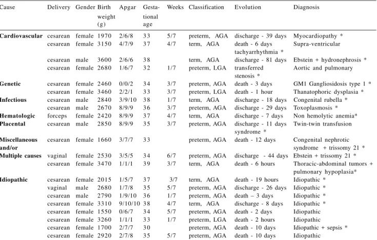
Table 2 - Characterization and etiopathic diagnosis of the study population.
Cause Delivery Gender Birth Apgar Gesta- Weeks Classification Evolution Diagnosis weight tional
(g) age
Cardiovascular cesarean female 1970 2/6/8 3 3 5/7 preterm, AGA discharge - 39 days Myocardiopathy * cesarean female 3150 4/7/9 3 7 4/7 term, AGA death - 6 days Supra-ventricular
tachyarrhythmia *
cesarean male 3600 2/6/6 3 8 term, AGA discharge - 81 days Ebstein + hydronephrosis * cesarean female 2680 1/6/7 3 2 1/7 preterm, LGA transferred Aortic and pulmonary
stenosis *
Genetic cesarean female 2460 0/0/2 3 4 3/7 preterm, AGA death - 3 days GM1 Gangliosidosis type 1 * cesarean female 3460 2/2/1 3 3 3/7 preterm, LGA death - 1 hour Thanatophoric dysplasia * Infectious cesarean male 2840 3/9/10 3 8 1/7 term, AGA discharge - 18 days Congenital rubella *
cesarean male 2670 8/9/9 3 6 3/7 preterm, AGA discharge - 29 days Toxoplasmosis * Hematologic forceps female 2420 8/9/9 3 7 4/7 term, AGA discharge - 7 days Non hemolytic anemia* Placental cesarean male 2850 8/9/9 3 5 3/7 preterm, AGA discharge - 11 days Twin-twin transfusion
syndrome *
Miscellaneous cesarean female 1660 3/7/7 3 3 preterm, AGA death - 12 days Congenital nephrotic
and/or syndrome + trissomy 21 *
Multiple causes vaginal female 2530 3/5/5 3 4 6/7 preterm, AGA discharge - 44 days Ebstein + trissomy 21 * cesarean female 3470 1/1/1 3 9 3/7 term, AGA death - 6 hours Thoracic-abdominal tumors +
pulmonary hypoplasia* Idiopathic cesarean female 2015 1/5/7 3 7 3/7 term, AGA death - 19 hours Idiopathic *
vaginal male 2680 1/7/8 3 5 5/7 preterm, AGA discharge - 26 days Idiopathic * cesarean male 2790 1/9/10 3 6 1/7 preterm, AGA death – 3 days Idiopathic * cesarean female 3310 9/10/10 3 8 4/7 term, AGA discharge - 8 days Idiopathic * cesarean female 1550 0/6/7 3 4 5/7 preterm, AGA death - 2 days Idiopathic cesarean female 3260 1/1/1 3 3 1/7 preterm, LGA death - 2 hours Idiopathic
cesarean female 1700 2/7/7 3 0 preterm, AGA death - 10 days Idiopathic + sepsis * cesarean female 2920 2/7/8 3 5 5/7 preterm, AGA death - 10 days Idiopathic
* Antenatal diagnosis for nonimmune hydrops fetalis, AGA = adequate for gestational age, LGA = large for gestational age, Apgar-1st, 2nd and 5th minute of life.
Ramos’ intrauterine growth curve14,
using the 10th and 90th percentiles as limits, was used to assess the nutri-tional adequacy of the birth weight to GA. The newborn with an inferior
birth weight (under the 10th
percen-tile), was classified as small for GA (SGA), the newborn with birth weight between the percentiles 10 and 90 was considered adequate for GA (AGA), and the ones with a birth weight higher than the 90th percentile of the reference
curve were considered large for GA (LGA).
An Apgar score of less than or equal to 6 in the 5th minute of life
character-ized the presence of perinatal asphyxia. Regarding the etiopathic diagno-sis, the cases of NHF were classified in the following categories: cardiovascu-lar, infectious, genetic, hematological,
placental, miscellaneous and/or asso-ciated causes, and idiopathic.
RESULTS
During the period of study, 11,190 live births occurred in the service. In this population, the diagnosis of hy-drops fetalis was made in 47 newborns (0.42% of the live births); 18 (38.3%) of whom presented an immune form and 29 (61.7%) with a nonimmune form, with an incidence of nonimmune hydrops fetalis (NHF) of 1 per 414 live births during this period. The data ob-tained was derived from 21 newborns, of whom 19 (90.5%) had been diag-nosed prenatally (Table 2).
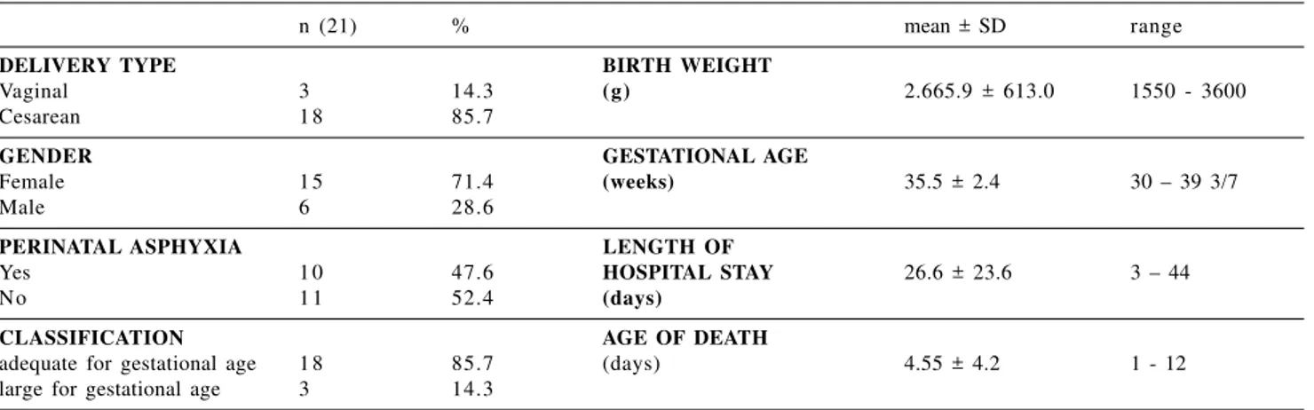
Regarding the characteristics of the newborns (Table 3), 18 (85.7%) were de-livered by cesarean, 15 (71.4%) were
fe-male, and 10 (47.6%) presented perina-tal asphyxia. The average birth weight was 2665.9 g ± 613 (1550 – 3600 g), and the average GA was 35.5 weeks ± 2.4 (30 – 39 3/7 weeks), with 14 of these (66.6%) being born preterm. Regarding the nutritional classification, 18 (85.7%) were adequate and 3 (14.3%) were large for GA. The average length of hospital stay was 26 ± 23.6 days, the mortality rate was 52.4% (11 newborns), and the age at death was 4.55 ± 4.2 days of life.
Table 4 - Data from the review of the literature regarding the etiopathic classification of nonimmune hydrops fetalis.
Causes for POESCHAMANN et al# PROENÇA et al#
Nonimmune hydrops fetalis modified 21 cases
529 cases
Cardiovascular n=120 (23%) n=4 (19%)
· malformations 50 (42%) 2 (50%)
· arrhythmia 37 (31%) 1 (25%)
· other 33 (27%) 1 (25%)
Genetics n=70 (14%) n=2 (9.5%)
· trisomy 21 16 (23%)
-· Turner’s syndrome 18 (26%)
-· skeletal 20 (28%) 1 (50%)
· other 18 (25%) 1 (50%)
Infectious n=17 (3%) n=2 (9.5%)
· CMV 7 (41%)
-· toxoplasmosis 3 (18%) 1 (50%)
· other 7 (41%) 1 (50%)
Placental n=38 (7%) n=1 (4.76%)
· twin-twin transfusion syndrome 32 (84%) 1 (100%)
· other 6 (16%)
-Hematologic n=24(4.5%) n=1 (4.76%)
· alpha-thalassemia 12 (50%) 1 (100%)
· nonhemolytic anemia 11 (46%)
-· other 1 (4%)
-Miscellaneous n=107 (20.5%) n=3 (14.3%)
· respiratory 30 (28%)
-· gastrointestinal 26 (24%)
-· genitourinary 22 (21%)
-· other 30 (28%) 3 (100%)
Idiopathic n=150 (28%) n=8 (38%)
#Adapted from the work of Poeschamann et al.4, using literature data gathered by the author between 1967-1987.
Table 3 - Characterization of the newborn infants with nonimmune hydrops fetalis.
n (21) % mean ± SD range
DELIVERY TYPE BIRTH WEIGHT
Vaginal 3 14.3 (g) 2.665.9 ± 613.0 1550 - 3600
Cesarean 1 8 85.7
GENDER GESTATIONAL AGE
Female 1 5 71.4 (weeks) 35.5 ± 2.4 30 – 39 3/7
Male 6 28.6
PERINATAL ASPHYXIA LENGTH OF
Yes 1 0 47.6 HOSPITAL STAY 26.6 ± 23.6 3 – 44
No 1 1 52.4 (days)
CLASSIFICATION AGE OF DEATH
adequate for gestational age 1 8 85.7 (days) 4.55 ± 4.2 1 - 12
large for gestational age 3 14.3
type until 1943, when Potter described 17 hydrops fetalis cases without asso-ciated isoimmunization4. The most
fre-quently reported etiology of hydrops fetalis until the 1960s was the isoimmunization by the Rh factor1.
Nowadays, the prenatal treatment of immune hydrops is so efficient that in many situations it can revert the dis-ease course through prophylaxis with
anti-D immunoglobulin and the
through intrauterine transfusion. Therefore, NHF appears to be the most important cause of perinatal hydrops fetalis, being responsible for 75% of the cases15. In this study, the immune
etiology occurred in 38% of the newborns, which shows that these rates in our environment still remain high, probably because ours is a reference service for hemolytic disease related to the Rh system, as well as because of a deficiency in prenatal monitoring of those pregnancies at risk for develop-ing immune hydrops.
The reported incidence of NHF is relatively low compared with this study, at approximately 1 per 3000 live births2. In this study, 1 case of
NHF was observed for every 414 live births, a frequency 7.6 times higher than in the general population. The explanation of this fact is quite sim-ple: since this medical center is a
ter-tiary and quaternaryreference in the
DISCUSSION
The first report concerning hy-drops fetalis was made by Diamond in
1932, who described a newborn with erythroblastosis fetalis in a terminal
stage of generalized edema2. The
health system for risk pregnancy, it therefore selects for a larger number of hydropic fetuses.
As previously mentioned, strict control during the prenatal period of pregnancies involving hydropic fe-tuses and the precise indication of the type of delivery required to improve the fetal prognosis led to a high fre-quency of cesarean deliveries (85.7%) and prematurity (67%).
Prenatal diagnosis of NHF was ac-complished in more than 90% of the cases, and the etiology was determined in 62%. The better prognosis and a
lower morbidity and mortalityare
re-lated to these facts because follow-up of the pregnancy in a specialized center permits the creation of better delivery conditions.
The analysis of the study popula-tion showed that the average birth weight was 2.665 ± 613 g, and regard-ing the nutritional classification, 18 (85.6%) of the newborns were classi-fied as adequate for GA, 3 (14.3%) as large for GA, and none as small for GA. The presence of fluids in the intersti-tial space results in newborns with a weight higher than expected for GA because of the increase in the total body fluid, therefore explaining the higher frequency of adequate and large newborns for GA.
This fact deserves to be emphasized because it is very difficult to evaluate nutritional status of hydropic newborns. The anthropometric param-eters (weight, length, head and arm cir-cumference, and skinfold thickness) widely used in the neonatal period must be avoided in those children. Ad-ditionally, biochemical parameters such as the visceral protein determina-tion do not apply either. The use of the fetal growth curve (birth weight x GA) will tend to result in classifications that are at a higher level than the ac-tual nutritional status, as previously mentioned16,17.
Imaging methods, such as
ultra-sonography and sectional computer-ized tomography of the arm, could have some utility in the nutritional evaluation, since they can be used to estimate the mass. Nevertheless, these methods are not yet well defined for neonatals18.
Regarding perinatal asphyxia, the frequency found in this population of 52% reinforces the hypothesis that the presence of hydrops is a relevant fac-tor in the worsening of the fetal wel-fare because, in addition to the unfavorable condition during the in-trauterine life, the generalized edema causes a more difficult delivery.
The predominance of females (71.4%) occurring in our study is a find-ing that is not reported in the literature, because those studies did not focus on gender differences in the incidence. However, some of the etiologies of hy-drops are more prevalent in females.
When we compared our etiopathic diagnosis data with that in the literature (Table 4), we found that the main causes of NHF were variable in every studied population. This variation is due to a variety of factors; the two primary ones are 1) the influence of genetics, where an example is the higher frequency of cases due to hematologic causes in southeastern Asian populations where there are higher numbers of alpha-thalassemia carriers5,19; and 2) the
vari-ation in the occurrence of infectious diseases, where an example is the in-crease in the diagnosis of hydrops that is secondary to infection by parvovirus B195,20,21 in the last decade, due to a
real increase in the number of cases or an improvement in the diagnosis of this viral disease.
The primary cause of NHF in this study arose from cardiovascular origin (19%), which is comparable to other studies in which cardiovascular dis-eases appear in 15% to 27% of the hy-dropic newborns, mainly due to ar-rhythmia and structural defects9,23.
Genetic abnormalities, primarily
the chromosomopathies including tri-somy 21 and 18 and Turner syn-drome24,25, have been associated with
hydropic fetuses in which this chromosomopathy can be the only cause of edema or it can be associated with other pathologies. In this study, genetic causes were responsible for 9.5% of the cases, but when evaluated for association with other causes, an increase of the incidence to 19% was observed. This increased incidence probably occurs because of the com-bination of the different pathophysi-ologic mechanisms, causing a fetus with a chromosomal abnormality evolve to hydrops.
The percentage of the idiopathic cases varies in the literature, depending primarily on the diagnostic methods available in each service. In this study, the etiopathic diagnosis of NHF was not achieved in 8 (38%) newborns.
The reported mortality rate for fetal hydrops is very high, around 50% to 100%10, depending on the etiology22. In
this study, 11 (52.4%) evolved to death, showing that the mortality rate, al-though high, was lower than in other major studies. Moreover, the deaths oc-curred primarily in the first week of life (average of 4.55 ± 4.2 days), arising out of complications associated with hypervolemia, such as cardiac, renal, and respiratory insufficiencies.
It has been reported that just 20% to 25% of the newborns with idi-opathic NHF survive in the neonatal period2. This study’s findings concur,
The newborns that survived re-mained hospitalized for a long period (average of 26.6 ± 23.6 days), either because of the need for a prolonged treatment or the need to control the etiopathic causes and the secondary complications of prematurity and hy-drops.
In summary, when confronted with the possibility of hydrops fetalis dur-ing the gestation, all possible effort should be made to achieve an etiopathic diagnosis. First it must be determined whether the hydrops are immune or nonimmune so treatment may be initiated. The establishment of
a correct etiopathic diagnosis associ-ated with precocious nonimmune hy-drops is fundamental to an adequate selection of the prenatal and neonatal therapeutic approach, reducing as a consequence the morbidity and mor-tality risks associated with this serious dysfunction.
RESUMO
MASCARETTI RS e col. – Caracteri-zação dos recém-nascidos com hidropisia fetal não imune admiti-dos em uma unidade neonatal de terapia intensiva. Rev. Hosp. Clín. Fac. Med. S. Paulo 58(3):125-132, 2003.
OBJETIVOS: Determinar a inci-dência e caracterizar a população de recém-nascidos com hidropisia fetal não imune.
MÉTODO: Estudo retrospectivo, referente ao período de 1996 a 2000, incluindo todos os recém-nascidos com diagnóstico antenatal ou neo-natal, com base na história clínica,
exame físico e avaliação laboratorial. Foram analisados: seguimento pré-na-tal, tipo de parto, sexo, peso de nasci-mento, idade gestacional, presença de asfixia perinatal, classificação nutri-cional, diagnóstico etiopatogênico, tempo de internação, mortalidade, ida-de do óbito.
RESULTADOS: Foram seleciona-dos 47 recém-nasciseleciona-dos com hidropisia fetal (0,42% dos nascidos vivos), 18 (38,3%) com a forma imune e 29(61,7%) com a não imune. A incidên-cia de hidropisia fetal não imune foi de 1:414 nascidos vivos. Obtiveram-se da-dos de 21 recém-nascida-dos destes, 19 (90,5%) apresentavam suspeita
a mortalidade de 52,4%.
CONCLUSÕES: O estabelecimen-to de um correestabelecimen-to diagnóstico etiopaestabelecimen-to- etiopato-gênico, associado à detecção antenatal
10. WY CA, SAJOUS CH, LOBERIZA F et al. - Outcome of infants with a diagnosis of hydrops fetalis in the 1990s. Am J Perinatol 1999; 16(10): 561-7.
11. CAPURRO H et al. - A simplified method for diagnosis of gestational age in the newborn infants. J Pediatric 1978, 93:120-2.
12. DUBOWITZ IM, DUBOWITZ V, GOLDBERG C - Clinical assessment of gestational age in the newborn infants. J Pediatric 1970, 77:1-10.
13. BALLARD JL, KHOURY IC, WEDIG K et al. - New Ballard Score, expanded to include extremely premature infants. J Pediatric 1991; 19:417-23.
14. RAMOS, JLA - Avaliação do crescimento intra-uterino por medidas antropométricas do recém-nascido. São Paulo, 1983. (Tese Doutorado – Faculdade de Medicina, Universidade de São Paulo).
15. MCMAHAN MJ, DONOVAN EF - The delivery room resuscitation of the hydropic neonate. Semin Perinatol 1995; 19(6): 474-82. 16. FALCÃO MC, CARDOSO LEMB - Avaliação nutricional do
recém-nascido pré-termo. Rev Bras Nutri Clin 2001; 16:144-7. 17. FALCÃO MC - Avaliação nutricional do recém-nascido. Pediatria
(São Paulo) 2000; 22:235-9.
18. HADLOCK FP et al. - Estimated age: computer assisted analysis of multiple fetal growth parameters. Radiology 1984; 152:497-501.
1. APKON M - Pathophysiology of hydrops fetalis. Semin Perinatol 1995;19(6):437-46.
2. PHIBBS R - Hydrops fetalis. In: SPITZER AR - Intensive care of the fetus and neonate. eds. St Louis, Mosby-Year Book, 1996. p. 149.
3. SANTOLAYA J, ALLEY D, JAFFE R et al. - Antenatal classification hydrops fetalis. Obstet Gynecol 1992; 79(2): 256-9.
4. POESCHMANN RP, VERHEIJEN RH, VAN DONGEN WJ -Differential diagnosis and causes of nonimmunological hydrops fetalis: a review. Obst and Gynecol Survey 1991; 46(4): 223-231.
5. YANG YH, TENG RJ, TANG JR et al. - Etiology and outcome of hydrops fetalis. J Formos Med Assoc 1998; 97(1):16-20. 6. JONES DC - Nonimmune fetal hydrops: diagnosis and obstetrical
management. Semin Perinatol 1995; 19(6):447-61. 7. STEPHENSON T, ZUCCOLLO J, MOHAJER M - Diagnosis and
management of non-immune hydrops in the newborn. Arch Dis Child 1994; 70:F151-4.
8. BULLARD KM, HARRISON MR - Before the horse is out of the barn: fetal surgery for hydrops. Semin Perinatol 1995; 19(6): 462-73.
9. SAMUELS P, LUDMIR J - Nonimmune hydrops fetalis: a heterogeneous disorder and therapeutic challenge. Semin Roentgenol 1990; 25(4): 353-60.
da hidropisia fetal não imune, consti-tui elemento importante para uma re-dução da mortalidade neonatal decor-rente desta grave doença.
DESCRITORES: Hidropisia fetal. Hidropisia fetal não imune. Recém-nascido. Anasarca. Hidrópico.
19. ARCASOY MO, GALLAGHER PG - Hematologic disorders and nonimmune hydrops fetalis. Semin Perinatol 1995; 19(6): 502-515.
20. BARRON SD, PASS RF - Infectious causes of hydrops fetalis. Semin Perinatol 1995; 19(6): 493-501.
21. YAEGASHI N, OKAMURA K, YAJIMA A et al. - The frequency of human parvovirus B19 infection in nonimmune hydrops fetalis. J Perinat Med 1994; 22(2) 159-63.
22. PAL A, GEMBRUCH U, BALD R et al. - The diagnosis and treatment of the nonimmune hydrops fetalis. Acta Paediatr Hung 1991; 31(2): 169-86.
23. KNILANS TK - Cardiac abnormalities associated with hydrops fetalis. Semin Perinatol 1995; 19(6): 483-92.
24. JAUNIAUX E, VAN MALDERGEM L, DE MUNTER C et al. -Nonimmune hydrops fetalis associated with genetic abnormalities. Obstet Gynecol 1990; 753: 568-72. 25. STEINER RD - Hydrops fetalis: Role of the geneticist. Semin
Perinatol 1995; 19(6): 516-24.
26. ISMAIL KM, MARTIN WL, GHOSH S et al. - Etiology and outcome of hydrops fetalis. J Matern Fetal Med 2001; 10(3): 175-81.